In the medical literature there are various definitions of lipoatrophy syndrome, or lipodystrophy. This term combines heterogeneous acquired or congenital pathological conditions, common to which are abnormal changes in adipose tissue in combination with laboratory signs of disorders, mainly lipid metabolism, and in some forms without laboratory symptoms.
…
General concept of pathology
Pathological conditions of the body and the development of subcutaneous fat can be expressed in the following forms:
- Atrophic, which consists of loss of subcutaneous tissue volume in selective areas of the body. In this case, destruction of muscle tissue with a decrease in its mass, in contrast to ordinary dystrophic changes in the body, does not occur. Lipodystrophy of the face, limbs and buttocks is especially pronounced. Facial lipoatrophy occurs in many older people, but, unlike a pathological condition, it is a cosmetic drawback, since it is associated with age-related tissue degradation, including adipose tissue.
- Hypertrophic, the meaning of which is the excessive accumulation of adipose tissue in such parts of the body as the abdomen, mammary glands, back of the neck and upper back, sometimes muscles and liver.
- Combined - depletion of adipose tissue in some areas of the body and its accumulation in others.
These clinical signs often have much in common with complex metabolic disorders, for example, with resistance to the influence of insulin in the liver and peripheral tissues, impaired resistance of the latter to glucose with the possible development of type 2 diabetes mellitus, with an increase in the blood levels of triglycerides, cholesterol with a predominance of an increase in low lipoproteins and very low density. Sometimes deviations from normal laboratory parameters precede clinical symptoms. At the same time, metabolic changes reach their greatest severity among patients who also have a pronounced clinical picture.
Of course, metabolic syndrome is combined with visceral weight gain, but, although this is paradoxical, loss of fat volume (lipodystrophy) can also be accompanied by metabolic disorders. They are similar to those in metabolic syndrome, that is, they are accompanied by an increase in the level of free fatty acids and liver synthesis of very low-density lipoproteins, accumulation of fat in muscles, fatty liver, tissue insulin resistance, increased risk of cardiovascular pathology, etc.
Manifestations of lipoatrophy in diabetic patients
Treatment
Diet is a necessary basis for the treatment and prevention of metabolic complications in lipodystrophies. Metreleptin therapy is effective in treating metabolic complications in patients with generalized lipodystrophy and low leptin levels, as well as in some patients with familial partial lipodystrophy. Other drugs can also be used for the symptomatic treatment of metabolic complications, such as metformin for the treatment of diabetes, statins and fibrates for the treatment of dyslipidemia. Taking oral estrogen preparations is contraindicated [2].
Manifestations of lipodystrophy and causes of development
In accordance with the type of distribution of changes in adipose tissue in the body, lipodystrophies are divided into partial (segmental), generalized, limited (local), and depending on the reasons, which are not always possible to establish, they are divided into:
- Purchased
- Hereditary
Each type of pathological condition is characterized by a specific development mechanism and clinical picture.
Acquired forms of the disease
Partial lipodystrophy in people infected with human immunodeficiency virus (HIV)
It is currently the most common type of pathology under consideration. The incidence of the latter among these patients is 10-80%. This “dispersion” of frequency indicators is associated with the age of patients, the state of the body preceding the disease, the level of immunosuppression, and the type and duration of treatment with antiretroviral drugs. All of them have the property of causing certain phenomena of lipodystrophy, but their nature and severity are different.
Facial lipodystrophy in an HIV patient
For example, atrophy of adipose tissue during the treatment of HIV-infected patients is explained mainly by the influence of Stavudine, Zinovudine and some other drugs that are nucleoside analogues of chemical compounds that inhibit the enzyme revertase. Disorders of lipid metabolism are possible as a result of taking almost any of the antiretroviral drugs, but still more often in individuals who receive therapeutic regimens that include protease inhibitors. The latter contribute to the development of insulin resistance and, accordingly, an increased risk of developing diabetes, which, in turn, aggravates metabolic disorders. However, the specific mechanisms that contribute to the development of lipoatrophy during treatment with drugs that are protease inhibitors have not been elucidated.
HIV-infected individuals very often develop lipoatrophy of the face, upper and lower extremities. These changes can sometimes be combined with excess deposition of body fat (in the back of the neck and upper back), its redistribution against the background of general body weight loss, impaired liver function, lactic acidosis and ascites (fluid in the abdominal cavity). But at the same time, excessive concentrations of insulin and lipids in the blood are quite rare.
Correction of facial volume in an HIV patient by administering calcium hydroxyapatite (Radiesse)
Barraquer-Simons syndrome
Or segmental, progressive lipodystrophy. In terms of frequency, it ranks second among acquired partial forms of this pathology. It occurs in a complete or incomplete form (only in the upper half of the body). Women get sick 4 times more often. The incomplete form occurs mainly in men.
The disease develops in childhood or adolescence. It is characterized by the disappearance of subcutaneous fatty tissue mainly in the face, neck, upper chest, upper extremities, and pit of the stomach. At the same time, it is possible, especially in women, to increase the volume of adipose tissue in the lower parts of the anterior abdominal wall, lumbar region, buttocks and thighs. Tissue resistance to insulin and associated complications are extremely rare. Approximately 8 years from the onset of the disease, 20% of patients develop membranoproliferative glomerulonephritis.
The cause of the pathology has not been established. The possibility of dysfunction of the hypothalamus of the brain, thyroid gland, etc. is assumed. But preference is given to autoimmune etiology. Often, patients with this pathology also develop autoimmune diseases such as juvenile dermatomyositis and systemic lupus erythematosus.
Acquired generalized form
Or Lawrence syndrome. It is rare (only 80 cases have been described, of which the male gender is about 33%). Loss of subcutaneous fat, as in the previous case, occurs in childhood or adolescence, less often in middle age. This occurs in large areas, mainly of the face and limbs, less often in the area of the palms and feet and, as a rule, bone marrow adipose tissue and retro-orbital adipose tissue are preserved. The amount of intraperitoneal fat loss varies. Most patients develop acanthosis nigricans, fatty liver, and 20% develop cirrhosis from childhood.
The cause of the acquired generalized form of lipodystrophy in 25% of patients is considered to be a rare and poorly understood disease, panniculitis, which occurs in various forms, which is a progressive inflammatory lesion of subcutaneous fatty tissue in the form of inflammatory nodules, in another 25% - autoimmune diseases, mainly juvenile dermatomyositis, in the remaining 50% - idiopathic form. In patients with lipoatrophy, which is caused by panniculitis, compared with other patients with the acquired generalized form, there is less severity of loss of adipose tissue, a lower incidence of type 2 diabetes mellitus and hypertriglyceridemia.
Limited forms of lipodystrophy
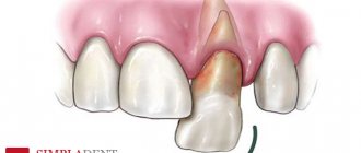
They are characterized by loss or, conversely, accumulation (in the form of lipomas) of adipose tissue, mainly in small areas. Much less often, large areas of the torso, upper or lower extremities are involved in the process. Pathological changes, as a rule, develop at the sites of injection of insulin (lipodystrophy in diabetes) or corticosteroid drugs, after prolonged mechanical compression, panniculitis, and sometimes for no apparent reason. Limited lipodystrophy is most often associated with injection treatments for diabetes.
Limited accumulation of adipose tissue or, conversely, lipoatrophy after insulin injections is a specific complication of therapy for insulin-dependent diabetes mellitus. It used to happen quite often. However, highly purified biosynthetic human insulins are now widely used for the treatment of diabetes. In this regard, cases of lipoatrophy have become a very rare complication, but the formation of lipomas, that is, the accumulation of adipose tissue at the sites of insulin injections, is a common complication among patients with insulin-dependent diabetes, even with a short period of illness. They complicate the absorption of administered insulin into the blood and its metabolism, which complicates the dosing of the latter and increases the need for the drug, complicating the possibility of compensating for diabetes.
Areas of lipodystrophy formed due to insulin administration
It is assumed that the cause is incorrect insulin injection technique in the form of a violation of the pattern of alternating quadrants or injection sites, failure to maintain distances (at least 10 mm) between injections to avoid repeated tissue injuries, repeated use of disposable syringes, damage to blood vessels and nerve endings. Sometimes the cause cannot be determined.
Gynoid lipodystrophy
Or cellulite, which WHO regards not as a disease, but as natural age-related changes in the tissues of certain areas of the body, mainly in women. These changes of varying severity occur on average in 90% of middle-aged women.
Tissue changes in the gynoid form occur in 4 stages. The development mechanism is the formation of high-density collagen fibers in the skin, which form septa (septa). The latter disrupt microcirculation in small blood and lymphatic vessels, which over time leads to stagnation of fluid and swelling of soft tissues.
The most pronounced changes are observed in the third stage, which is determined by the pronounced development of coarse collagen fibers and excessive uneven growth of adipose tissue, significant disruption of the skin relief, formation of nodes, etc. At the last stages, significant tissue pain appears on palpation, infectious and inflammatory processes are possible, tissue necrosis. Before this stage, cellulite develops very rarely.
Gynoid lipodystrophy
LIPOHYPERTROPHY
Lipohypertrophy is a thickened, rubbery lesion of the subcutaneous tissue that occurs in up to half of patients using insulin. In some patients, the lesion may be firm or scar-like. (Fig. 2 Example of lipohypertrophy in the abdominal area).
Detection of lipohypertrophy requires both inspection and palpation of injection sites, as many lesions can be detected by touch rather than visually. Normal skin can be pinched tightly, but with lipohypertrophic lesions it cannot be pinched. Lipohypertrophy occurs when used as (with repeated insertions of a catheter into the same place). Published data support an association between the occurrence of lipohypertrophy and the use of older, less pure insulin solutions, lack of rotation of injection sites, use of a small area of skin for injection, repeated injection of the drug into the same area, and repeated use of needles. Injections into the area of lipohypertrophy can increase its severity. Patients should not inject into areas of lipohypertrophy as insulin absorption may be slow or uneven, potentially worsening diabetes control.
For the prevention and treatment of lipodystrophy it is necessary:
1. Regularly independently inspect the insulin injection sites.
2. Show the injection sites to your doctor or nurse at least once a year (and preferably at every visit).
3. Avoid injections into hypertrophied areas until the pathologically altered tissues become normal again (this can take from several months to several years).
4. Change injection areas. Remember! When moving from a lipodystrophic area to normal tissues, adjustment of the dose of insulin administered is usually required. The extent of dose changes varies and varies from person to person, so it should be based on frequent blood glucose testing and consultation with your doctor.
5. Today, the optimal therapeutic approach for lipodystrophy includes: the use of high-quality insulin preparations, constant rotation of injection areas, expansion of injection zones and refusal to reuse needles.
Hereditary lipodystrophies
Hereditary lipodystrophies are much less common than acquired lipodystrophies. Of these, the most common forms are:
- congenital generalized form, or Berardinelli-Seip syndrome, which is a rare autosomal recessive disease with a prevalence of 1:10 million people. The syndrome is characterized mainly by an almost complete absence of subcutaneous fat with a well-defined muscle contour already at birth, accelerated growth, increased appetite, and later - acanthosis nigricans in the cervical region, on the trunk and in areas of skin friction; in addition, enlargement of the liver and spleen, acromegaly are often observed;
- familial partial lipodystrophy caused by point mutations in the PPAR-gamma gene - several cases have been described;
- lipodystrophy, which is associated with mandibular (mandibular) dysplasia, is an extremely rare pathology;
- some others.