- home
- Lipofilling
- Facial lipofilling in a clinic in Moscow
Facial lipofilling is a popular procedure for correcting appearance and rejuvenation. The uniqueness of lipofilling is that it is a volumetric method. The transplant replenishes age-related volume loss in certain areas, providing aesthetic correction of appearance and rejuvenation of surrounding tissues due to the high content of growth factors in adipocytes. The essence of the procedure is to transplant the patient’s fat tissue into the areas requiring correction.
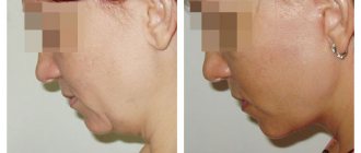
The patient underwent complete facial lipofilling, with correction of the area of the lower eyelids, lips, nasolabial triangle and skin rejuvenation using the nanofat procedure.
Go to photo gallery
Benefits of lipofilling
— Lipofilling provides deep rejuvenation and correction of appearance. — Allows the use of the body’s own tissues, which completely eliminates the risk of allergic reactions or rejection. — Lipofilling is a minimally invasive and minimally traumatic procedure.
Who performs facial lipofilling surgery?
Grishkyan David Rubenovich , certified plastic surgeon, Doctor of Medical Sciences, full member of the Society of Plastic Reconstructive and Aesthetic Surgeons of Russia. Member of the American Academy of Facial and Reconstructive Surgery (AAFPRS), full member of the European Society of Rhinology (ERS) Certified as a plastic surgeon (new type).
David Rubenovich is the founder of lipofilling in Russia. He is the author of his own method of facial lipofilling, which allows for up to 95% survival rate of fat autograft. He has more than 11 years of scientific and practical experience in lipofilling, made more than 35 reports at international scientific congresses and symposia on the topic of lipofilling, has publications in scientific journals, and is an international consultant on the topic of lipofilling.
1.General information
Microsurgery is a branch of medicine that uses powerful magnifying optics and special precision instruments to carry out high-precision interventions. As a rule, we are talking about operations on structurally complex organs and tissues, where the range of possible and necessary movements is limited to fractions of a millimeter, and any technical error or inaccuracy threatens damage to neighboring areas, which are equally saturated with microscopic anatomical formations. Simply put, microsurgery is the pinnacle of operational excellence; it is the work of a surgeon, jeweler and sapper rolled into one.
A real breakthrough in this area has been observed only in the last 30-50 years: microsurgical departments have been opened in large clinics, methodology has been improved, increasingly complex operations that were previously pure fantasy are being successfully carried out (today, say, the technical aspects of head transplants are being discussed quite seriously, and there is no doubt that in the near future such an operation will not only be carried out, but will be crowned with long-term success). One of the main factors in the flourishing of microsurgery is, of course, the permanent scientific and technological revolution and rapid technological progress, primarily in the areas of microminiaturization, imaging diagnostics and computer assistance. However, we should not forget that the foundations of today's microsurgery were laid much earlier - in fact, throughout the entire history of medicine, the history of desperate and almost doomed attempts (made again and again with the accumulation of invaluable experience of success) to return what was lost to the body, restore what was destroyed and compensate for what was defective.
Reconstructive medicine is far from the only, but one of the main fields of application of modern microsurgery. The method of autologous tissue transplantation, like many other microsurgical methods, did not originate and arise today, but only now has it entered everyday clinical reality from “distant prospects”.
A must read! Help with treatment and hospitalization!
Possibilities of facial lipofilling
Lipofilling is used both as an independent rejuvenating procedure and in combination with other methods of aesthetic correction of appearance. Including in combination with other plastic surgeries (facial plastic surgery, blepharoplasty, breast augmentation, etc.). During plastic surgery, it is necessary to recreate the lost volume so that the face does not look “flat”, overtightened or skeletal. Corrective lipofilling gives a natural appearance and rejuvenates the skin. In young patients, lipofilling replaces cosmetic procedures using filler and provides a lifelong effect.
- Chin and cheekbone correction
- Facial contouring
- Volumetric facial rejuvenation
- Neck lipofilling
- Lipofilling of the nasolacrimal groove, upper and lower eyelids
- Lipofilling of nasolabial folds
- Lipofilling of lips
- Lipofling of the chest, dorsum of the hands, legs, buttocks
- Scar lipofilling
general information
Curvature of the legs is a problem that creates a strong psychological complex in women.
A woman with such a complex always tries to wear trousers, and wears dresses only when necessary, and then long ones - to the floor. Curvature of the legs can be true , when the basis is a violation of the axis of the limb, caused by the structure of the bones and joints, and false , when the main reason is the underdevelopment of the muscles of the leg or their atrophy due to past diseases. If you put your feet together, then with a false curvature your legs touch each other at the knees.
Unfortunately, correcting the shape of the lower leg through physical exercise is impossible.
Treatment of true curvature of the leg requires surgery on the bones of the leg and falls under the competence of traumatologists - orthopedists.
To treat false curvature of the tibia, we use two methods of surgery: lipofilling and endoprosthetics.
Lipofilling of the lower leg is a surgical operation to increase the volume and correct its shape by transplanting your own fat.
Surgery planning and preoperative preparation
Correction of the curvature of the legs is performed under general anesthesia or epidural anesthesia and takes about an hour.
The duration of the operation may be longer due to the fact that in addition to correcting the curvature of the legs, operations are simultaneously performed to improve the shape of the hips, sometimes the buttocks and waist. This is very reasonable, as it significantly improves the aesthetic proportions of the body.
When assessing the beauty of legs, we evaluate the situation as a whole, paying attention not only to the straightness and smoothness of the lines, but also to the ratio of the volumes of the buttocks, thighs and legs. Straight but thick legs are far from aesthetic standards.
Two weeks before surgery, you should not take aspirin, due to possible increased tissue bleeding.
The day before surgery, you should shower with antibacterial soap. There is no need to shave your legs before surgery.
If one-stage liposuction is planned, then for 2-3 weeks after the operation you will need to wear compression garments, which must be selected and purchased in advance at the clinic.
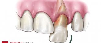
Facial lipofilling consists of 4 main stages, these are:
Stage 1 . Infiltration of the area of fat collection in order to reduce the density and moisturize the adipose tissue. At the same stage, the donor area is anesthetized.
Stage 2 . Fat sampling (we manage to obtain an autograft of the minimum size, which has the maximum chance of complete engraftment). Preparation of fat autograft - purification and isolation of the most viable fat cells. Adipose tissue is removed from donor areas by liposuction, centrifuged, filtered and processed to increase subsequent vascularization, and therefore survival. In some cases, the transplant is additionally saturated with platelet mass obtained from the patient’s blood (PRP mass) for better survival.
Stage 3 . Introduction of fat autograft. The procedure for introducing a strictly dosed volume of fat is carried out through small holes using a disposable blunt microcannula. The microcannula size does not exceed 0.8 mm, which minimizes damage to tissues and blood vessels. (multi-level administration of fat, high survival rate due to a greater number of blood vessels located in different layers of tissue than in one. The combined distribution of fat - on different layers, allows you to fill deep voids and not create excessive surface volume.
Each stage must be carried out clearly in accordance with the rules that were formulated in the process of long-term experience in conducting operations and which form the basis of the author’s method of David Rubenovich Grishkyan.
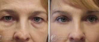
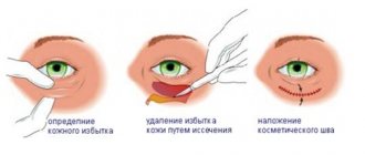
The patient underwent comprehensive facial rejuvenation, namely blepharoplasty of the upper eyelids with skin excision and lipofilling of the eyelids and face. Nanolipofilling was performed. Even the photo shows a change in the quality of the skin and the overall impression of rejuvenation and lifting, which occurs due to the work of fat cells in the facial tissues.
Main part
The use of adipose tissue in reconstructive plastic surgery has been known for a long time; in 1883, Neuber first proposed the idea of transplanting the patient’s own adipose tissue to replace the volume of subcutaneous soft tissue. In 1889, Van Der Meulen used the greater omentum, removed during surgical treatment of a diaphragmatic hernia, as a fat graft. T. Czerny in 1895, performed breast augmentation through lipoma transplantation. Charles C. Miller of Chicago, a birth defects specialist, expressed his positive views on the concept of fat grafting in his book Cosmetic Surgery: Correction of Congenital Defects. Following the example of Czerny, A. Bier in 1910 used adipose tissue from a removed lipoma to correct facial hemiatrophy. Lexer in 1920 performed breast augmentation using fat pads as a filler, and he also used this method to correct atrophic scars in the periorbital area, noting that the technique not only serves to fill the defect, but also prevents re-fixation of the skin to the bone by scar tissue fabrics. Erich Lexer also owns a two-volume treatise, Die Freien Transplantationen (free transplantation), published in 1919, containing almost 300 pages on the technique of fat tissue transplantation. However, by this time the disadvantages of fat tissue grafting had already been noted. Thus, F. Verderame noted that the transplanted fat has a tendency to be reabsorbed, and therefore recommended transplantation with hypercorrection. Since the 20s of the last century, a large number of works have been published on fat tissue transplantation. Louis Placide Mauclaire in 1922 (Paris) published the monograph “les Greffes Chirurgicales”, which summarized the experience of fat transplantation around the tendons of the hand to restore tissue glide and transplantation of the greater omentum to close a large bladder defect. And in 1925, Davis and Traut published data on methods for increasing the effectiveness of fat tissue transplantation. Charles C. Miller in 1926 proposed the syringe method of fat tissue transplantation for the correction of nasolabial folds, periorbital area and saddle nose. In 1929, O. Loewe proposed including dermis in the fat graft, which, according to the author, not only gave it density, but also provided better vascularization. By the beginning of the 40s of last year, the number of works devoted to the problem under study increased. Errors and complications were analyzed, new methods of using fat were proposed, and a study was carried out on the speed and volume of fat loss after transplantation. In 1940, Lyndon A. Peer wrote in his writings: “...fat grafts lose approximately 45% of their mass and volume after 1 year.” Bames in 1953 reported on the possibility of breast augmentation using the patient's own adipose tissue as a filler. Of course, the result of these “experimental” operations was unsatisfactory, because At that time, there was no modern understanding of the anatomical and physiological properties of adipose tissue. Since the 80s of the last century, many authors have reported on the successful use of autologous adipose tissue in the correction of facial wrinkles, aging hands, etc. The first publication reflecting retrospective results of the use of adipose tissue in plastic surgery of the mammary glands dates back to 1987 and is devoted to the “microinjection technique of breast augmentation.” glands using lipoaspirate obtained after liposuction” (Bircoll et al). In 1988, the American Society of Plastic Surgeons published an article, “Banned the Procedure,” which detailed the “dark side” of using autologous fat tissue and defined the technique as “vicious.”
However, the accumulation of experience and knowledge in the field of using adipose tissue as a plastic material and the definition of the lipografting procedure from the perspective of transplantology made it possible to formulate a modern and effective concept of using a fat graft. In the late nineties, Aiache and a number of other authors reported positive long-term results of lipografting. Nechajev pointed out the need to separate the resulting aspirate in order to separate the active fat portion from the blood, supernatant and oil. He reported 40-50% adipocyte survival. The scope of application of lipofilling in aesthetic surgery has expanded (Adant J., Bluth F., 2000). In 1989, Abel Chajchir reported a 90% success rate for fat grafting. Based on the experience of fat tissue transplantation in 500 patients, he formulated his concept of this technique: a ban on the use of local anesthetics; gentle sampling of adipose tissue; ban on fat washing; injection of lipoaspirate in three layers: skin, fascia, muscle.
In 1994, Carpaneta published the results of the dependence of the resorption of transplanted fat on the volume of injected tissue. His research showed that the thickness of the fat graft should not exceed 3 mm. The culmination of almost 80 years of research into fat tissue transplantation was the work of Coleman SR, which essentially became the basis of the modern method of transplanting one’s own fat tissue. In the 1990s, Sidney R. Coleman generalized the lipografting technique. He pioneered the use of multi-level cross-injection of fat grafts. He also recommended centrifuging the lipoaspirate before its administration in order to divide the resulting graft into the necessary fractions during collection. His concept of effective fat engraftment was as follows:
1) It is necessary to use blunt-ended cannulas with a diameter of no more than 17 mm, connected to a 10 ml syringe for collecting fat;
2) Lipoaspirate must be purified to specific, strict technical specifications;
3) The injection of fat should be performed with microgranules to increase the area of contact of the graft with surrounding tissues and improve diffuse trophism of adipocytes until revascularization occurs. The culmination of his work on fat tissue transplantation and the achievement of lasting results after this procedure were the works “Structural Fat grafting” in 2004 and “Fat injections: from filling to regeneration” in 2009.
Due to the widespread introduction of adipose tissue transplantation into clinical practice, new methods and algorithms for the collection, processing and injection of fat are being developed. So Dr. Pierre F. Fournier proposed a new method of vacuum aspiration of adipose tissue using disposable Felman canulas. This cannula allows for the harvesting of individual columns of adipose tissue while minimizing damage to adipocytes, which helps ensure long-term aesthetic results. Since individual columns of adipose tissue are collected, there is no need to centrifuge the lipoaspirate, which reduces surgical time. However, this method of harvesting adipose tissue is very aggressive in relation to donor areas, and, as a rule, is accompanied by quite pronounced hematomas in the postoperative period. Also, the use of a monoblock method of sampling adipose tissue is possible only in patients with a sufficiently pronounced fat depot. The use of lipografting in aesthetic facial surgery is considered in the concept of “volumetric rejuvenation”. Despite the fact that tissue augmentation through fat injection has been used for more than 100 years, the search for optimal methods is still ongoing. One of Roger Amar's techniques is called "autogenous fat injection" (AFI) aimed at achieving the most long-lasting aesthetic results. This technique involves injecting centrifuged fat into or directly next to the facial muscles. This method is based on the work of Guerrosantos, who in 1996 proved the five-year persistence of fat in the muscle tissue of rats.
Most volume grafting procedures using adipose tissue are aimed at replacing lost subcutaneous fat. However, as we age, all facial tissues undergo atrophy: fat, muscle, bone. Significant loss of subcutaneous fat can lead to noticeable age-related changes. Coleman and others have described a flattening of the facial contour due to fat loss.
IMAGE is indicated for patients with reduced facial tissue volume, but sufficient skin elasticity. In this procedure, fat is not used to fill deep wrinkles. The method involves restoring muscle volume, contours and function. Of the individual areas of the face, the lower third most often needs correction. Restoration of this area can be performed using IMAGE, replenishing lost lip volume, adding volume to the chin and emphasizing the edge of the lower jaw. In the periorbital region, IMAGE can be used to restore a deformed tear trough. The procedure is also ideal for post-facelift patients whose neck appears skeletonized. IMAGE is not suitable for patients with significant skin laxity, very deep nasolabial folds and ptosis of the cheeks and neck.
IMAGE involves the injection of a small amount of fat along the well-vascularized bed of the facial muscles. Injecting fat along the muscle enhances its function, which is used in various fields of medicine, for example in otorhinolaryngology, urology and gastroeterology, when fat is injected into weak vocal cords or sphincters for therapeutic purposes. Due to fat, not only the thickening of the muscle bundle occurs, but also the hypertrophy of the muscle tissue itself. Correcting volume and enhancing muscle function during IMAGE provides a facelift, giving it the contours characteristic of a young age.
Recently, in the literature, along with reports concerning the use of adipose tissue as a kind of “filler” or plastic material, works have appeared devoted to the use of autotransplantation of adipose tissue enriched with stromal vascular cell fraction (SVCF), obtained using the classical P. Zuk method - “manually” » – in the laboratory, where lipoaspirate is supplied immediately after surgery; Cytori Celution device since 2008 and GID SVF-1.
Intact adipose tissue is a vascular, self-renewing structure consisting of adipocytes, a stromal vascular cell fraction (SVCF), and a supporting fibrous stroma. SVCF is a unique cellular complex containing adipose tissue stem cells - SCAT (which are a key component of SVCF), endothelial and smooth muscle cells of blood vessels and their precursors, pericytes, fibroblasts, blood cells, including B and T lymphocytes. It should be noted that adult adipose tissue is the richest in stem cells compared to other sources (in particular, 1 cm3 of this tissue contains 100–1000 times more stem cells than 1 cm3 of bone marrow). The positive effect of SVCF on reparative processes in the transplantation area is due to the cumulative interaction of stem cells that are part of SVCF. This feature of adipose tissue stem cells is based on significant endocrine activity (FGF-fibroblast growth factor, VEGF-vascular endothelial growth factor, TGFb-transforming growth factor, IGF-insulin-like growth factor, PDGF-platelet growth factor), as well as the ability of the latter to carry out neoangiogenesis and adipocyte regeneration. Unlike adipocytes, the cells that make up the SVCF are resistant to oxygen deficiency. Moreover, according to H. Suga and H. Thangarajah (2009)[13], hypoxia helps stimulate the differentiation of SCAT in the angio- and adipogenic directions. Thanks to this, during the first 2–3 months after transplantation of the SVCF-enriched lipograft, its renewal is observed, which, accordingly, significantly improves its quality.
Transplantation of one's own adipose tissue, as a separate technique for breast reconstruction, was described in 2000 in the article “Reconstruction after mastectomy and lumpectomy; Delay/ Rigotti.” However, in the same year, a work was published: “the method of fat tissue transplantation is good as an addition to breast reconstruction, unsatisfactory as a method of primary breast augmentation...” (American Society of Plastic Surgeons), which describes in detail and justifiably a number of unsolved problems that limit its use this technique as a mono-tool for reconstructive and aesthetic surgery of the mammary glands. In reconstructive surgery of the mammary glands, such tasks are: deficiency of recipient capacity; fibrous changes in the area of interest after radiation therapy; lack of skeletal properties in adipose tissue. The main obstacles to the use of one's own adipose tissue as a monomethod for primary mammary gland augmentation is the shortage of donor and recipient areas, because Most patients seeking treatment for breast enlargement have an asthenic body type. One of the solutions to the problem of deficiency of recipient zones during primary augmentation and reconstruction of mammary glands through lipografting is the use of external tissue expanders. As an example of the technique of using adipose tissue in combination with external tissue expansion in reconstructive plastic surgery of the mammary glands, see Roger Khouri in 2002. A system for reconstruction and augmentation of mammary glands with own fat, based on the principles of tissue engineering in vivo. The BRAVA system is a modified external expander that produces a gentle three-dimensional stretching effect. As a result, the glandular tissue hypertrophies. The tissues are more intensively supplied with blood, creating favorable conditions for the engraftment of fat grafts.
Research into the metabolism of adipose tissue in vivo and in vitro is currently ongoing. The dependence of the degree of adipocyte survival on the technique and area of fat harvesting is studied. A search is underway for a universal donor area. Morphological studies of the transplanted adipose tissue are carried out and the degree of its resorption is determined. The process of vascularization of the fat graft and its effect on the survival rate of transplanted adipocytes is being studied. Methods for pre-injection treatment of adipose tissue are being developed in order to increase its viability. Despite great achievements in these areas, many issues still remain unresolved.
So, taking into account all of the above, the question naturally arises: “Has the role and consistent method of fat tissue transplantation been determined?” Is it possible to widely and actively use this technique? All of the above suggests that it is necessary to clearly formulate the indications, contraindications and target audience of patients for whom the lipofilling method is not only possible, but also safe. Based on the information received, our center began active work to introduce this technique into clinical practice.
Where does fat come from for facial lipofilling?
Ideal areas are the inner thigh, knee area, stomach, outer thigh. These areas with the most stable fat are “fat traps”. But also from those areas that the patient wants to correct.
There are no stitches after lipofilling; the holes are closed with flesh-colored strips, which do not interfere with immediately assessing the result.
As a result, we guarantee:
— No lumps or compactions; — High degree of fat survival; - Lifelong results.
Lipofilling of the lower leg. Risks and complications.
Patient dissatisfaction with the treatment result is a possible risk.
In cases where the deficiency of soft tissue along the inner surface of the leg is significant, the task of replenishing it through lipofilling is not easy in terms of guaranteeing the result.
The disadvantage of lipofilling is the incomplete engraftment of fat cells. However, if the correction is incomplete, lipofilling can be repeated.
The engraftment of fat cells depends on compliance with the surgical technology, the individual characteristics of the patient and ranges from 30 to 50%. That is, out of 200-300 ml of fat transplanted to each lower leg, only half can perform its function. This poses particular challenges to obtaining symmetry and accurately correcting contour deficits. Overfilling the site of tissue deficiency with fat leads to the destruction of fat cells, and if an infection occurs, then to suppuration at the site of surgery.
Suppuration is manifested by severe swelling, local redness, pain at the operation site and symptoms of general malaise. Treatment of this complication requires surgery to open and drain the inflammation. Fortunately, this complication occurs quite rarely.
In my opinion, if the tissue deficiency in the lower leg area is significant, it is more rational to use another treatment method - lower leg replacement.
Facial lipofilling contraindications
Contraindications to surgery exist, both general and specific, related to the patient’s health. The patient's health status before surgery is determined on the basis of tests and opinions of specialists, anesthesiologist, therapist, cardiologist. If the patient has chronic diseases, then an opinion from a specialized specialist on the possibility of undergoing surgery is also required. In addition, there are contraindications related to the anatomical characteristics of the patient. For example, as a general rule, a patient may be denied lipofilling due to very thin skin, since there is a possibility of contouring of the transplanted fat.
Some time ago in my practice, fearing complications, I also injected only a small amount of fat into the lower eyelid area - the total volume did not exceed 1.5 ml. At the same time, I carefully selected patients based on the “thickness” of the skin of the lower eyelid area and did not recommend lipofilling to patients with “thin” skin at all. But as I gained experience, I began to increase the amount of fat injected into this area. Today, injection of approximately 5 ml of fat into the lower eyelid area alone is routine. And I accept patients with thin skin without any problems. The rest of the list of contraindications is standard: diseases of the cardiovascular system, infectious diseases, acute and chronic pathologies of connective tissue, diabetes mellitus, oncological and mental diseases, blood clotting disorders.
2.The essence of the method
The purpose of microsurgical tissue autotransplantation is to replace integumentary defects with minimal postoperative traces; Often such problems have to be solved not in isolation, but as part of a more complex and complex operation, for example, replantation of a traumatically amputated limb.
Several types of autografts are used:
- simple (a single fragment, tissue or organ is transplanted, for example, a section of bone or muscle);
- complex (several interconnected anatomical structures with a common blood supply circuit);
- combined (the substitution fragment is made up of two or more different structures that together replace the defect);
- modeling (the autograft is “constructed” artificially from various fragments).
One of the main microsurgical problems is the preservation of the so-called. vascular pedicle - so to speak, a connecting fitting through which the autograft will then be “connected” to the new blood supply circuit.
It is important to note that there are many areas on the human body that could theoretically be a source of autotransplant material (without any damage to the body as a whole), but at this stage this is only partially used in practice. Research in this direction continues, and periodically published results indicate the successful expansion and development of the autotransplantation method.
Visit our Microsurgery page
Guaranteed long-lasting results
The patient underwent facial fat grafting as a rejuvenation procedure after significant weight loss. A sharp decrease in weight led to sagging and ptosis of the face, as can be seen in the photo before the operation. Lipofilling not only leveled and recreated volume in the area of the eyelids, cheekbones, lips and chin, but also worked on the skin.
The graft survival rate is not 100%; part of the adipocytes are removed from the transplantation zone with lymph flow within a month, but the engrafted cells remain in the lipofilling zone for life. Since the number of adipocytes in the body is constant unless fat is removed through liposuction, the appearance of the corrected area depends only on whether the patient gains or loses weight after surgery. The fatty tissues in the lipofilling area age and experience the effects of gravity, like the rest of the patient’s non-operated tissues.
How long does it take to see results?
Immediately after surgery, patients can see positive changes in appearance, and even the presence of slight swelling after lipofilling does not spoil this impression. The final result is usually formed after 3 months. During these three months, the fat autograft grows with vessels that keep it in its new place forever.
How long will the effect of facial lipofilling last?
For life, forever! All the fat that has taken root within 3 months from the moment of surgery remains in the tissues forever!
Own experience of using lipografting in contour surgery of the mammary glands
From June 2014 to November 2014 at the Department of Breast Tumors of the Federal State Budgetary Institution Research Institute of Oncology named after. N.N. Petrov performed 27 operations aimed at correcting the shape of the mammary glands through fat tissue transplantation. All patients underwent careful selection and all had clear indications for the specified surgical treatment. Lipografting was not used in immunosuppressed patients or those scheduled for radiation therapy. In all cases, lipografting was used as an additional technique to the main reconstructive plastic surgery.
The main goals of using lipografting as an additional technique were: 1) increasing the projection; 2) filling the upper slope; 3) restoration of symmetry; 4) adding volume to the mammary glands after reconstructive operations using full-thickness complex autografts (flaps).
The main way to achieve these goals was to solve the following tasks:
1) Obtain a sufficient volume of lipoaspirate. 2) Correlate the volume of lipoaspirate with the reserve capacity of the recipient zone. 3) Select the introduction layer. 4) Determine the frequency of operations.
Based on the principles of transplantology, the following stages of lipografting can be determined:
1) Determination of the area of interest (recipient area).
2) Selection of donor area. Preference is given to areas with a sufficient amount of fat: paraumbilical area, abdominal flanks, inner thighs, riding breeches, back, shoulders.
3) Anesthesia. Depending on the volume of fat removed, we use local anesthesia with intravenous potentiation or ETN. To collect fat, a tumescent technique is used: Sol. Lidocaini 10%-1ml+ Sol. NaCl 0.9%-400ml+ Sol. Adrenalini 0.5 ml. To prevent flooding of lipocytes, we abandoned the use of glucose and bicarbonate in the solution.
4) Fat removal. To obtain lipoaspirate with the maximum number of living adipocytes and minimize the content of blood in the lipoaspirate, we use: delicate cannulas, minimal vacuum, minimal aggression in the donor area.
5) Preparation for introduction. After taking the fat, the latter is centrifuged for 1.5 minutes at 1300 rpm; This ensures minimal contact of lipoaspirate with air and minimal transfer from system to system.
6) Introduction of fat. To inject fat, we use the micro-grafts technique, layer-by-layer retrograde injection without resistance. Hypercorrection is unacceptable, because The greater the volume of fat, the higher the risk of necrosis. At the introduction stage, strict adherence to the FTF (fat to fat) principle. To determine the permissible volume of transplanted fat into the recipient area, we are guided by the concept of “recipient capacity”; the amount of transplanted fat in one horizontal layer is limited by the capacity of 2 mm channels and the need to maintain no less distance between them, so as not to compromise the vascularization of the recipient bed. For a simplified calculation, we will take the average distance between the axes of adjacent channels to be 5 mm. Then for a square recipient zone 5 x 5cm. the permissible number of channels will be 10, and the permissible volume of transplantation = 10 channels x 50mm x 2mm x 3.14 (p number) = 3140 cubic millimeters, i.e. about 3 ml. These are the inevitable limits of reliable volumetric growth. The boundaries of the injection zone can be expanded if lipofilling is carried out not in one, but in two or more tiers. You just need to remember that the vertical distance between the layers should be sufficient to maintain the same vascularized layer of recipient tissue between them.
7) Volume distribution. All stages of the operation are aimed at improving the survival of fat. In the postoperative period, bandages were applied to the puncture sites and antibiotic therapy was prescribed for two days. In this case, cold, pressure and massage of the recipient area were completely excluded.
Example #1:
Patient O., 33 years old. Diagnosis: cancer of the right breast, condition after complex treatment in 2012. BRCA mutation. Condition after bilateral subcutaneous mastectomy with simultaneous reconstruction with implants. Ripping in the area of the upper slope of the right breast.
Rice. 1 (a,b)
Example #2:
Patient X. 40 years old. Diagnosis: cancer of the left breast. Condition after complex treatment in 2013. Condition after subcutaneous mastectomy on the left with simultaneous reconstruction with an implant in combination with TDL, prophylactic subcutaneous mastectomy on the right with simultaneous reconstruction with an implant dated July 26, 2013. Deficiency of the upper slope of the mammary glands, cicatricial deformation of the lower slope of the right mammary gland.
Rice. 2 (a,b)
Depending on the clinical case, patients underwent one to three lipografting sessions. It should be noted that over the eleven-month follow-up period, the loss of adipose tissue in the recipient area ranged from 0 to 40%.
Lipofilling at Dr. Grishkyan’s clinic in Moscow
Facial lipofilling
| Operation name | Cost in rubles |
| Lipofilling of the upper eyelids | 160000 |
| Lipofilling of the lower eyelids | 160000 |
| Lipofilling of the lower eyelids with transblepharoplasty | 180 000 |
| Lipofilling of the midface, including the lower eyelids | 180000 |
| Lipofilling of the face, including the lower eyelids | 250000 |
| Facial lipofilling, including upper and lower eyelids | 310 000 |
| Lipofilling of the lower third of the face | 120000 |
| Chin lipofilling | 90000 |
| Facial rejuvenation with growth factors (“Nanofat”) | 60000 |
Benefits of Fat Transfer
After the first successful attempts at lipofilling, plastic surgeons paid attention to improving the quality of skin with age-related changes or signs of photoaging. Later after fat grafting, many doctors noted the disappearance of pathological changes such as scarring, tissue damage from radiation therapy, and steroid-induced atrophy. It has now been proven that stem cells or messenger cells present in all fatty tissues promote tissue regeneration by forming new blood vessels or act directly on damaged or aging structures - restore and rejuvenate the areas of lipofilling. Therefore, adipose tissue is nature’s source of regeneration of the human body. Thanks to lipofilling, the term “volumetric rejuvenation” is increasingly heard.
In recent years, research into the nature of the extracellular matrix has begun to actively develop, which has shown that, in addition to adipocytes, adipose tissue consists of several types of cells, such as preadipocytes, endothelial cells, smooth muscle cells, fibroblasts, and adipose-derived stem cells (ADSCs). ). ADSCs present in the stromal fraction of lipoaspirate are capable of differentiating into various tissue types, such as bone, muscle, cartilage, nerves, blood vessels, and so on.
Dr. Gino Rigotti made a great contribution to the development of the theory and practice of lipofilling. In his articles and reports, he demonstrated not only the restoration of soft tissue defects by lipofilling, but also the improvement of the texture of irradiated tissue. At the same time, Rigotti noted the important therapeutic role in tissue treatment processes precisely due to stem cells (ADSCs) present in adipose tissue. The author gave examples of patients who could not be healed with conventional treatments and demonstrated healing after transplantation of their own fat.
The discovery of stem cells in adipose tissue has prompted researchers from around the world to develop devices that can isolate these cells from fat during liposuction. It is expected that transplanted adipose tissue enriched with stem cells will not only achieve almost 100% cell engraftment, but will also promote true rejuvenation of surrounding tissues. Currently, such devices operate almost throughout Europe and Asian countries. But until now, there has not been a randomized, controlled, double-blind clinical trial to prove whether this process is superior to standard fat grafting.