Services Doctors Results Reviews
Expert tested
Gutsenko Liliya Anatolyevna Cosmetologist
Publication date: December 05, 2022
Review date: November 03, 2022
You can look young at any age! And not thanks to good makeup, stylish hairstyle or fashionable clothes! And because of maintaining the body’s own resources at a young level. Today the aging process of the body can be controlled and even stopped!
This is no longer fantasy, but reality!
How does human skin age?
Skin aging, like the body as a whole, is a complex biological process that involves many factors, including genetic and environmental factors. In this case, ultraviolet radiation plays a special role. There are two main types of skin aging: internal (chronological) and external (photoaging). Each of them has its own clinical and morphological features.
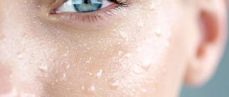
Skin that ages over the years and has been protected from the sun becomes thinner, paler, its elasticity and firmness decrease, and fine superficial wrinkles appear. The sun acts differently: the skin thickens, coarsens, becomes drier, deep wrinkles and individual areas with dark pigmentation form in it - the so-called solar lentigo.
Photoaging can occur much earlier than chronological aging - it was not for nothing that in the 19th century ladies carefully hid their faces from the sun under hats and umbrellas. And if chronological aging, alas, is inevitable, then photoaging directly depends on the time spent in the sun and in solariums, as well as on the genetically predetermined degree of skin pigmentation. Open areas (face, neck, arms) are especially affected, since external factors interfere with the chronological process and accelerate it. People living in hot countries are especially unlucky. Their skin is usually rough. And signs of aging appear earlier than in people living in temperate climates.
During the aging process, the content of collagen in the skin, the main structural component, decreases. The changes are dramatic: older people (80 years and older) not only produce about 75% less collagen than young people (18-29 years), but the level of its degradation increases by 75%. At the same time, the amount of collagen types I and III decreases. These are some of the main types of skin collagen.
Sunbathers should remember: a single exposure to UV rays on the skin in medium doses (until it becomes slightly reddened) reduces collagen production by 80%, and it will return to normal only after two to three days. If you regularly expose your skin to the sun, collagen production is suppressed for a very long time and over time the changes can become irreversible.
Avdeyuk Elena Vladimirovna
Dermatovenerologist, cosmetologist
Collagen not only becomes smaller over the years and under the influence of the sun, but its structure and organization also change. Because of this, the supporting frame of the skin is disrupted and wrinkles form. It must be said that in areas of the skin that have been exposed to prolonged UV exposure, specific changes also develop that are associated with impaired formation of elastin fibers (so-called elastosis).
Collagen is produced by fibroblast cells, which means they are responsible for those fundamental processes that upset us so much. Fibroblasts decrease by about 35% with age, but the question is not only their number. The morphology of fibroblasts and their ability to divide also change; most of them perform their productive functions worse.
As a result, the balance between the synthesis and degradation of the intercellular matrix is disrupted, and the collagen content decreases. The skin becomes thinner, its hydration and elasticity decrease, and wrinkles form.
Dermal fibroblasts can be stimulated to actively work and replenish the lost volume of the intercellular matrix in various ways. In aesthetic medicine today, various injection and hardware techniques are actively used for this purpose. For example, intradermal injections of various drugs are made that are related in their biochemical composition to the intercellular substance (this could be, for example, hyaluronic acid). They act on the skin with a laser - this causes dosed microdamage to the layers of the dermis, which activates regeneration. Radio wave therapy, dermabrasion and other methods are also used that force fibroblasts to get to work and begin to synthesize young collagen again.
What is SPRS therapy?
Regenerative medicine uses a fundamentally different approach. It involves introducing a drug into the tissues, which itself restores these tissues, or triggers their own physiological mechanisms and regeneration in them, or does both at once.
Effective biotechnological approaches already exist. For example, Apligraf (USA), a skin equivalent artificially grown in a special environment, is successfully used to restore skin for various ulcers. It consists of allogeneic keratinocytes (allogeneic means donor; keratinocytes make up the bulk of the epidermis - the upper layer of the skin) and dermal fibroblasts. The effectiveness of the drug has been tested in more than 250 thousand clinical observations.
Another American drug, Dermagraft, contains only allogeneic dermal fibroblasts (more than 50 thousand observations). And in the complex treatment of severe burns, the drug Epicel with the patient’s own keratinocytes (USA, approximately 2 thousand observations) has performed well.
In 1994, the American company Isolagen (now called Fibrocell Science) first proposed a new method for correcting wrinkles - introducing autologous, that is, his own, fibroblasts into the patient’s skin. It turned out that dermal cells taken from the patient and multiplied actively synthesize collagen and other components of the intercellular matrix. But the main thing is that after transplantation into the dermis, their synthetic activity remains.
In 1999, Isolagen published the results of a large-scale clinical study conducted in 1995-1999. It involved 1,450 patients with severe facial wrinkles. The method has been shown to be safe (follow-up period 48 months) and effective. Later, in 2003-2008, the company conducted other clinical studies involving more than 800 patients. Twelve months of observation again confirmed the safety of the technology and that wrinkles were indeed reduced by almost half. Moreover, the effect lasts for a very long time.
Scientists at the Russian Institute of Human Stem Cells began working on this problem in 2003. As a result, a Russian technology was created (its official name is SPRS therapy), similar to the American one, but with a number of significant differences. It has been officially approved for use for the correction of age-related and scarring skin changes in Russian clinics since July 2010. A year later, Fibrocell Science also received approval from the US Food and Drug Administration (FDA) to use a similar technology to correct wrinkles in the nasolabial folds. In general, over almost twenty years, both Russian and American scientists have conducted many clinical studies, which have confirmed that the method indeed gives very good results - all patients and doctors are satisfied with it.
How is SPRS therapy performed?
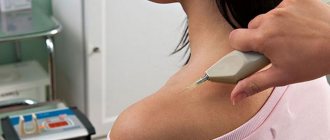
A piece of skin with a diameter of 4 mm is taken from the patient from behind the ear (where the skin is least damaged by UV rays) and a fibroblast culture is obtained from it.
Then, under GMP (Good Manufacturing Practice) laboratory conditions, a cell preparation (SPRS preparation) containing cultured fibroblasts is obtained. During cellular processing, only young functionally active fibroblasts are selected and stimulated, which have retained a high ability to divide and synthesize components important for the skin.
All procedures are carried out in accordance with the medical technology approved for use by Roszdravnadzor of the Russian Federation “Collection, transportation, isolation, cultivation, cryopreservation, storage and use of autologous fibroblasts for the correction of age-related and scarring skin defects” (FS permit No. 2009/398).
The SPRS drug is delivered to the clinic under strictly controlled conditions (in a thermal container that constantly maintains a temperature of +4–8°C), where the patient is given a course of therapy consisting of two procedures at an interval of a month.
The cellular material is introduced using a special technique: intradermally - into the papillary layer of the dermis, in a tunnel way using mesotherapy needles (30G, 13 mm). This injection method allows the drug to be administered evenly and with adequate density throughout the entire area requiring correction, and to replenish the pool of resident fibroblasts in the patient’s skin with functionally active cells.
An hour before the procedure, EMLA anesthetic cream is applied to the skin, which makes the administration of the drug painless.
Materials and methods
A primary culture of dermal fibroblasts was obtained from skin biopsies through mechanical disaggregation and subsequent enzymatic treatment. To do this, the biopsy specimen was washed with phosphate-buffered saline containing an antibiotic (gentomycin) and treated with a trypsin solution. Then they pipetted, as a result of which the cells were freed from the matrix, centrifuged, washed from the trypsin solution and resuspended in a DMEM/F12 culture medium containing 10% serum. They were seeded onto Petri dishes at a density of 1 × 10⁵ – 2 × 10⁵ cells/cm2 and placed under standard incubation conditions (+ 37 0C, 5% CO2). They were cultured until the 4th passage, characterized by the expression of characteristic markers and cryopreserved, creating a bank of a primary culture of human dermal fibroblasts, on which all further studies were carried out. The use of the same primary cell culture in the study allows us to compare and contrast the data obtained.
To obtain an “aging” culture, cells from the previously obtained bank were thawed by rapid thawing at +37 0C, washed from serum residues and DMSO with Hanks solution using centrifugation for 7 minutes. at 1000 g. The supernatant was removed, the resulting cell sediment was resuspended in complete growth medium, plated on Petri dishes and placed under standard incubation conditions (+ 37 0C, 5% CO2). They were cultured until the 18th passage, when all signs of replicative aging of dermal fibroblasts became clearly visible. The thawed, unpassaged cell culture of the 4th passage (P4) was considered a control and corresponded in its characteristics to fibroblasts of young skin.
To study the dose dependence of Meso-Wharton P199™ in 2D culture, aliquots (500 μl) of P199 peptide dissolved in saline buffer were mixed with complete growth medium in the ratio of 1:2, 1:10, 1:100, 1:1000. Cytotoxicity assay was performed on a 2D culture of passage 18 dermal fibroblasts (P18). Cells were plated in 12-well plates at a density of 10,000 cells per well. As a control, the behavior of cells was studied in complete growth medium without the addition of peptide. Cells were cultured in the presence of the peptide for 72 hours, then stained with solutions of Hoechst 33258 (0.004 mg/ml) and propidium iodide PI (0.001 mg/ml), with which cell viability was assessed by penetration of PI into their nucleus under an Olympus inverted fluorescence microscope CKX41 (Olympus, Japan).
The bioactivity assay was performed on a 2D culture of dermal fibroblasts and placed on coverslips in Petri dishes (35 mm). Cell morphology and expression of markers characteristic of fibroblasts were studied: cytokeratin 19, elastin, α-smooth muscle actin (αSMA), PCNA (proliferation marker), collagen types I, III and IV. As a control, passage 18 cells were cultured in complete growth medium without the addition of the drug. To carry out immunocytochemical analysis, cells were cultured in the presence of the peptide for 72 hours, then fixed in a 4% paraformaldehyde solution (4°C, 20 minutes). The slides were then incubated with primary antibodies to cytokeratin 19, elastin, α-smooth muscle actin (αSMA), PCNA (a marker of proliferation), collagens I, III and IV. The result was visualized using secondary antibodies conjugated to FITC (E=525 nm) and DyLight594 (E=617 nm) fluorochromes. Nuclei were counterstained with Hoechst 33258 solution (0.004 mg/ml). The analysis was carried out using an Olympus Fluoview FV10 laser scanning confocal microscope (Olympus, Japan).
To study the effect of Meso-Wharton P199™ on the formation of spheroids from dermal fibroblasts, monolayer cell cultures of P18 fibroblasts were studied, which were cultured with the addition of Meso-Wharton P199™ to the growth medium and cultures of P18 fibroblasts, cultured without the addition of Meso-Wharton P199™. For this stage of the study, aliquots (500 μl) of p199 peptide dissolved in saline buffer and mixed with complete growth medium in a ratio of 1:10 were used. Fibroblasts were cultured for 72 hours, washed from the growth medium with Versene solution, then treated with a 0.25% trypsin solution and removed from the dishes. The number of cells in the suspension was counted and centrifuged. The pellet was resuspended in complete growth medium and plated on agarose plates at a concentration of 1 × 10 cells per well. Agarose plates were placed in 12-well culture plates and intravital monitoring was carried out under standard chamber conditions of a Cell-IQ device (CM Technologies, Finland) for seven days. Photo recording was carried out every 20 minutes. using the Cell-IQ Imagen program; image processing was carried out in the Cell-IQ Analyzer software package. The formation of spheroids from fibroblasts of the 4th passage (P4) in complete growth medium without the addition of peptide was considered a positive control.
When will the results appear after SPRS therapy?
The result is visible 10-14 days after the end of the procedures: the skin becomes firmer, more elastic, the color and contours of the face improve, the number and depth of wrinkles decrease, as well as the severity of age spots.
Another important indicator is that the thickness of the skin increases (after a year by an average of 65%). After a month, 88% of patients assessed the effect as “good” or “excellent”, after 3, 6, 12 and 24 months - 100% of patients. Wrinkles decrease after a month by 14%, and after 12 months - by 46% compared to the initial state.
More than a third of patients who underwent correction of one area returned for treatment in other areas - neck, décolleté, and arms.
The effect lasts for at least two years and increases over time.
Required number of drug administrations
The most important thing in the procedure is an individual approach to each patient. Most often, the course includes 1-3-5-7-10 injections of fibroblasts. The number of times depends on the amount of drug administered. However, regardless of the number of injections, the tissue sample only needs to be taken once, since cell cultures are cryogenically preserved for subsequent treatment.
If you have any questions about the use of the drug FIBROBLASTS or would like to make an appointment for a consultation, call or.
| № | SPRS therapy, SKIN TREATMENT WITH AUTOFIBROBLASTS | Prices, rub. |
| 1 | The doctor's consultation | 3 000 |
| 2 | Creation of a bank of your own skin fibroblasts (collection of biopsy material, production of cellular material for the production of SPRS-preparation (12 or more courses of 120 million cells) Skin passport (morpho-functional characteristics of skin fibroblasts, determination of the regenerative potential of the patient’s skin) SPRS program (data interpretation Skin passports and drawing up recommendations for the development of an individual program of cosmetic procedures Course of SPRS - therapy for any one area (face/neck/décolleté) - production of SPRS - drug: 2 doses of 60 million cells / 1 dose of 120 million cells | 450 000 |
| 3 | A course of SPRS therapy, without creating a bank For any one area (face/neck/décolleté) - biopsy specimen collection, patient serum collection, 2 doses of 60 million cells / 1 dose of 120 million cells) | 250 000 |
| 4 | Production of SPRS-DRUG with repeated treatment 2 doses of 60 million cells / 1 dose of 120 million cells | 220 000 |
| 5 | Production of SPRS-DRUG with repeated treatment 2 doses of 40 million cells / 1 dose of 80 million cells | 190 000 |
| 6 | Production of SPRS-DRUG with repeated treatment 2 doses of 80 million cells / 1 dose of 160 million cells | 260 000 |
| 7 | Production of SPRS-DRUG upon repeated application to any one area 1 dose 60 million cells | 180 000 |
| 8 | Skin Passport SPRS program - interpretation of Skin Passport data and drawing up recommendations for the development of an individual program of cosmetic procedures, Creation of a Skin Passport - biopsy specimen collection, SPRS - diagnostics (morphofunctional characteristics of skin fibroblasts, determination of the regenerative potential of the patient's skin) | 80 000 |
| 9 | Only bank SPRS-program - interpretation of Skin Passport data and drawing up recommendations for the development of an individual program of cosmetic procedures, Creation of a bank of your own skin fibroblasts (sampling of biopsy material, production of cellular material for the production of SPRS drug (12 or more courses of 120 million cells) Skin Passport ( morpho-functional characteristics of skin fibroblasts, determination of the regenerative potential of the patient’s skin) | 150 000 |
Bank of the patient's own fibroblasts
It should be noted that the medical technology for the use of autologous fibroblasts has another feature - it allows the banking of patient’s skin fibroblasts. They can be thawed at any time and used both to correct age-related changes in the skin, and in case of an unforeseen situation - in case of burns and other injuries that require the immediate use of fibroblasts to restore the integrity of the skin. This banking of autologous skin fibroblasts represents a kind of “biological insurance system.”
The patient’s “Skin Passport”, compiled during the process of collecting and culturing skin cells, will remind the cosmetologist about how often one can resort to repeated skin correction with synthesized fibroblasts.
Only 3 visits to the clinic!
1st VISIT
- Consultation, medical history, determination of the area of therapy;
- Taking a biopsy sample;
- Serum collection;
- Creation of a bank of your own skin fibroblasts;
- Creation of a skin passport;
- Storage of autofibroblasts for 12 months.
2nd VISIT
- Administration of the first dose of SPRS medication;
- Setting the date for administration of the 2nd dose of the drug (in 30-45 days);
- Recommendations for rehabilitation after the procedure;
- Interpretation of Skin Passport data.
3rd VISIT
- Administration of the second dose of the SPRS drug;
- Recommendations for rehabilitation after the procedure