New moles - are they dangerous?
Moles are the most common harmless formations on the skin. They usually appear during childhood and adolescence, but new moles can appear in adulthood.
A new mole appears when melanocytes begin to multiply and create a characteristic appearance on the surface of the skin that is different in color from the rest of the skin. New moles may be harmless but may have a malignant origin, such as melanoma, caused by a mutation.
Possible reasons for the appearance of a new mole:
- exposure to ultraviolet (UV) radiation – exposure to the sun;
- bright light;
- genetics;
- weakened immune system.
Reasons for the appearance of moles in adulthood
The appearance of moles in adulthood may be due to the reasons mentioned earlier, as well as additional factors:
- Metabolism and surface renewal of the skin can provoke the appearance of chloasma - age-related hyperpigmentation with a predominant localization on the face.
- With age, the skin loses elasticity and firmness, and the protective barrier also decreases. All this makes it easier for the human papillomavirus (HPV) to enter the body, which leads to the formation of tumors (warts, condylomas).
- A lack of vitamin K in the body, found in green vegetables, liver, dairy products and eggs, causes blood clotting problems and a weakened immune system. In turn, this can cause the formation of new birthmarks.
Types of moles
Regular stain
They occur during childhood, mainly in areas exposed to the sun. Round, symmetrical in appearance, with a smooth and clearly defined edge, up to 5 millimeters in diameter. They rarely become cancerous, but people who have 50 or more moles in common are at increased risk of developing melanoma.
Congenital spot
They are present from birth and can vary greatly in size and be quite large in area. They are usually benign, but have a predisposition to the development of melanoma, especially very large congenital moles.
Atypical spot
Or dysplastic moles, these can develop anywhere on the body and are usually larger than normal moles. They differ in color and texture, irregular edges with many colors. Some atypical moles can become cancerous, so regular checkups with a dermatologist are necessary.
A rare type of mole that looks like melanoma, but is not cancerous. It usually occurs in fair-skinned children and young adults under 20 years of age. They grow quickly and vary in size from a few millimeters to centimeters.
Which moles on the face should be removed?
Moles on a person’s face can become a highlight of one’s appearance, or, on the contrary, they can be a source of complexes. It all depends on their size, shape and color. You can try to remove really ugly large nevi, but doctors recommend subjecting them to mechanical stress only if they can be accidentally injured. For example, men at risk have moles located in places where they can be easily shaved off: the chin, cheeks, and neck.
Hair on a mole doesn't look very attractive, but it doesn't mean anything bad. However, it is not recommended to pull them out by the roots, so as not to injure the nevus.
Another serious reason for removing benign moles is the risk of them turning malignant. In most cases, nevi in women and men are safe for health, but as a result of a malfunction in the body, the cells can mutate, forming a cancerous tumor - melanoma. Moles that are more than 10 cm in diameter, begin to grow, itch, become inflamed, change color from light to darker, even almost black, have torn edges and/or asymmetrical sides are a health hazard. Doctors also always attach serious importance to swollen moles that hurt, including when pressed, but specialists prohibit rubbing them.
Having surgery to remove a mole on the face and body is not a problem, and it’s inexpensive. But it is important to note that you cannot decide about this on your own. Just like trusting this to an ordinary cosmetologist. According to doctors, exposure to a nevus can provoke a mutation of pigmented cells. It is especially risky to choose the wrong removal method without taking into account the type of mole. That is why they should be removed only after consultation with an oncologist, especially if the nevus looks unhealthy.
No one can guarantee the burning of absolutely all pigmented cells, despite the fact that missed melanocytes as a result of certain factors can provoke the development of melanoma.
Methods for removing moles on the face:
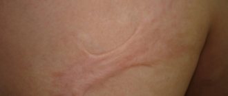
- Laser removal is the most popular procedure today, as it gets rid of nevus quickly, under local anesthesia and without scars, which is very important for appearance, especially for girls. With this method, a mole, including a dark one, is removed layer by layer. After using the laser, a depression may remain, but over time it levels out. Recovery time is up to 14 days.
- Surgery is the oldest and least expensive method, which may leave a scar. Nevertheless, it has a number of advantages: the possibility of histological examination of tissue, prevention of relapse and safety.
- Electrocoagulation - allows you to remove moles using direct and alternating current, in most cases it leaves a scar on the skin, but the risk of infection is minimal, there is no bleeding and recovery occurs in 7-10 days.
- Burning with liquid nitrogen is the longest procedure in terms of recovery time, taking about 1.5 months. In addition, when it is carried out, there is a high risk of burning out healthy skin tissue located next to the mole. External nevi are removed using a regular swab soaked in liquid nitrogen, and internal nevi are removed by injecting this substance under the skin using a thin needle.
Any of these methods has its advantages and disadvantages, but for permanent removal of moles on the face for cosmetic purposes, the advantages of the laser procedure are given due to the absence of scars after it. If there is a suspicion of oncology, experts recommend surgery to take a tissue sample.
Getting rid of a mole on your own at home can be dangerous and fraught with inflammatory processes and the risk of infection, but the most serious consequence is the mutation of cells from benign to malignant.
Skin care after mole removal
Caring for the area of skin that has undergone surgery and treatment resulting from the removal of a nevus depends on the method of eliminating the spot. Medicines and detailed instructions should only be given by the specialist who performed the operation. In any case, on the first day it is recommended to refrain from washing and using any cosmetics. After surgical removal, the wound should not be wetted until it heals. The crust formed as a result of using some methods should not be peeled off under any circumstances. Also, until complete recovery, it is important to avoid tanning, intense thermal effects on the skin and the body as a whole, and cosmetic procedures. All this is possible only after consultation with a specialist. Tattooing and tattooing are not recommended.
Mole removal methods
Surgical removal of moles is the safest option against melanoma-type malignancies, and the surgically removed tissue is submitted for pathological analysis to definitively confirm the diagnosis and ensure that the skin edges are free of malignant cells.
For some types of moles, other removal options are possible, such as dermabrasion, excision of the superficial layers of the skin, or laser ablation.
Doing mole removal yourself using alternative methods is not recommended or medically approved.
People prone to the appearance of moles (usually people with fair skin, blue eyes, blond hair) should undergo regular preventive examinations by a dermatologist, preferably once a year. It is also recommended to avoid prolonged sun exposure and use sunscreen, from sunscreen to hats or light shirts that cover most of the body.
Mole enlargement with age
An enlargement of a mole in adulthood and a change in its shape are reasons to consult a dermatologist. Such signs may indicate serious disorders in the body, the degeneration of a benign formation into melanoma - a malignant tumor.
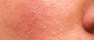
Causes for concern may be the rapid growth of a mole without any reason, an asymmetrical form of formation (blurred or uneven edges), bleeding, the appearance of cracks, bumps or roughness on the surface, a change in color (darkening, redness). In addition, itching, pain or other unpleasant sensations in the mole area are an alarming sign. During the consultation, the specialist will conduct a full diagnosis, including a blood test and, if necessary, a biopsy of the tumor, after which he will be able to accurately determine whether the mole is benign.
It is important to remember that with age, the likelihood of degeneration of education increases significantly. It is not recommended to start the process and ignore these signs, since skin metabolism worsens in adulthood.
Diagnostics
In the process of diagnosing nevi, the doctor uses both standard methods (history collection, visual examination) and instrumental diagnostic methods. In this case, studies such as dermatoscopy, biopsy and histological analysis of the mole are highly informative.
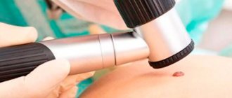
Dermatoscopy
The essence of the dermatoscopy method is to examine the skin under magnification. During the examination, the doctor evaluates the color and structure of the epidermis, identifies the main characteristics of skin rashes, examines the epidermal-dermal junction and the papillary layer of the dermis.
To perform the study, a special device is used - a dermatoscope. This device may resemble a small magnifying glass (manual dermatoscopy) or be a special device that can be equipped with a digital photography function with the possibility of subsequent detailed, even computerized, analysis of various skin structures.
The dermatoscopy method is simple and accessible, but at the same time very informative. Examination of moles by a doctor using a dermatoscope significantly increases the chance of detecting malignant skin tumors, primarily melanoma.
Injury to nevi
Another factor that can trigger the onset of malignancy is trauma to the mole. Moreover, the formation can be injured either over a long period of time, for example, due to constant friction or squeezing by clothing, or as a result of a more serious external influence, for example, when a mole is cut.
If a mole is accidentally damaged, it must be thoroughly treated with a disinfectant solution. If bleeding occurs, it should be stopped using a sterile bandage or gauze. The injured formation should be examined by a doctor as soon as possible in order to determine a further course of action. The specialist may recommend removing the remaining part of the mole or undergoing routine examinations for malignancy over a period of time.
Relationship between the number of moles and the risk of melanoma
There is a lot of research on this issue. If you start reviewing everything, you will end up with a long and boring article that you will not be interested in reading, and I will not be interested in writing.
I have selected some of the largest studies that will help us form our own opinion on this issue.
However, before considering research data, let’s move from the folk concept of “mole” to the medical concept of “nevus”.