What is atheroma
Despite the fact that the pathology looks similar to a skin tumor, it is a sebaceous cyst. The appearance of a lump is associated with a violation of the outflow of glandular secretions and its deposition under the skin layer. The secretion usually accumulates in the duct; it is not removed from the gland and makes its walls stretchable. The greasy contents gradually mix with dead organic substances, cholesterol inclusions, drops of fat, and dead keratinized epidermis. The inside of the neoplasm is lined with flat epithelium.
The structure of skin atheroma can be divided into several elements:
- cavity with contents;
- capsule;
- hole (does not always happen);
- leather;
- subcutaneous fat tissue.
The disease is equally common in men and women. It is a common pathology in skin appendage surgery. Most often, a lump is found on the head due to the presence of a large number of sebaceous glands there.
Removal of atheroma on the head
Removal of an atheroma cyst is carried out when inflammatory processes are completely absent. If there is redness, swelling, or suppuration, the cyst is opened and its contents are removed. And only after the inflammatory process has stopped, you can begin to completely remove the formation and its capsule. Removal of atheromas on the head is usually performed on an outpatient basis using local anesthesia. The operation can be performed in any of three ways. In the traditional way, with a laser scalpel or using a radio wave electrode. At the earliest stages of formation, laser cauterization is applicable. Large cysts on the head are opened and removed by traditional surgery, or a radio wave scalpel is used to perform the operation, which leaves no traces.
Causes of atheroma
The specific reason for the appearance of atheroma is the disruption of the sebaceous glands, but a number of predisposing factors lead to this. They are the ones who provoke problems with the separation of sebum, its increased production and blockage of the duct to the outside, which can lead to the appearance of skin atheroma. Factors in the development of the disease include:
- heredity;
- hormonal imbalance (especially in adolescence, when taking hormonal drugs, etc.);
- hyperhidrosis – increased sweating;
- untimely exfoliation of dead skin;
- high viscosity of sebum (individual feature);
- problems with excretory function;
- increased skin contamination;
- narrowness of the gland ducts;
- insufficient hygiene;
- chronic skin trauma;
- negative impact of the environment;
- the use of antiperspirants, which narrow the ducts and clog the gland.
Proper care
If the operation was performed using a scalpel, the surgeon will apply stitches. When using the laser or radio wave method, no stitches are applied. Stitches will be removed approximately a week after the procedure, and sometimes sooner.
If the removal of atheroma involved a serious surgical intervention, for example, a procedure was carried out for enucleation of a giant cyst located on the head under the hairline, bandages will be required.
After removing a large purulent cyst or atheroma, the wound is treated with spruce to prevent inflammation of the scar.
For two weeks after surgery (this is how long the healing process lasts), you must follow the doctor’s recommendations:
- Do not wet the removal area for at least two days.
- If indicated, use anti-inflammatory absorbable agents topically.
- If necessary, treat the wound daily with an antiseptic.
- Wear a bandage or a clean hat (if the operation was performed on the scalp). This will prevent infection from entering the wound.
The speed of wound healing and the likelihood of a scar depends not only on the methods used by the surgeon, but also on the characteristics of the patient’s skin.
Symptoms and localization
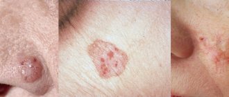
It is not difficult to recognize atheroma by visual examination; additional diagnostics, as a rule, are not required. It looks like a neoplasm located under the skin and comes in various sizes - from a button to a chicken egg. When palpated, the lump is dense, covered with skin, since it is fixed in the area of the gland and does not move freely during palpation. A black dot can be found on the surface - this is a clogged duct of the sebaceous gland. When pressed, fat masses come out.
When palpated, the neoplasm is dense and painless if there is no inflammatory process. Sometimes, in places where visibility is difficult, patients cannot even assume the presence of pathology. When the cyst is small, it does not cause discomfort, and its enlargement occurs quite slowly. For tumors 3–4 cm in diameter, the neoplasm causes discomfort and is a significant cosmetic defect. If we talk about the face, where the skin is thin, patients prefer to remove even small tumors up to 1 cm in diameter, since the lump becomes noticeable.
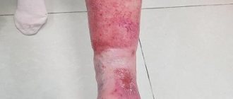
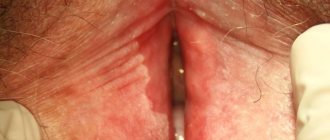
Atheroma is located in places where sebaceous glands accumulate, actively secreting secretions. Most often, a tumor appears in the head area, growing to large lumps. Skin atheromas are also diagnosed on the back between the shoulder blades, in the neck, on the face, near the tailbone and even on the genitals. They can become encysted with connective tissue, remaining the same painless bumps for a long time.
Symptoms of skin atheroma become more obvious if it suppurates. This usually happens with long-existing large tumors. Typical signs of an inflamed cyst:
- pain at the site of the tumor;
- redness and local hyperthermia;
- subcutaneous abscess;
- swelling of the skin;
- increased body temperature;
- involvement of surrounding structures, such as lymph nodes, in the inflammatory process.
Atheroma during inflammation can open on its own, especially if there was a blocked duct. Purulent discharge and the contents of the gland are released through it. It is better to remove the cyst in a timely manner so as not to worsen the situation. The transformation of pathology into a malignant neoplasm occurs extremely rarely.
Diagnostics
Diagnosis of the disease is carried out on the basis of an individual examination, according to the clinical picture:
- localization;
- appearance;
- symptoms;
- visual observation, the presence of a (black dot) excretory duct;
- the presence of clear boundaries of the tumor;
- no pain.
A more accurate diagnosis is obtained using morphological and histological studies. In order to exclude lipoma and malignant tumors.
Treatment
Treatment of skin atheroma is not required if it is small in size and not inflamed. In this case, it is enough to observe the neoplasm. As the size of the tumor increases, if it causes discomfort and is a cosmetic defect, it is removed surgically. Surgery is performed under local anesthesia. There is no conservative treatment for the disease.
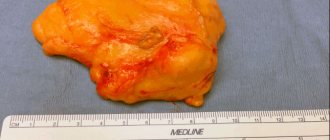
The intervention is performed in a clinic and does not require hospitalization if the skin atheroma is small and located in a place convenient for the intervention. The tumor is excised along with the capsule, as this gives a better effect and reduces the risk of relapse in the same place. An uncomplicated tumor is operated on in one of the following ways:
- an incision is made at the site of the neoplasm in its center, the contents are removed, and then the capsule is excised;
- an incision is made along the edge, the cyst is moved along with the intact capsule and removed with a surgical spoon;
- enucleation using two incisions and jaws, which remove the neoplasm along with the capsule.
The latter method of surgical intervention is the most effective. After removal, self-absorbing internal sutures and atraumatic thread sutures are applied, which must be removed after a week.
Possible complications
A very common complication of atheroma is its suppuration. After the addition of a bacterial infection, the tumor increases in size with the appearance of:
- Secondary inflammation of atheroma
- Development of suppurative processes in the skin and subcutaneous tissue
- Re-formation of atheroma after its spontaneous opening or poor-quality removal (relapse)
- Scars after elimination of a neoplasm
- In very rare cases, it is possible to transform an atheroma (epidermoid) into a malignant tumor; retention atheromas never become malignant.
If left untreated, the atheroma membrane may rupture on its own, which is accompanied by the release of thick pus with the sebaceous contents of the tumor. Further spread of infection to nearby tissues threatens the development of phlegmon. In such a case, the patient requires immediate surgical intervention with drainage of the affected tissue area.
Specialists at the Shifa clinic recommend removing atheroma in Moscow even before complications develop. Elimination of a blocked sebaceous gland at an early stage of development prevents deterioration of the patient's condition and guarantees a good cosmetic effect.
Prevention
There are no effective methods of prevention. If there is increased secretion of the sebaceous glands, atheromas may appear in places near removed bumps or in new locations. As a preventative measure, you can eat less fatty and high-carbohydrate foods so as not to stimulate the glandular apparatus. Use antiperspirants as little as possible; your skin is oily. Use the right skincare products for your skin type to ensure that your pores are free of sebum.
Removal of atheromas on the face
Self-treatment by squeezing is unacceptable. You can get infected and have serious complications. Neoplasms in the form of atheromas are localized in areas where many sebaceous glands are located. There are many such zones on the face. Most often it can form on the forehead, eyebrows, nasolabial triangle and cheeks. Inflammation, pain, suppuration, signs of fluctuation (formation of liquid masses under the skin) provide an indication for removing the cause of discomfort - the cyst. It can only be removed through surgery. Traditionally, using laser surgery and using radio wave excision. During the operation, using traditional surgery, an incision is made in the skin over the cyst, the contents are removed and the capsule is peeled out. When using the laser method of surgery, a laser scalpel is used, and when using the radio wave method, a surgical electrode is used that emits a high-frequency current. When using alternative methods, secondary formations of cysts in the same place are excluded.
Features of the procedure
The duration of the operation is from 60 to 90 minutes, depending on the complexity of the particular case. First, an antiseptic treatment of the required area of skin is carried out and drugs for local anesthesia are administered.
The specialist, directing the laser beam to the area of skin located directly above the atheroma, dissects the skin.
Next comes the stage of evaporating the accumulated liquid. Then the beam destroys the remains of the capsule and seals the vessels. If a large tumor is to be removed, a cosmetic suture is placed at the site of the skin incision.
In most cases, this manipulation is not necessary - after 5-10 days, the hole formed at the site of the atheroma becomes invisible.
In the case when an active inflammatory process has begun inside the cyst and it has increased to its maximum volume, a combination of surgical and laser techniques is recommended. In this case, general anesthesia is used.
The skin is cut with a laser, and the capsule with liquid itself is removed mechanically. Then laser treatment of the resulting cavity follows again in order to seal the vessels and burn out the remaining tumor. In this case, the postoperative recovery period increases to 3 weeks.
Preparation for the procedure
After the patient has been diagnosed with atheroma, he is indicated for surgery to remove it. Preliminary preparation for the procedure includes passing the necessary list of general tests and ultrasound examination of the area in which the cyst is located. This will allow the specialist to see the full picture and determine how difficult the upcoming operation will be. The main purpose of laser exposure to an encapsulated sebaceous gland is to completely remove the fluid inside and destroy the cyst shell. In the case when even a small fragment of the shell remains inside, there is a high probability of relapse of the disease.
3 days before the date of surgery, the patient is advised to completely stop drinking alcohol and smoking. Additionally, there are restrictions on taking certain medications—blood thinning medications are prohibited. If it is impossible to completely refuse drugs from this group, the patient must notify the specialist who will perform laser removal of the atheroma.