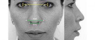
A saddle nose is a nose with a recessed dorsum. A concave nose creates an aesthetic defect - the face looks ugly, simple, the profile causes aesthetic discomfort.
A saddle nose can be accompanied by various dysfunctions of the nose - difficulty in smelling, nasal breathing, nosebleeds often occur, and headaches.
A saddle nose defect can be congenital or acquired as a result of injury. Boxers often suffer from nasal injuries that result in saddle defects.
Degrees of saddle nose deformity
The saddle nose defect has several degrees of severity:
- Normal projection of the tip of the nose and slight concavity of the back of the organ.
- The tip of the nose is turned up, the concavity of the nasal bridge is moderate.
- There is a structural defect of the tip of the nose with a pronounced retraction of the back of the organ.
- A pronounced concavity of the nasal dorsum is accompanied by pronounced defects in the parts of the organ.
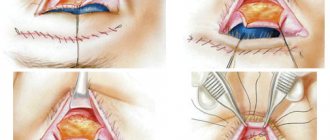
Concavity of the nose can be observed not only in the bone part, but also in the area of the nasal septum; combined nasal concavity is more common. When performing saddle nose rhinoplasty, the concave area of the nose is filled with a graft, restoring the normal shape of the organ.
The following implants are used to fill the concavity of the nose:
- Allografts. To make a transplant, plastic materials are used that easily take the desired shape - silicone, polytetrafluoroethylene and other materials.
- Autografts. The material that is taken from the patient during surgery is cartilage, skin and fat grafts. The material is taken from the tibia, ear cartilage, nasal septum cartilage, rib, and ilium.
The method of correction and the extent of surgical intervention depend on the degree of nasal concavity. At a consultation with a plastic surgeon after a full examination, a decision will be made on the method of correction.
Contour plastic
How to remove a cavity on the nose without surgery? A small depression on the bridge of the nose can be corrected using contouring methods - injections of special fillers based on hyaluronic acid or calcium hydroxysapatite. This is possible only with 1st degree of saddle nose deformity.
When introducing fillers based on hyaluronic acid, the effect lasts up to six months, then the procedure must be repeated, as the filler is absorbed. If Radiesse (a preparation based on calcium hydroxysapatite) was used, the effect lasts from one to two years. But the drug must be administered in minimal doses – tenths of a milliliter, often in more than one procedure, so as not to introduce too much.
If you want to get rid of a hollow nose forever, it is better to undergo rhinoplasty.
Stages of preparation and performance of rhinoplasty
The first step in preparing for surgery is visiting a plastic surgeon. The surgeon will refer you for tests and consultation with an otolaryngologist. Test results are valid for 10 days. Preparation and performance of the operation consists of several stages:
- On the day of surgery, the doctor makes marks on the skin in the patient’s nose area, then takes photographs in order to later compare the results before and after rhinoplasty.
- Anesthesia is administered.
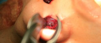
- Rhinoplasty is performed, the duration of which is from one to two hours.
- The patient is transferred to a ward, where he can stay from one to several days, depending on the condition.
Recovery period
The patient spends 2-3 days in the hospital under 24-hour medical supervision.
- Patients have 1-2 bed rooms equipped with showers.
- Each one has an individual monitor for vital functions, a functional bed with oxygen supply, and a personal TV with headphones.
- Delicious food takes into account the doctor’s prescriptions and recommendations.
After discharge, the patient visits the doctor according to his recommendations, observes the medical and protective regime:
- excludes physical activity for 3 months after surgery;
- does not use glasses for 2-3 weeks, replacing them with lenses. The temple of glasses can injure the operated nose;
- Do not eat food that is too hot or cold to avoid causing bleeding.
The final result of rhinoplasty can only be assessed six months or even a year after the operation, when the skin has healed and all traces of the intervention have disappeared.
Diagnosis of the disease
At the department of our medical center GMS Hospital, you will undergo a thorough diagnosis, which includes:
- general clinical and physical examination;
- collection of life history data, diseases, allergy history, questioning of complaints;
- laboratory research methods - general and biochemical blood tests, blood clotting indicators (coagulogram);
- instrumental diagnostic methods - rhinoscopy, CT, radiography of the skull bones;
- additional methods – MRI, endoscopy;
- consultation with other specialists.
Dystrophies (stigmas)
The occurrence of a number of dystrophies in congenital syphilis is not associated with exposure to Treponema pallidum (the causative agent of syphilis) and does not have any diagnostic value. They develop with many infectious diseases and intoxications, for example, with parental alcoholism. Stigmas can indicate that a child may be affected by syphilis and help in making a diagnosis.
Rice. 16. Enlarged and protruding frontal and parietal tubercles without a dividing groove (“Olympic forehead”). The anomaly occurs in 36% of patients.
Rice. 17. A high hard palate (“lancet” or “Gothic”) occurs in 7% of cases.
Rice. 18. Diastema (distance, gap) between the central incisors. Most often found on the upper jaw.
Rice. 19. A thickened sternal end (usually the right) of the clavicle (Ausitidian-Igumenakis symptom) occurs in patients with congenital syphilis in 25% of cases. The cause of the pathology is hyperostosis. In 13 - 20% of cases with congenital syphilis, there is an absence of the xiphoid process (Queir's axiphodia).
Rice. 20. A shortened (infantile) little finger (Dubois symptom) is recorded in 12% of cases with congenital syphilis. The little finger may be curved and turned towards the other fingers (Hissar's symptom).
Rice. 21. Stigmas indicating congenital syphilis may include spider fingers—abnormally long and narrow fingers (arachnodactyly).
Rice. 22. Girls and boys with congenital syphilis may experience hypertrichosis - growth of hair on the forehead (Tarkovsky hypertrichosis).
Symptoms of the disease
Often, patients with deformation of the external nose complain of:
- change in shape;
- acute or chronic sinusitis;
- nasality;
- snore;
- frequent vasomotor rhinitis;
- violation of nasal breathing;
- cosmetic defect;
- nosebleeds.
Symptoms can appear in combination or individually.
The specialists of our medical center will conduct a thorough diagnosis and select the necessary method of surgical treatment of defects.
Reliable signs of late congenital syphilis
Jonathan Getchinson, an English dermatologist, surgeon, syphilidologist and ophthalmologist in 1852 described the symptoms of late congenital syphilis - labyrinthine deafness, parenchymal keratitis and dental damage. At the suggestion of the French dermatologist and venereologist A. Fournier, these signs began to be called Hutchinson’s triad. Some symptoms of tabes dorsalis are also named after this scientist.
Rice. 2. Pictured is Jonathan Getchinson.
Abnormalities of dental development in congenital syphilis
The triad of congenital syphilis includes developmental anomalies (hypoplasia) of teeth. In children with congenital syphilis, pathologies such as Hutchinson, Fournier and Pfluger teeth are recorded. The reason for the development of these hypoplasias is the effect of syphilitic infection on the metabolic processes in the rudiments of the teeth, resulting in the formation of a malformation of the organ.
- D. Getchinson was the first to describe a special form of pathology of the central incisors, in which a semilunar notch of the incisal edge was determined. However, this sign of congenital syphilis, even by D. Getchinson himself, was recognized as reliable only in the presence of 2 more signs - deafness and parenchymal keratitis.
- A. Fournier pointed out that congenital syphilis is characterized not by a semilunar notch, but by a barrel-shaped crown, when the neck of the tooth is larger in size than the cutting edge in the absence of a semilunar notch.
- Another anomaly of dental development in congenital syphilis is Pfluger’s teeth. The pathology is characterized by damage exclusively to the first large molars - a wide neck of the tooth (wider than that of the chewing surface) and significantly underdeveloped cusps. In this case, the tooth takes on a kidney-shaped appearance.
- Pfluger's teeth, an additional tubercle on the side of the tongue on the first molars (Carabelli tubercle), thinning of the free edge of the canine (Fournier's pike tooth), purse-shaped fangs, widely spaced upper teeth, dwarf teeth and the growth of teeth on the hard palate are probable signs of congenital syphilis.
The formation of pathology of permanent teeth occurs when they are formed - at 6 - 7 months of pregnancy, when the placental blood circulation is already functioning and treponema pallidums penetrate into the fetus, exerting their negative effect. The formation of baby teeth occurs in the fetus even before the transition to placental blood circulation, so this pathology is not observed in them.
Rice. 3. In the photo: a) Fournier’s teeth, b) Pflueger’s teeth.
Rice. 4. Anomalies of dental development in congenital syphilis.
Hutchinson's teeth
The triad of symptoms of congenital syphilis in children includes such a symptom as Hutchinson's teeth. This pathology occurs in 5 - 20% of cases. Hutchinson's teeth are a special form of hypoplasia, in which changes are recorded in the upper incisors:
- the area of the neck of the teeth is wider than the area of the cutting edge within 2 mm, so the crowns of the teeth take on the shape of a screwdriver or barrel-shaped;
- along the lower edge of the incisors there are semilunar notches;
- the semilunar recess is sometimes covered with enamel, sometimes the enamel is present only at the corners of the tooth, sometimes there is no enamel at all, often the enamel covers the entire recess, but quickly wears off;
- as soon as the teeth have erupted, 3-4 spines can be seen on the cutting edge in the middle, which quickly break off;
- gradually the incisors wear down and by the age of 20 the teeth become short and wide, often with carious edges.
Treatment of dental pathology consists of restoring the size and anatomical shape of the organ using artificial crowns or composite materials after the final formation of the permanent bite.
Rice. 5. The photo shows Hutchinson’s teeth. Robinson-Fournier scars are clearly visible along the edge of the lower lip.
Syphilitic parenchymal keratitis
Parenchymal keratitis is the most common among Hutchinson's triad and accounts for 48% of cases. The disease affects the middle layer of the cornea (middle stroma). Lacrimation, photophobia, pain, blepharospasm and corneal opacification are the main signs of syphilitic parenchymal keratitis. The disease leads to decreased or complete loss of vision. Bilateral lesions are observed in half of the patients. Often parenchymal keratitis is the only sign of late congenital syphilis.
Initially, specific inflammation develops in one eye. The second eye is affected after weeks, more often after 6-10 months, but maybe after years.
Parenchymal keratitis can manifest itself in limbal, central, annular and avascular forms.
- The disease begins with clouding of the cornea, which is focal or diffuse. In the diffuse version, the clouding covers the entire cornea; it has a milky color and greater intensity in the center. In the focal version, the turbidity has the appearance of cloud-like spots.
- After 4 - 6 weeks, a ciliary or ciliary injection (vasodilatation) appears around the edge of the cornea (limbus), which has a purple color. Newly formed vessels grow deep into the cornea, sometimes there are so many of them that the cornea takes on the appearance of a ripe cherry. The vessels of the outermost layer of the eye, the conjunctiva, dilate. The process takes 6 - 8 weeks. Often, along with parenchymal keratitis, patients develop inflammation of the iris and vascular membranes of the eye, the ciliary body (iritis, chorioretinitis, iridocyclitis) and optic nerve atrophy.
- The period of reverse development proceeds slowly. The cornea brightens along the periphery, and the cloudiness in the center of the eye resolves. Vision is restored. Photophobia and pain decrease. The recovery continues for more than a year.
The inflammatory process takes a long time and often ends in clouding of the cornea, which manifests itself in the form of weakened visual acuity or complete blindness. A significant degree of vision loss is observed in 3-4 patients. No earlier than one year after the disease, relapses of parenchymal keratitis may occur, often occurring in the form of an avascular form. Empty vessels are always detected during ophthalmoscopy, so the diagnosis of previous syphilitic chorioretinitis can be made retrospectively. All patients have positive specific serological reactions.
Rice. 6. Parenchymal keratitis in late congenital syphilis.
Syphilitic labyrinthitis (labyrinthine deafness)
Labyrinthine deafness is rarely recorded - in 3 - 6% of cases, between the ages of 5 and 15 years, mainly in girls. When the disease occurs in the labyrinth (usually on both sides), hemorrhagic inflammation develops, which is often accompanied by noise and ringing in the ears. Sometimes the disease is asymptomatic and ends with sudden deafness.
If damage to the labyrinths develops in children under four years of age, the child may become deaf and mute. Syphilitic labyrinthitis is difficult to treat.
Rice. 7. Inflammation of the labyrinth, periostitis and damage to the auditory nerve during syphilis lead to deafness.
The detection of at least one reliable sign from Hutchinson's triad and the receipt of positive serological reactions indicates the presence of late congenital syphilis in the child.
Other signs of late congenital syphilis
Lesions of the skeletal system
Osteoperiostitis and periostitis, gummous osteomyelitis and osteosclerosis are the main types of bone lesions, which occur in 40 - 50% of congenital syphilis. The lower legs (59%), nasal bones (18%), forearms (10%), skull bones (5%), and hard palate (4%) are affected.
Lesions of internal organs
Pathology of internal organs with congenital syphilis is registered in 20 - 25% of cases. The liver, spleen and kidneys are most often affected. With syphilitic damage to the heart, all its membranes, valves and vessels are affected. There is dysfunction of the thyroid, pancreas, thymus and gonads, pituitary gland and adrenal glands.
Nervous system lesions
Pathology of the nervous system with congenital syphilis occurs in 27 - 43% of cases. Of these, more than 50% are caused by damage to the brain, 32% to the spinal cord, and 11% to the tabes dorsalis. In 23% of cases, mental disability develops. With congenital syphilis, mental retardation, speech disorder, hemiplegia and hemiparesis, tabes dorsalis, and Jacksonian epilepsy are recorded. The child constantly suffers from headaches. Secondary atrophy of the optic nerves develops.
Syphilitic chorioretinitis
Syphilitic chorioretinitis leads to changes in the retina and choroid of the eye. Visual acuity is not reduced. Optic nerve atrophy leads to vision loss. With syphilis in children, a combination of chorioretinitis and damage to the optic nerve is more common.
Rice. 23. The photo shows chorioretinitis in early congenital syphilis. The disease is characterized by the “salt and pepper” symptom, which is characterized by the appearance of lumps of pigment and zones of depigmentation along the periphery of the fundus.