For most women, sagging breasts are a serious aesthetic problem that prevents them from loving their own body and causes complexes, so the issue of treating mammary ptosis is especially pressing. Involutional aging processes, genetic predisposition and the minute-to-minute impact of the Earth's gravitational forces contribute to natural ptosis of the skin, body tissues and some internal organs. If we are talking about prolapse of the mammary glands, then in medicine it is called “ mastoptosis.”
" With mastoptosis, a woman’s breasts lose their elastic shape and look saggy. Special care for preventive purposes only helps to slow down the process of breast ptosis. This includes: contrast showers, massages, wraps, cosmetics, mesotherapy, biorevitalization of breast skin, as well as some other salon procedures. However, with severe mastoptosis, conservative methods are no longer effective. In this article we will talk about possible ways to treat mammary ptosis. The fastest and most effective of them is still mastopexy (mammopexy) - plastic surgery to tighten the mammary glands with or without the use of implants. But first, let’s look at why breasts sag in the first place: what factors contribute to this?
Causes of breast ptosis
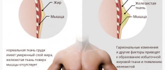
The volume and appearance of a woman’s bust is determined by: the condition of the pectoralis major muscle and ligaments, the ratio of fatty and glandular tissue formed into lobules.
- Age-related changes - over time, after about 45 years, the process of fibro-fatty involution of the mammary glands begins in a woman’s body. It consists in the degradation of glandular tissue and its gradual replacement with adipose tissue. This occurs against the background of hormonal changes, which are especially active during menopause. These changes lead to a deficiency of the sex hormones estrogen and the gradual “fading” of the body. The synthesis of collagen and elastane, which are structural components of connective tissue, dermis and ligaments, slows down. As a result of fibrofatty involution, a woman’s breasts lose their aesthetically attractive appearance and, under the influence of the natural gravitational forces of the Earth, droop.
- Hereditary factor - congenital features of the synthesis of collagen and elastin determine the rate of tissue aging and predisposition to gigantomastia, and therefore to mastoptosis.
- Pregnancy and breastfeeding - during pregnancy, a woman’s breasts increase in volume due to the filling of the glandular lobules with milk. At the same time, the load on the ligaments increases, and the skin stretches. After completion of lactation, the volume of the gland gradually decreases. Due to stretching of the skin, the breasts look “empty” and saggy. In addition, uneven distribution of milk, as well as feeding a baby from one breast, can lead to noticeable asymmetry.
- Rapid weight loss or weight gain - fatty tissue not only actually determines the shape of the mammary glands. In case of rapid weight loss, part of the breast volume is lost, and the skin does not have time to recover. Adipocytes in adipose tissue store and synthesize hormones that are responsible for metabolism. On the contrary, rapid weight gain leads to an increase in the volume of the mammary glands and stretching of the skin.
- Endoprosthetics of the mammary glands with implants that are too large - breast ptosis after mammoplasty occurs the faster, the larger the prosthesis and the less elastic the skin.
- Smoking and alcohol abuse.
Lipoma
Breast lipoma is a benign disease characterized by the growth of fatty tissue in the breast area. The appearance of such tumors is typical for women after 40 years of age. Lipoma itself does not pose a threat to the health and life of a woman. It creates only a cosmetic defect: due to its ability to reach a large size, the lipoma deforms the mammary gland.
The only treatment option is surgery. Malignancy is extremely rare.
Breast lipoma can be either single or multiple (lipomatosis). It can include not only fat cells, but also other tissues. The following types of disease are distinguished:
- lipofibroma – predominance of adipose tissue;
- fibrolipoma – the presence of fibrous (connective) tissue;
- angiolipoma - riddled with small blood vessels;
- myxolipoma – the presence of mucus cells;
- myolipoma - muscle fibers are detected.
Typically, lipoma does not manifest itself with characteristic symptoms. It can be detected independently by palpation (palpation). It is inactive, painless and has a dough-like consistency. Often the lipoma reaches up to 2 cm in diameter, and can also be large in size - up to 10 cm. In some forms of the disease, a woman may feel pain and discomfort in the chest.
The following causes of the disease are identified:
- blockage of the outlet duct of the sebaceous glands;
- metabolic disorders;
- excess body weight;
- hereditary predisposition.
The following factors are predisposing:
- sedentary lifestyle (lymph movement is impaired);
- unbalanced diet, which is dominated by fats and carbohydrates;
- changes in hormonal levels;
- mechanical injuries of the mammary gland;
- long-term use of hormonal contraceptives;
- the appearance of stretch marks on the chest due to pregnancy and lactation;
- bad habits (smoking, drinking alcohol, drugs);
- X-ray and ultraviolet irradiation;
- incorrectly selected underwear;
- unfavorable environmental conditions.
Stages of mastoptosis
According to the classification proposed by American plastic surgeons, there are 3 degrees of mastoptosis.
Breast ptosis of the 1st degree is the initial stage of breast ptosis, in which the nipple is at the level of the submammary (inframammary) fold or slightly lower.
Breast ptosis of the 2nd degree - the nipple falls less than 3 cm below the inframammary fold.
Breast ptosis of the 3rd degree - the nipple is located more than 3 cm below the level of the fold and looks down.
False ptosis (mastoptosis) is characterized by the normal position of the nipples and sagging of the lower pole of the mammary glands - it is located under the submammary fold.
Prevention
The main preventive measure is regular and systematic breast examination by a mammologist or gynecologist:
- women 30-35 years old - ultrasound (annually), mammography (every 2-3 years);
- after 40 years – ultrasound of the mammary glands, mammography (annually);
- after 50 years - ultrasound, mammography (2 times a year).
The following measures will help prevent the disease:
- timely and complete treatment of inflammation of the genital organs;
- maintaining hormonal balance;
- timely implementation of childbearing function;
- absence of abortions;
- maintaining an active and healthy lifestyle;
- correct selection of underwear;
- avoiding stress, nervous tension and negative emotions;
- moderate physical activity;
- maintaining weight within recommended limits;
- balanced diet;
- complete rest;
- moderate passion for solarium and sunbathing;
- careful and attentive attitude towards your body.
Each woman is independently responsible for her health. It is always worth remembering that it is easier to prevent a disease than to start it and subsequently undergo hormonal therapy or, what is much worse, surgical intervention.
Treatment of breast ptosis
With the help of physical exercise and diet, you can reduce the volume of fatty tissue, but this does not affect the condition of the connective and glandular tissue, which also gives the shape of the breast. In addition, with mastoptosis, the problem is also stretching of the skin.
On the contrary, breast lifting in the salon (mesotherapy, biorevitalization, massage and other cosmetic procedures) is aimed at improving the condition of connective tissue. These non-surgical methods require regular repetition and will not correct severe breast ptosis. Injections of vitamin complexes, hyaluronic acid and other active substances will help achieve a qualitative improvement in the condition of the skin, restoring the synthesis of collagen and elastane fibers. Cosmetic procedures can slow down the aging process and improve the appearance of the breasts, but they cannot correct breast ptosis or breast shape.
Fibroadenoma
Fibroadenoma (fibrosis) is a benign neoplasm that develops from connective tissue. The tumor occurs against the background of the production of hormones - estrogens. The disease is predominantly observed in women 20-30 and 40-50 years old, sometimes occurring at a young age (12-20 years). It does not pose a threat to the health and life of a woman. In most cases, fibroadenoma is not prone to progression and development of complications.
Sometimes it can reach large sizes and thereby deform the mammary glands. Only fibrosis of gigantic size is prone to malignancy. As a rule, the method of treating a tumor is surgical, but if it is small in size (up to 5-8 mm), therapeutic treatment is carried out aimed at its resorption.
Pathology is of the following types:
- Nodular - deformation of connective tissues near the glandular ducts or their ingrowth into the ducts;
- Leaf-shaped - characterized by an extreme speed of development and an increased threat of degeneration into sarcoma. This is a terrible disease that leads to breast amputation and even death.
Fibroadenoma is asymptomatic for a long time. The general symptoms are not pronounced. It is manifested by compactions in the body of the mammary glands, which have the following features:
- painlessness, density and sometimes mobility of the formation;
- clearly defined boundaries;
- variation in tumor size;
- lack of direct dependence of size changes on the phase of the menstrual cycle;
- intensive growth of education (sometimes).
In medicine, the exact causes of fibroadenoma are still unknown. There are a number of factors contributing to the development of pathology:
- genetic predisposition;
- puberty;
- diseases of the endocrine system and thyroid gland;
- pregnancy;
- frequent abortions;
- nervous and physical exhaustion;
- severe frequent stress;
- obesity;
- incorrect selection of oral contraceptives or their uncontrolled use;
- mammary gland injuries;
- overheating of the mammary glands;
- abuse of solarium and sunbathing.
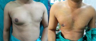
How to correct ptosis with a surgical lift (mastopexy)
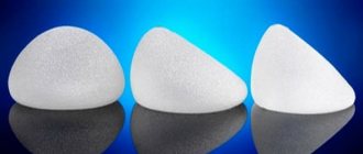
In case of breast ptosis of the 1st degree, the bust can be restored to its beautiful shape with the help of endoprosthetics - an operation to enlarge it with implants. The problem of more advanced ptosis of the 2nd or 3rd degree is solved by mastopexy (mammopexy), that is, a breast lift. The essence of this plastic surgery is the resection of excess skin, fat and glandular tissue, as a result of which it is possible to raise the nipple-areolar complex. If there is insufficient volume of breast tissue, mastopexy can be combined with endoprosthetics and thus increase breast size. If a woman has noticeable asymmetry, it can be eliminated by combining a lift with reduction mammoplasty or endoprosthetics. In general, mastopexy is performed under general anesthesia for 1-3 hours.
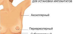
There are three main methods of breast lift:
Periareolar mastopexy
It is used to eliminate mild and uncomplicated ptosis of 1st or 2nd degree. The plastic surgeon makes an incision around the areola to remove excess skin.
Vertical mastopexy
The method is used for breast ptosis of 2 and 3 degrees. The plastic surgeon makes two incisions: one around the areola, and the other vertically downwards from the circumference by 7-8 cm.
Anchor (or “T-shaped”) mastopexy
The most effective way to eliminate advanced stage 3 breast ptosis. The technique involves a composition of three neat incisions: vertical, around the areola and along the contour of the inframammary fold.
Intraductal papilloma
Intraductal papilloma is a cyst-like formation with a disruption of the structure typical of secretory cells. Diagnosed in 10% of cases of breast diseases in women. It can develop at any age - from puberty to postmenopause. This is a benign tumor that can progress to malignant. Its size varies from 1 mm to 3 cm in diameter.
There are two main types of intraductal papilloma:
- central – location of cystadenomas in the areola area;
- peripheral - the occurrence of papillomas in any peripheral area of the mammary gland ducts, often multiple.
Often the disease is asymptomatic for a long period, and its diagnosis occurs by chance during an examination by a doctor. Symptoms of the disease include:
- discharge from the nipples of various types, increasing when pressing on the chest. May be transparent, bloody, whitish or greenish in color;
- pain in the area where the tumor develops, aggravated by pressure or when wearing tight clothing;
- palpation of a small elastic knot.
With the development of inflammation of the tumor and surrounding tissues, signs are observed:
- increased body temperature;
- severe tenderness of the mammary glands;
- swelling, breast enlargement;
- redness of the mammary glands;
- change in color and consistency of discharge.
The causes of intraductal papilloma are:
- hormonal imbalance;
- changes in hormonal homeostasis (ovarian dysfunction, oophoritis, adnexitis, abortion, obesity, stress, etc.);
- long-term uncontrolled use of hormonal contraceptives;
- smoking;
- influence of viruses;
- ionizing radiation;
- no history of pregnancy.
Why should a breast lift be done at the clinic? N.I. Pirogov?
Our plastic surgeons know how to correct breast ptosis, giving it a beautiful and toned shape again without noticeable post-operative scars. Look at the portfolio of our specialists and you will be convinced of this. Since 1999, doctors of the clinic named after. N.I. Pirogov in St. Petersburg successfully perform all types of operations on the mammary gland: from endoprosthetics to reconstruction after mastectomy. If, after some time, you are faced with the problem of breast sagging after primary mammoplasty, our plastic surgeons will not only solve this problem, but will take care of high-quality and long-term preservation of the result. We offer our patients:
- services of the city's leading plastic surgeons;
- medical offices equipped with modern equipment at the level of European private clinics;
- stay in comfortable wards for 1-2 people with meals, nursing care and round-the-clock supervision by a doctor on duty;
- special promotional offers (all inclusive) at affordable prices;
- own laboratory, which allows you to quickly obtain accurate diagnostic results, and intensive care;
- cosmetology department services, which can be useful for the prevention and treatment of mild breast ptosis;
- organizational assistance from a supervisor for patients from other cities.
Oleogranuloma
Oleogranuloma is a benign tissue compaction altered by inflammation as a result of a foreign body entering the mammary gland. As a rule, it occurs as a result of surgery. Silicone implants, synthetic threads, etc. can act as a foreign body.
The symptoms of oleogranuloma and cancer are similar to each other, so it is often mistaken for oncology. The final verdict can be made by the doctor after a thorough examination.
There are the following types of breast oleogranuloma:
- injection (artificial) – formed as a result of the introduction of various fats and oils;
- post-traumatic – occurs after trauma to the chest (blow, fall, improper massage). A frequent companion is necrosis of fatty fibers;
- periinflammatory – location near the source of the inflammatory process, gradual movement to fatty tissue, which turns out to be destructive;
- spontaneous (cutaneous) – causes unknown.
Oleogranuloma can be identified by the following signs:
- the appearance of voluminous nodules under the skin of the breast;
- lumpy and dense consistency of the skin surface;
- redness in the area of formation;
- pain and discomfort in the area of compaction;
- retraction of the skin over the tumor;
- increasing education over time;
- formation of ulcers, discharge of pus through fistulas (in advanced forms of the disease).
Causes of oleogranuloma:
- breast injuries;
- surgical interventions (removal of the mammary gland or its lobe, breast augmentation by introducing implants);
- a sharp decrease in body weight;
- ingress of foreign bodies (silicone, gels, ointments, synthetic threads);
- infectious and inflammatory diseases;
- hormonal imbalance;
- radiation exposure.
Diagnostics
In men
The doctor examines the patient, palpates the testicles and mammary glands, and collects anamnesis. Then tests are carried out for hormonal examination. This is a blood donation for FSH, estradol, hCG, testosterone, prolactin, LH, etc. To exclude tumors, CT, radiography, and ultrasound of the scrotum are prescribed. If cancer is suspected, an ultrasound and biopsy are performed.
Among women
A thorough examination and palpation of the patient is carried out in the supine, lateral, and standing positions. Next, mammography, ultrasound, ductography, and MRI are prescribed. If cancer is suspected, a biopsy is necessary.