A beautiful bust is the dream of many women!
The owner of beautiful breasts radiates confidence and sexuality. However, it is extremely important that not only external beauty, but also the health of the mammary glands, gives a woman joy of life and confidence in the future. Every representative of the fair sex needs to avoid pathological changes in the breasts that adversely affect the general condition of the female body. After all, breasts are nothing more than mammary glands - a paired organ of the reproductive system responsible for the secretion of milk - the main food product of future generations.
Women's milk, lac feminum.
Due to the presence of numerous fat globules, it appears as a pure white or bluish-white liquid, is odorless, has a weak sweetish taste and exhibits a neutral reaction.
Its specific gravity is from 1028 to 1034, and its temperature at the time of separation is 38.C. The milk secreted in the first days after birth is transitional milk and is called colostrum puerperarum
;
it is, for the most part, thicker, yellower, grayer, sometimes even thinner than later milk. The fluid released from the breasts during pregnancy is called colostrum of pregnant women, colostrum gravidarum
, and over time gradually takes on the properties of
colostrum puerperarum
. Both of these fluids represent unripe milk and are characterized by the presence of a large number of large nucleated cells loaded with fat globules; they are called Donne bodies, or colostrum bodies. In all likelihood, these are nothing more than lymphatic cells loaded with fat. “Similar relationships are observed in mammals.
Formal components of mature milk. are numerous rounded fat drops, ranging in size from 2 to 5 microns, called milk globules, corpuscula lactis
, and a few lymphatic bodies.
Apparently, these fat globules are surrounded by thin protein membranes. When the milk sits, some of the fat globules separate from the liquid, rise to the top and form cream, cremor lactis
.
Milk completely freed from fat globules represents milk plasma; it also contains casein in solution. Due to the action of enzymes and acidification, milk curdles, i.e. casein falls out. The liquid remaining after casein is removed is whey, serum lactis
.
The most important chemical components of milk can be expressed in the following average figures.
Water 87.79%, solids 12.21%; casein and albumin 2.11% fat 3.79%; milk sugar 5.71%; salts 0.24%. Its other organic components are peptone, urea, and lecithin. Of the inorganic salts, human milk, according to Bunge, contains: potassium 0.0703%, sodium 0.0257%; lime 0.0343%; magnesia 0.0005%; iron oxide 0.0006%; phosphoric acid 0.0468%; chlorine 0.0445%; together this amounts to 0.2287%, while cow's milk contains only 0.8404%.
According to Bouchut, a cubic millimeter of milk contains, on average, 1,026,000 large and small milk pellets.
At the end of the lactation period or if feeding is not carried out, the gland undergoes reverse development, whereby the alveoli become smaller again, and their cavities, as well as passages and epithelial cells, are filled with fat drops and granular decay. During menopausal involution, the mammary glands gradually obliterate, and this process can even extend to the excretory ducts.
It is interesting that even the mammary gland of newborns is already able to produce secretions; that's the name. witches milk, lac neonatorum
. According to some researchers, this secretion is not real milk; but, in any case, both chemical and microscopic examination indicate a significant similarity between these two secretions. Barfour is also inclined to consider this secretion to be milk.
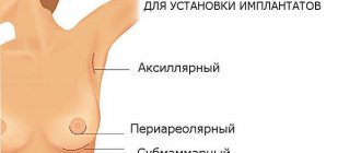
| Fig. 62. Network of lymphatic vessels on the anterior surface of the mamma; subareolar plexus; trunks . | |
Arteries originate from aa. intercostales
(
rr. mammarii mediales et latt.
), and also from
rr.
mammarii , belonging to
a.
mammaria interna [According to PNA -
a.
thoracica interna - IIH].
Arterial anastomoses, into which rr ultimately enter, are considered (without reason)
as an incidental reason for the stronger development of mamma .
mammarii , belonging to
mammaria interna
, with
aa.
uterinae (through aa. epigastrica superior and inferior).
Namely, a.
epigastrica inferior gives
a.
spermatica externa [According to PNA -
a.
cremasterica in men
and a.
lig. teretis uteri in women - IIH], and the latter anastomoses with a. uterina, as it goes to the uterus along lig. teres uteri.
The saphenous veins form the papilla mammae
polygonal network of anastomoses,
plexus venosus mamillae
(see section III.).
The saphenous veins stretch to neighboring large veins, even to v. cephalica
, while deep veins follow the arteries. Lymphatic vessels form a narrow-loop plexus in the skin covering the glands, especially in the areola. The interalveolar connective tissue also contains lymphatic vessels. Fig. 62.
Nerves are quite numerous in the outer skin, in the areola
and
papilla mammae
, but there are few of them inside the gland, and here they are mainly vascular nerves.
They start from nn.
supraclaviculares , from
rr.
cutanei antt. , belonging to
nn.
intercostales II-V-VI , pass through the skin radially to the nipple and in the form of
rami glandulares
, originating from
nn.
intercostales IV-VI , penetrate into the gland itself. Together with the arteries, the glands and their sympathetic plexuses penetrate inside.
In the papillae papilla mammae
tactile corpuscles are found; At the base of the nipple there are separate Vater-Pacinievsky bodies. Near the larger milky passages, V. Krause found end flasks.
The nipple is capable of erection and, when its cutaneous nerves are irritated, lengthens; but erection is determined only by the action of the corresponding smooth muscles, without any participation of the venous spaces.
How to properly prepare for research
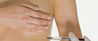
The test for antibodies to TPO is carried out from venous blood. We recommend:
- donate blood during the period from 8 to 11 am, on an empty stomach, after an 8-12 hour overnight fasting period;
- on the eve of the study - a light dinner with limited intake of fatty foods;
- on the day of the test, drink still water and it is better to avoid coffee and tea;
- 24 hours before the test, exclude alcoholic beverages;
- 1 hour before the test it is better to refrain from smoking;
- 24 hours before the study, exclude the use of medications (in consultation with the attending physician);
- 24 hours before the test, eliminate emotional and physical stress;
- do not donate blood immediately after radiography, ultrasound, massage, endoscopic and physiotherapeutic procedures;
- Rest for 10-20 minutes before donating blood.
Unusual phenomena
The nipple can be double, with the normal nature of the gland itself. This is the case when the third mamma
.
It is necessary to distinguish from this those conditions when the nipples and, probably, also the glands corresponding to them develop in two symmetrical longitudinal rows. Under the normal mamma
, there may be one extra one on each side, but there are also several such additional formations (
mammae accessoriae
).
In extreme cases, eight additional nipples are observed, of which three are located above the developed mamma, and two below it. All primates have only one pair of glands. Thus, the appearance of additional nipples is adjacent to the condition that we find in prosimians, etc. See Wiedersheim, Bau des Menschen.
4 Aufl. , 1908. Fig. 12-18.
| Fig. 63. Hypermastia in a 22-year-old girl From Wiedersheim. (According to Neugebaur). | |
Above we have already mentioned the Montgomery glands as intermediate forms between the ordinary sebaceous and mammary glands. Those cases where mammary glands appear in unusual places are quite in accordance with the morphological relationship of both groups of alveolar glands. Such heterotypic mammary glands were observed on the shoulder, in the armpit, on the thigh, etc. Without a doubt, we are dealing here, to a certain extent, with displacements, but still in all cases there was a morphological basis on the face in the form of sebaceous ( or sweat glands.
What do the results mean?
The normal level of antibodies to TPO is up to 34 IU/ml. If a significant increase is detected, then this is a sign of autoimmune inflammation of the thyroid gland:
- thyroiditis: postpartum, Hashimoto's, newborns;
- diffuse toxic goiter.
A moderate increase in the level of antibodies to TPO can also occur in other autoimmune diseases of the thyroid gland: subacute thyroiditis, single and multiple thyroid nodules, thyroid cancer. An increase in antibodies during treatment indicates its insufficient effectiveness, while a decrease indicates the success of therapy.
Embryological and comparative anatomical data.
The first rudiment of the milk secretion organs appears in the form of a paired epithelial strip on the ventral wall of the body. This rudiment is called the milky stripes. The milky stripes thicken and turn into milky plates (milky lines). The individual projections developing from them are called milk tubercles (primary nipples). These latter, becoming flatter and growing deeper like protrusions, give this name. milky points, or points; through the immersion of individual glandular areas, they lead to the formation of milky pockets.
The first germ of mamma
Guldberg (1899) found in dolphin embryos, 18 millimeters long, in the form of vaguely demarcated elevations of the epidermis, which in this place forms a crescent-shaped protrusion. The rudiment appears just at the time when the temporary hind limbs begin to show a tendency to disappear.
In cattle embryos, according to G. Burckhardt, “accessory” or “anal” nipples are found very often in both sexes (in approximately 37% of cases). They are always located between the normal nipples (on one or both sides), or behind the back pair of normal ones; but they are never ahead of the first pair. The mammary organs of modern cattle are subject to the process of caudocranial reduction.
Both in humans and in cattle, they distinguish between hypermastia, with all the signs of a normal, but only very small, mammary organ, and hyperthelia (false nipples).
False nipples and mikromammae
disappear, for the most part, already before birth.
In his article on the morphology and history of the development of normal and accessory mammary glands, G. Schickele
) (1889) comes to the following conclusions: in some mammals (mouse, rat, rabbit, cat) the nipples are arranged in 2 groups, which are characterized by cranial and caudal convergence, as well as the presence of a typical significant gap between the most caudal and the most cranial pair of glands.
Accessory areas of glands in these animals, of course, occur only to a limited extent. But broad-nosed monkeys have extra nipples more often than narrow-nosed monkeys. At Zebus
this condition seems to be observed very often.
In guinea pigs and mice, the milk line is the starting point for the formation of glands. Adult guinea pigs have never had more than 2 teats. But embryonic accessory rudiments of the mammary glands are found in varying numbers (from 2 to 10); but they are at a lower stage of development than the rudiments of the main glands ( Ztschr. fur Morph. u. Antroph. 1899
).
Schmidt, H, Uber normale Hyperthelie menschlicher Embryonen. Anat. Anz. XI, 1896; and Morphologische Arbeiten, herausg. v. G. Schwalbe. 7 Bd.
1896.
Humans do not have a milk line as noticeable from the outside as in mammalian embryos, or it is present only for a short distance. But microscopic examination reveals growths of the epithelium and the rudiments of the mammary glands. In one case there were 8 additional rudiments on each side, 4 above and 4 below the main rudiment. In other cases, a rudiment of 7 to 14 parts was found on one side. The cranial primordia were mostly oriented laterally, while the caudal primordia were oriented medially with respect to the normal primordium. In the lower abdomen, the rudiments were completely absent.
Callius ( Anat. Hefte, Bd. III
, 1897) found in a 15 mm human embryo a milky line 1.5 mm long.
Regarding the comparative anatomy of the mammary glands, the reader can find detailed data in the relevant textbooks. We will only point out here that Monotremata has a paired and “glandular area,” which generally corresponds to areola mammae
higher, is the only external apparatus; The glands here are built according to the tubular type. In echidna, such a device is hidden in a skin pocket. Alveolar mammary glands first appear in marsupials (Gegenbauer).
Bonnett, Die Mammaorgane. Ergebnisse der Anat. von Merkel und Bonnett. Bd. II and VII. — Breslau , E., Beitrage zur Entwicklungsgeschichte der Mammarorgane bei den Beuteltieren. Zeitschr. f. Morph. u. Antrop. IV
, 1902.
The pouch arises through the fusion of small pockets (marsupial pockets), each of which contains one lacteal rudiment (mammary pocket). Marsupialia bag
is homologous to the echidna bursa, in the same way the marsupial pocket of the first is homologous to the corresponding formation in the second;
the mamillary pouch of Marsupialia
corresponds to the glandular field of the echidna.
Marsupial pouches in marsupials can be demonstrated even in placental animals. They correspond to the pockets that surround the Murinae
nipples. In all mammals, the mammary glands arise in a homogeneous manner and should be classified as tubular skin glands. The possibility of diphyletic origin must be excluded.
Unger (1898) also subscribes to this conclusion. The mammary glands are derived from the glomerular glands. Normal, comparative and pathological anatomy, phylogeny and ontogeny unanimously point to precisely this origin of the mammary glands. Neither their structure nor their function gives us the right to compare them with the sebaceous glands.
H. Eggeling
) in his work on the skin glands of Monotremata (Verh. anat. Ges. 1900) criticizes previous attempts to subdivide the skin glands.
As a principle of division, one must use, firstly, the ratio of the epithelium to the lumen, and, then, the method of secretion formation. All glomerular glands, and together with them the mammary glands of higher mammals, should be considered as constantly canalized skin glands secreting in the intravital state; the sebaceous glands and peculiar glandular organs of reptiles should be considered temporarily canalized, secreting through necrobiosis by the skin glands; their secretion is released necrobiotically, as the secreting cells die. v.
Runge, G., Die zunehmende Unfahigkeit der Frauen, ihre Kinder zu stillen. Munchen., E. Reinhardt , 1900.
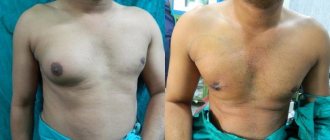
4.Treatment
In cases of a painless benign neoplasm, as a rule, patients are more concerned (or, conversely, not worried at all) about the cosmetic defect, sometimes reaching the degree of deformation of the chest. However, first of all, the real likelihood of tumor malignancy should be taken into account. Therefore, the method of choice is surgical removal of the chest wall tumor, regardless of its histological nature.
If the malignant nature of the tumor is confirmed, further supervision is provided (depending on a number of individual factors and indicators) according to standard protocols for combined oncological treatment, including surgical, radiation and chemotherapy methods. One of the decisive factors in success statistics is the timeliness of detection and initiation of treatment.
Male mammary gland, mamma virilis.
The mammary gland is formed equally in both sexes and develops equally until puberty. But then, the male gland, as a general rule, does not undergo further development. Areola
and
papilla mammae
, however, are present, but the isola is small in size, and the nipple reaches only 2-5 mm in height. In an adult male, the male gland is located in the fourth intercostal space, approximately 12 cm from the midline. The body of the gland reaches 1.5 cm in width and 0.5 cm in thickness and has a whitish color. The lobules and passages are small and short.
The blood and lymphatic vessels exhibit similar relationships as in the female gland. The nerves of the nipple are relatively numerous and partly end in tactile corpuscles. Vater-Pacinian corpuscles were also found at the base of the nipple and on the lower surface of the body of the gland.
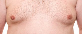
In rare cases, mamma
increases in a man on one side or both sides. This condition is called gynecomastia. Sometimes it is associated with improper development of the reproductive apparatus. There are observations regarding the actual secretion of milk in gynecomasts, but in general they cannot be considered completely reliable.
In men, there is also an excessive number of nipples, the so-called. hyperthelia ( "telia"
, nipple);
according to detailed research f. Bardeleben, hyperthelia is an even more common phenomenon than one might think. Bardeleben says the following about this. “Since Dr. Overweg and I, with the most attentive attitude to the matter and experience, always received higher percentages and since, of course, many cases last year were completely missed by us, and many were doubtful (especially when the case was going on about the axilla
and shoulders) were not taken into account by us, then the above figure of approximately 14% must be considered the most consistent with reality.
Thus, it turns out that every seventh man has one or more redundant nipples. Perhaps even more important than the unexpected frequency of such cases, I think the above instructions regarding their location; the fact is that these redundant “nipples” are not only located along a certain line from the shoulder and axilla
to the pubic region, but are also located in completely specific places, according to which we can assign each of them a corresponding number in order.
Our normal nipple and mamma
are fourth from the top."
Let us remember that, in the given extreme case of redundant mammae
in a woman, on each side, the normal
mamma
was also the 4th in the row.
According to A. Kirchner's observations of 890 men, in 6/7 of the subjects studied, both nipples were placed at the height of the same rib. Of the 763 cases when both nipples occupied the same position, in almost half they were at the height of the 5th rib, in more than 1/3 - at the height of the 4th intercostal space, in 89 cases at the height of the 4th rib and in 21 case - at the height of the 5th intercostal space.
Immunohistochemical analysis of breast cancer: interpretation and indications
Immunohistochemical study is a type of tissue examination using certain reagents. After a biopsy or surgery, the material taken is stained for histological examination.
In immunohistochemistry of breast cancer, the reagents used contain antibodies labeled with certain substances. An antibody-protein compound that forms a specific reaction when combined with other sites. As a result of this method, one can judge the presence of various substances in tissues.
Immunohistochemical reactions occur every day in the human body, the essence of which is based on antigen-antibody interaction. For example, during illness, when viruses and bacteria enter the human body, antibodies are formed in the blood that capture foreign substances. Vaccination also works on this principle, when in response to administered antigens, antibodies are produced, which, in the event of illness, capture foreign microorganisms.
The material for research is tumor tissue, which is collected during a biopsy or during surgery. It is worth noting that the material must be taken before starting the course of therapy, otherwise the result will be unreliable.
The collected material is placed in formaldehyde (disinfectant) and sent to the laboratory. There it is degreased and then filled with special paraffin. Next, the sample is thinly cut into slices up to 1 μm, laid out on glass slides and stained with reagents of the required concentration.
The color intensity of reagents with tumor cells depends on the content of receptors. The more of them there are in the material, the more intense the color will be. Histologists interpret the staining results using a special scale:
- 0-1+ indicates the absence of an oncological process;
- 2+ means average concentration of HER2 protein in the sample and negative neoplasm;
- 3+ indicates an increased content of HER2 protein, and the tumor is positive.
You can see results within one to two weeks.