Doctors Cost
Price list Doctors clinic
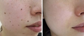
A mole, or nevus, is a benign skin formation that consists of cells filled with melanin pigment. This gives them their characteristic dark shade. Almost every person over 8–10 years old has moles. They appear in large numbers during puberty, which is explained by hormonal surges.
Pregnant women also often develop new moles, which is associated with endocrine changes. However, it is not at all necessary to resort to their removal during pregnancy if they do not bother you. It is necessary to show skin growths to a doctor; usually only observation is required.
Surgical removal of a mole is resorted to not only for cosmetic reasons. There are certain medical indications for removal.
When deletion is indicated
Epidermal nevi are benign formations and are harmless to health. However, any nevus carries a potential threat; there is a possibility of degeneration into a malignant tumor - melanoma. This is one of the most dangerous types of cancer. Therefore, it is important to monitor existing moles and consult a doctor if there is the slightest change.
You should visit a dermatologist as soon as possible in the following cases:
- the mole has increased in size, changed color, shape, outline;
- pain, itching, bleeding, burning, peeling, ulceration appeared, the surface became lumpy and uneven;
- the nevus was injured.
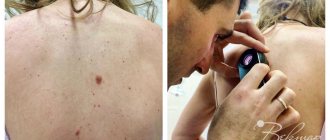
There is a high risk of degeneration for nevi located in open areas of the body. This is due to intense exposure to ultraviolet rays during the hot season. Also subject to removal are nevi that are constantly injured by elements of clothing - the handle of a bag, a collar, elastic bands of underwear, etc. Those formations that are located on the hairy areas of the body are also at risk - there is a possibility of tissue damage during shaving, waxing or sugaring.
General characteristics of formations
A mole is a pigment formation of one of the shades of the brown palette. The base contains melanin and the melanocyte cells that produce it. Melanins are high molecular weight pigments. They are characterized by a heterogeneous structure and complex chemical composition. Pigments are found not only in moles, but also in all skin, hair, iris, inner ear and even some parts of the brain.
Content:
- General characteristics of formations
- Indications/contraindications for the procedure
- What you need to know about melanoma?
- How to properly prepare for mole removal
- Technique of the operation
- Possible complications and side effects
- Features of rehabilitation
It is important not to confuse nevi and birthmarks. Moles do not form in the prenatal period, but as a person grows older and are closely related to his lifestyle. The cause of the formation of pigment spots can be a hereditary factor, exposure to ultraviolet radiation, or hormonal changes.
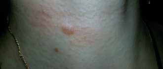
Each mole is a separate formation with a unique life cycle. At first it is a flat spot, which can increase and rise slightly above the skin as it grows and develops. The size and location of the formation depends on the level of melanocytes. If they are in the epidermis (top layer of skin), the mole will be flat. The deeper the melanocyte is immersed in the dermis, the higher the formation is located on the surface of the human body.
Convex nevi, which rise slightly above the skin, should not frighten their owners. This condition has no negative consequences and is considered a variant of the norm. The main thing is that the mole is miniature, round and uniform (in terms of color and structure) throughout its life cycle.
Can a mole disappear on its own and how safe is it? Yes, the formation can disappear without the influence of mechanical factors. This happens gradually and can alert a person. First, a white outline is formed around the formation, which resembles an orbit. This contour begins to increase and gradually covers the surface of the entire formation. The nevus disappears, and a small white spot remains in its place. Most often this occurs after severe sunburn. The disappearance of moles can also be a harbinger of the rare disease vitiligo.
If you notice changes in a mole (increase/decrease, uneven edges, change in shade or structure), be sure to consult a specialist.
Selecting a removal method
So, an oncologist or dermato-oncologist recommended removing the mole, or you decided to get rid of the formation. Consult with a specialist which removal method is best for your situation.
In many cases, histological examination of a removed mole is a mandatory procedure. It is important to evaluate the structure of the nevus tissue and make sure that the formation is truly benign and that nothing threatens your health. In this case, traditional mole removal with a scalpel is one of the most suitable methods. Some modern instruments do not leave a trace of tissue, so it will not be possible to study them in the laboratory.
If a large mole is located on the face or open areas of the body, discuss with a specialist the prospects of using a laser or radio wave removal method. These methods have fewer cosmetic consequences - they do not leave noticeable scars, so it is often advisable to resort to them. Remember that health is more important - if the doctor insists on conventional surgical removal, you should listen, because the prevention and early diagnosis of cancer depends on this.
What you need to know about melanoma?
Every mole on the human body can degenerate into a malignant melanoma. What it is? Melanoma is one of the types of malignant tumors. It also contains melanocyte cells (producing melanin), but in melanoma the cells are more aggressive.
They constantly divide, displace healthy cells, enter the vascular bed and spread throughout the body. As soon as the first aggressive cell approaches the brain or, for example, the lungs, it settles inside the organ and begins to rapidly divide. This process is called metastasis.
Melanomas are extremely dangerous because they quickly metastasize and divide at an incredible rate. The main thing is to detect the pathology in time. Surgical removal of melanoma will prevent the process from spreading throughout the body and guarantee a complete cure. Failure to see a doctor in a timely manner or complete refusal of treatment can result in death.
Preparing for the intervention
No preparation is required for surgery: you can eat and drink water without restrictions, and you do not need to take medications. However, it is important for women to plan the day of surgery taking into account the menstrual cycle; it is better to do this after menstruation. The recommendation applies to any surgical intervention. Also, in cases where the nevus is located on open areas of the body, it is better to remove it from October to April. This will eliminate the need to hide the wound and protect it from ultraviolet rays.
If you are taking medications that affect blood clotting, tell your surgeon. You should not stop taking medications on your own—check with your doctor.
Examination before mole removal
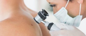
All patients undergo a dermoscopic examination before removal of nevi. The essence of dermatoscopy is to repeatedly enlarge the area under study, down to the cellular level. The doctor determines what cells the neoplasm consists of and whether there is dysplasia. As a rule, no tests are required before removal. However, if a large area of skin is affected or the mole has signs of malignancy, in addition to the histological analysis of the nevus itself, you will need to submit the following:
- clinical blood and urine test
- blood chemistry
- blood for infections, clotting
- blood group and Rh factor
- and undergo fluorography
Features of surgical removal
Surgical removal of nevi has several advantages:
- the mole is removed forever - there is no risk of reappearance;
- the operation requires only one visit to the surgeon;
- minimum contraindications;
- possibility of histological study of tissues.
However, there are also disadvantages - the need for reliable local anesthesia, the likelihood of a mark/scar forming at the site of the removed mole, and a relatively longer recovery period.
Removal steps:
- Pain relief with local anesthetics.
- Excision of nevus within normal tissue.
- Stitches (if the mole is small, this may not be necessary).
- Application of a special bandage.
Sutures are removed on average on the 5th–7th day. The overall healing period ranges from 7 days to 21 days depending on the health condition. The surgeon will give recommendations on how to care for the postoperative area at home. You can go home immediately after the intervention. As a rule, only the use of antiseptic solutions is required. You can also come to the clinic for dressing changes.
The doctor sends the material obtained as a result of excision to the laboratory for histological examination. A laboratory assistant examines it under a microscope for any atypical structure. You will receive the results within a few days, and if suspicious cells are detected, the specialist will definitely refer you to a dermato-oncologist.
As a measure to prevent skin cancer, your doctor may recommend giving up tanning beds and reducing the time you spend in direct sunlight. It is necessary to use sunscreen with a suitable protection factor, do not drink alcohol in excess and do not smoke. Patients with a large number of moles may be advised to visit a dermatologist for preventive purposes once a year.
Possible complications after surgical excision of nevi
After surgical removal of a mole, the following side effects may occur:
- Itching, pain, discomfort at the surgical site;
- Allergic reaction to a drug for local anesthesia;
- Prolonged bleeding;
- The appearance of keloid scars;
- Swelling in the area of intervention.
An increase in body temperature may indicate the presence of an inflammatory process, so in this case you should consult a doctor.
Surgical removal of moles is one of the effective methods of getting rid of nevi, which helps prevent the possible development of cancer pathologies. Our clinic employs qualified and experienced doctors who will help you efficiently and quickly get rid of not only nevi, but also other neoplasms.
Attention!
This article is posted for informational purposes only and under no circumstances constitutes scientific material or medical advice and should not serve as a substitute for an in-person consultation with a professional physician.
For diagnostics, diagnosis and treatment, contact qualified doctors! Number of reads: 4854 Date of publication: 08/06/2018
Dermatologists - search service and appointment with dermatologists in Moscow
Who is contraindicated for
Surgical removal of a nevus is contraindicated in the presence of the following diseases:
- exacerbation of chronic inflammatory diseases;
- infectious disease - viral, fungal, bacterial;
- fever of unknown origin, increased body temperature and other symptoms of intoxication.
Also, surgery may be postponed during pregnancy or breastfeeding - in each case, the doctor makes a decision taking into account the symptoms and condition of the formation.
Cryodestruction
Removing moles and warts using liquid nitrogen does not require pain relief. As a result of cold exposure, nerve fibers freeze and sensitivity is switched off. Liquid nitrogen is charged into a mini-cylinder with removable nozzles. The surgeon selects a tip 1 mm larger than the diameter of the tumor and applies it to the mole. The mole is frozen and rejected on its own. The disadvantage of this procedure is that it is difficult to calculate the depth of nitrogen penetration. If the exposure is too strong, healthy tissues are frozen, and if the exposure is weak, the nevus is partially removed. Damage to healthy tissue provokes cosmetic defects and increases the rehabilitation period.
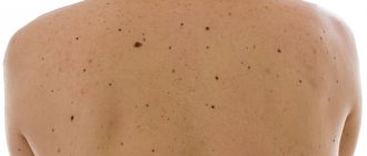
Large moles on the body
Moles appear in the first year after birth or throughout life.
The size also varies, and the location on the body can be anywhere. There are several types of large moles:
- fibroepithelial - benign formations up to one and a half centimeters in size;
- Sutton's or halonevus - characterized by the presence of a colorless ring;
- melanocintar – flat moles that appear during hormonal changes;
- Becker - are more often observed in men, consist of epidermal cells;
- blue – nevus is bluish in color and small in size;
- basal – characterized by inactive pigmentation, colorless;
- warty - have a bumpy surface, consisting of tiny nodules;
- papillomatous – formed by the papilloma virus;
- red – associated with the growth of the capillary network;
- nevus of the sebaceous glands - formed due to an embryonic anomaly of tissue development;
- Port-wine stains are a vascular pathology inherent in newborns.
Some springs are completely harmless, while others can pose a danger, which is expressed in the risk of malignancy. Most of the formations must be removed. But only a doctor can determine what to do with a large mole.
Prevention of degeneration
In order to prevent the transformation of an ordinary nevus into a malignant one, it is important to promptly contact a specialist with a request for diagnosis. The question of whether large moles are removed will be clarified at the first appointment. Doctors usually recommend getting rid of pigment formation as early as possible from the point of view of aesthetics and safety.
But measures to prevent degeneration are still worth taking:
- do not get carried away with tanning;
- in the hot season, go outside, applying cream with SPF 30 - 50 to areas of the skin not protected by clothing;
- protect yourself from injury;
- Do not take hormonal drugs uncontrollably.
And most importantly, do not ignore scheduled examinations by specialists. Especially during periods of hormonal changes in the body, it is necessary to constantly see a doctor.
The specialists at the Desir clinic specialize in various areas of medicine. We have some of the best dermatologists and highly specialized specialists: dermatologists-oncologists. Make an appointment and get your moles examined. If you are concerned about the presence of a large mole, we will provide qualified assistance. Our innovative equipment allows us to conduct research as accurately as possible and use the latest techniques to remove formations of various types.
Reasons for appearance
The first stage of an education survey is to determine the source of its formation. There may be several reasons for the appearance of large moles:
- genetic predisposition;
- anomalies of intrauterine development of the fetus;
- Excessive tanning in the sun and solarium - negative effects of ultraviolet radiation;
- skin injuries;
- viruses, for example papillomavirus;
- frequent stress;
- diseases of internal organs - pancreas, liver;
- bowel dysfunction;
- exposure to radiation, toxins;
- taking certain oral contraceptives;
- after surgical interventions;
- pathologies associated with lipid metabolism.
Most often, patients complain that a large mole has grown due to hormonal changes. This can happen during pregnancy, during menopause, and during puberty.
How to remove more moles?
There are several ways to get rid of large nevi:
- 1. Radio wave surgery. Using a special knife, the doctor excises the formation without affecting healthy tissue. There is no risk of bleeding, disinfection is ensured, and no scars are left. The technique is safe and painless.
- 2. Electrocoagulation. Heat treatment with electric current. There may be some slight discomfort. The healing process is fast.
- 3. Laser coagulation. Modern safe and painless technique. The equipment is equipped with cooling systems, so the patient feels only a slight tingling sensation. The formation of scars is excluded.
- 4. Cryodestruction. Exposure of nevus to liquid nitrogen at extremely low temperatures. Regeneration takes longer than with other methods.
All methods, except cryodestruction, allow you to save tissue particles for sending for histology. Surgical removal is indicated for very large moles and suspected cancer. The doctor excises the pigment spot with further sutures or tissue replacement with a deep incision.
Are they dangerous?
After the dermatologist explains to the patient why large moles appear and what is the reason for the specific formation, the doctor finds out the nature of the pigment spot and its type.
An increase in size and the appearance of new moles is usually observed after harmful exposure to UV rays and hormonal changes. Whether large moles are dangerous can be determined by the following factors:
- cessation of growth and loss of hairs on the surface of the spot;
- change in outline;
- the appearance of pronounced asymmetry;
- an increase in size of more than 5 mm in diameter;
- itching and burning in the area where the nevus is located;
- redness and swelling of the tissues around the spot;
- discharge;
- bleeding
For an experienced specialist, there is no problem in determining at first glance whether large moles can be removed and how dangerous they pose. But even the most expert dermatologist can evaluate a spot subjectively. Therefore, if there are more than three signs of danger, the doctor prescribes a test.
Diagnostics
A visual examination allows you to determine the type of birthmark. At the first appointment, the doctor asks the patient a number of questions:
- how often does one take sunbathing or tan in a solarium?
- were there any burns from ultraviolet radiation;
- whether there were cases of malignant moles in the family;
- whether the patient took hormonal medications;
- whether he works in a specific industry related to the processing of toxic substances and investment;
- suffers from chronic diseases of internal organs;
- were there any cases of injury?
After this, dermatoscopy is performed - a method for examining suspicious pigmented formations. The specialist applies immersion oil to the spot, then uses a magnifying device to carefully examine the condition of the nevus. Using a three-point rating system, the dermatologist checks the following parameters:
- asymmetry of structure;
- coloring;
- the presence of an atypical pigment network;
- presence of light blue areas.
Dermatoscopy is considered an effective technique that gives a completely reliable result - up to 95%. But in some cases, this research method is not enough, so the question of whether large springs can be removed is decided after additional tests:
- biopsy - analysis of tissue particles of the formation;
- cytology - a test for the growth of malignant cells;
- histology – determination of the degree of pathological processes, assessment of the condition and structure of cells;
- epiluminescence microscopy is a clarifying technique for benign formations.
In some cases, an additional examination using ultrasound, radiography, or MRI is prescribed. If necessary, the doctor sends the patient for clinical urine and blood tests to assess the general health before surgery to excise the tumor.