The problem of the appearance of tumors on the skin of the face worries every person: tumor lesions cause discomfort and inconvenience and require cosmetic or therapeutic treatment. The capabilities of high-precision medical equipment and innovative techniques make it possible to effectively and safely remove tumors on the face. The most common, gentle and low-traumatic method that does not leave noticeable marks is laser removal of facial lesions.
Why do benign formations appear on the skin?
Cosmetologists and dermatologists do not know the exact mechanism of their formation. Most often the cause is:
- injuries;
- viruses;
- systemic diseases of the body, for example xanthomas, occur due to an excess of fat in the blood;
- long-term skin diseases;
- exposure to aggressive substances;
- excessive exposure to ultraviolet radiation;
- x-rays;
- heredity (for example, seborrheic dermatosis).
Most skin lesions are benign
general information
A common wart is a growth on the skin that has a rough surface and protrudes to a certain height above the surface of the epidermis. The color of warts is lighter or darker than the surrounding tissues; on large formations, internal vessels can be seen. The main cause of warts is infection with the human papillomavirus. HPV enters the body through damaged or water-soaked skin. Sometimes infection is possible even if there is no damage to the body. The difficulty of identifying the cause of infection is due to the fact that some warts in adults appear on the body only six months after infection.
HPV mainly affects children and young people. Patients who have a history of chronic skin diseases are susceptible to the problem:
- Psoriasis;
- Dermatitis;
- Eczema, etc.
Patients who have a weakened immune system are also at risk. We are mainly talking about people infected with HIV or those who have undergone organ transplantation. If a patient has problems with immunity, treatment difficulties may arise in the future. In this case, a viral wart most often recurs after removal.
Warts can be found on the skin anywhere on the body. Moreover, most often they are localized in the area of the fingers, elbows, knees, and hands.
Benign and malignant neoplasms on the skin: what are the differences?
Benign pathologies do not pose a threat to human life. If they reach large sizes, they can interfere with the adequate functioning of various body systems. In contrast, malignant ones grow quickly and aggressively, penetrate into surrounding tissues, and form metastases over time. Some damage vital organs and cause death.
Sometimes benign skin tumors change due to external or hereditary causes. They acquire the ability to degenerate into malignant pathologies. Such conditions are called borderline or precancerous. They pose a great danger to health and life, although they do not always have pronounced symptoms.
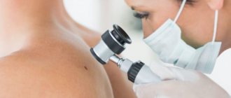
Diagnostics
The differential diagnosis is based on the clinical picture of the specific type of neoplasm, as well as on the study of the anamnesis. The size of the papule, the rate at which it increases in size, the number of formations and other factors are taken into account.
To make a final diagnosis, the patient may be referred for histological examination.
If the appearance of warts is planned to be excluded using destructive methods, it is first important to be tested for HIV, hepatitis and syphilis.
What is the structure of benign neoplasms
The growths consist of cells that have partially retained their original functions and are capable of growing slowly. They are similar in structure to the tissues from which they originated. They can put pressure on nearby tissues, but do not penetrate them, since they have a capsule in their structure. They respond well to hardware and surgical treatment and, as a rule, do not cause relapses.
There are always congenital formations on the skin - moles or warts, as well as acquired ones. The latter are formed on the surface or in the subcutaneous layer as a result of metabolic disorders, decreased immunity, or under the influence of a virus.
Types of warts and specific symptoms:
- Molluscum contagiosum
. Localization - on the body or in the genital area. There are depressed areas on the surface. If you press on the wart, a mushy substance may begin to ooze. - Nevi
. May be a congenital problem, raised significantly above the surface of the skin. Characterized by a dark brown color, as well as the presence of hair. - Basalioma
. It has a crust on the surface; if it is removed intentionally or accidentally, bleeding begins.
Types of benign formations
- Warts and papillomas
Papilloma is a small tumor on a stalk or broad base, which has clear boundaries. Flint is visible on an uneven, grainy surface. The growth is painted in any color - from white to dark brown. It is found both individually and in large quantities.
Both papillomas and warts appear as a result of the papilloma virus entering the body, live in different parts of the body, and reach a diameter of several centimeters. They are activated on the skin as a result of nervous tension, stress, decreased immunity or vegetative disorders.
Papillomas and warts are not dangerous if they are not injured
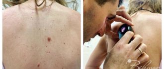
- Birthmarks
They are not prone to degeneration, but such cases do occur, so it is necessary to pay attention to changes in contour, color, and size, especially if the area is constantly injured.
- Lipoma
A round, soft-to-the-touch tumor of adipose tissue that does not disrupt the functions of the body, but only creates a cosmetic defect. Wen appears in various areas where there is a fat layer. Reaching large sizes, they grow into tissue and approach bone surfaces, often spreading to muscles and blood vessels.
- Atheroma
Atheroma is a residual cyst caused by blockage of the ducts of the sebaceous glands. It contains products produced by the sebaceous glands in the form of an odorless curd mass. The pathology has clear contours and a dense consistency. Superficial atheromas occur mainly on the back, head, neck, face, and limbs - due to poor hygiene, metabolic disorders and unprofessional depilation.
When subjected to mechanical action, the atheroma becomes inflamed, swells and becomes red. When an infection gets inside, pus is formed and a greasy consistency is erupted. There is a risk of degeneration into liposarcoma, so it is recommended to remove this pathology.
A seemingly harmless atheroma can degenerate into liposarcoma
- Nevus
More often than others, melanoma degenerates into a malignant formation, especially with prolonged exposure to negative external factors or hereditary predisposition.
- Lymphangioma
The pathology is mainly congenital, developing from lymph nodes in the skin, but can also spread to fiber or muscles. It is most often localized on the head, face and upper body. The blue tumor-like growth rises above the skin, has a dense consistency and clear boundaries, dimensions are 1-5 mm. Since it puts pressure on vital organs (lungs, larynx, trachea), surgical removal is resorted to.
- Hemangioma
It is formed on the basis of blood vessel cells in the subcutaneous layer, rises above the surface, and is characterized by rapid spontaneous growth. When pressed, the growth decreases. It is mainly localized in the head and neck area, mainly in young children. Cavernous hemangioma is colored blue and resembles a node, located deep in the tissues. Capillary - in the epithelium, has a red or blue color. If it becomes inflamed, it can provoke open bleeding, and if it is close to vital organs, it disrupts their functions.
Hemangiomas are diagnosed more often in children under three years of age.
- Fibroma (dermafibroma)
A tumor of dense consistency, light pink in color, is formed from connective tissue. Mild varieties occur more often on the neck and chest, in the groin folds and armpits in women. Hard - in different areas near the upper layers of the skin. Dermatologists recommend removing fibromas, since under favorable conditions they can transform into fibrosarcoma.
- Keratoma
This skin tumor is formed from keratinocytes that make up the stratum corneum. A growth in the form of a spot or node is formed from dead cells and is localized on the back, head, face, and limbs. Such a benign neoplasm requires urgent treatment.
- Neurofibroma
Formed from nerve sheath cells, it looks like a hard tubercle up to three centimeters in diameter. Causes discomfort and pain because it puts pressure on nerve endings. Most often located on the face, back, abdomen, arms and legs. If there are many formations, they talk about neurofibromatosis - a disease that is mainly inherited.
Causes of warts
Viral warts are most common on the face.
The main reason for their formation is the papillomavirus.
It enters the skin and causes excessive cell division.
As a result, small benign tumors form.
This virus is transmitted by contact.
For infection, an important condition is the presence of damaged epidermis.
Therefore, warts and papillomas on the face appear more often in men.
After all, they regularly shave their beard hair.
The blade causes microdamage to the skin.
Through these defects, a virus enters it, causing warts to grow on the face.
They look unattractive.
In addition, they can be damaged during shaving.
Other types of warts on the face are less common.
There are hereditary diseases that cause their multiple occurrence.
Also, a number of pathologies lead to the formation of a rash that looks like warts.
Removal of benign skin tumors
Dermatologists are convinced: it is necessary to get rid of benign formations, with the exception of small scatterings of moles or other minor defects throughout the body. This is especially true for the face, because the growth attracts attention, spoils the overall impression and gives a lot of unpleasant emotions to its owner.
There are several methods for removing benign skin tumors:
- Electrocoagulation
Under local anesthesia, the growth is cut off with a special surgical coagulator, which creates a high-frequency current. Simultaneously with removal, the tissues are soldered together, which avoids bleeding and infection. The crust at the treatment site disappears after 7-10 days, sometimes leaving a slightly noticeable scar. The method is effective if the defect is small.
- Cryodestruction
Liquid nitrogen can only be applied to formations in the upper layers of the epidermis. If it is flat, apply applications with liquid nitrogen. In case of deeper occurrence, a cryodestructor is used. After the procedure, the body begins a reaction of rejection of the treated defective tissue. The resulting crust disappears and heals within a month and a half.
- Laser removal
A powerful beam of light destroys neoplasm cells and evaporates them from the skin. The laser does not affect healthy neighboring tissues. The manipulation is carried out under local anesthesia, it is low-traumatic and bloodless. Laser treatment parameters are selected individually. Damaged tissues are evaporated layer by layer until the beam reaches healthy skin. The formed crust disappears on its own after 1-2 weeks.
- Radio wave method
High frequency radio waves cut and coagulate tissue. The crust disappears after a week. The method is contraindicated in patients with a pacemaker, herpes, or elevated body temperature.
- Surgical removal of benign skin tumors
This method is used when the growths are too large and other technologies cannot cope. The formation is excised with a scalpel and removed, capturing a small area of healthy skin. The scar after surgery takes several weeks to heal, but the wound requires long-term careful care. The description of this traumatic technology is not encouraging, so it is better to choose another method to eliminate defects on the face.
On the face, neoplasms are removed using laser or cryodestruction
Laser removal mechanism
The laser beam is able to carefully remove the formation without damaging adjacent tissue. The laser radiation beam penetrates the skin to a depth of 1 micron and works in a targeted manner, which prevents the occurrence of burns and microtraumas. The method is especially suitable for small areas of skin, for example, for removing tumors on the nose. The doctor prescribes from 2 to 4 sessions, the number of procedures for each case individually. Additional correction may be required after a month. The duration of one procedure is about 20 minutes and depends on the severity of the lesion.
Prevention of skin tumors
Unfortunately, medicine has not yet learned to prevent the appearance of various formations on the skin. But dermatologists give their patients the following preventive recommendations:
- do not delay contacting a doctor if a tumor appears on the skin;
- remove formations only after a specialist and diagnostics confirm their benign nature;
- avoid excessive exposure to the open sun;
- use sunscreen, especially if you are prone to moles and hyperpigmentation;
- do not come into contact with chemically active and carcinogenic substances;
- do not eat foods that contribute to the development of cancer (smoked meats, sausages, animal fats, meat products with food stabilizers).
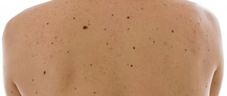
Do you have a lot of moles? Forget about tanning in the open sun
Preparatory activities
Beginning the course of restoring beautiful and elastic skin requires additional measures. Need to:
- avoid prolonged sun exposure before and after treatment
- do not visit saunas, baths, do not swim in hot water
- refrain from cosmetic peelings
- treat affected areas with sunscreen
- do not use medications that affect the blood clotting system
What should you do after a laser therapy session?
The procedure is performed on an outpatient basis and does not require a hospital stay. During the rehabilitation period, you should refrain from exposure to the sun or solarium. At the site of the tumor, a crust forms, which should not be injured, as this can lead to the formation of a residual mark, which, however, can be easily removed by dermabrasion or laser resurfacing. Under no circumstances should the wound be wetted or allowed to soak the crust covering it. It is advisable to treat it with alcohol-based antiseptics.
The result of laser treatment and compliance with all the measures described above will be clear and smooth skin without scars. The price of laser removal of tumors on the face depends on the area and type of lesion, and is calculated individually for those wishing to receive high-quality treatment. General prices are listed below.
You can ask additional questions to a specialist at the medical center, and make an appointment with a dermatologist. ABC Clinic specialists perform effective removal of tumors of various etiologies using new equipment and guaranteeing the absence of relapses.
Primary source honey / September 2018
Do benign formations hide the danger?
Benign neoplasms are unpredictable structures that can manifest themselves at any time or not at all. The process of their transformation into malignant ones has not been fully studied. There is no clear answer to the question of what exactly activates this process. It is believed that mechanical trauma, excess ultraviolet radiation, metabolic disorders and other factors contribute to degeneration. One way or another, if you have a benign skin lesion, you should not experiment and rely on chance. Moreover, today removal does not cause difficulties.
Contraindications for laser treatment of tumors
Laser removal of formations on the face cannot be used for the following pathological conditions:
- increase in tumor size
- changes in color, shape, structure of the tumor focus
- formation of cracks, ulcers, skin hyperemia, bleeding
- inflammation
- the appearance of pain
With such symptoms, it is appropriate to assert that the formation is oncological in nature and intervention is necessary in other ways, for example, with a radio wave device, followed by histology of the tumor.
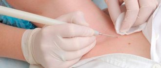
Diagnostics and examinations
Pigmented nevi, keratomas, condylomatous growths, and papillomas deserve special attention. Removing tumors on the face with a laser requires a microscopic analysis of the tissue.
Important! If malignant degeneration is suspected, the doctor performs dermatoscopy. It is forbidden to take a tissue biopsy before removal, so as not to provoke the development of a cancerous tumor.
What is the best way to treat?
A large number of papillomas certainly causes inconvenience and does not look aesthetically pleasing. However, this is not a reason to use old-fashioned methods for removal with thread, pliers or a blowtorch.
For single small (1x1 mm) formations, celandine juice
- a proven folk remedy.
When there are many papillomas or they are large, this method is not suitable
, as it can last for months and lead to severe inflammation.
Removal using radio wave surgery is much more convenient and effective
The operation does not require special preparation, is practically painless, and after it you can drive a car.
Once I had the opportunity to remove about 70 papillomas from one patient at once. It is difficult to convey all the joy of a person who got rid of them, whom they tormented for several years.