Today, the question of the possibilities of scar correction is relevant for both dermatocosmetologists and surgeons. It is extremely important to understand the stage of scarring and prescribe therapy based on the “age” of the scar tissue.
As a rule, if a skin injury occurs only in the epidermis layer, then during healing there are practically no traces, due to the ability of the cells of the basal layer of the epidermis to regenerate throughout life. Damage to the deeper layers of the skin is filled with granulation tissue, which is further transformed into a scar. Gradually, the scar tissue flattens and takes on the color of the surrounding tissues, that is, it becomes a normotrophic scar. In some cases, this process is disrupted, leading to the formation of pathological scar tissue.
Scar formation is divided into four successive phases:
I - stage of inflammation and epithelization
Occurs from 7 to 10 days from the moment of injury. The severity of swelling and inflammation of the damaged skin area gradually decreases. Granulation tissue begins to form. There is no scar at this stage. During normal healing, the wound heals with the formation of a barely noticeable scar. To prevent complications, atraumatic sutures can be applied, and daily dressings with an antiseptic effect are performed. It is necessary to limit the patient's physical activity to reduce the risk of wound dehiscence.
II - stage of formation of a “young” scar
From 10 to 30 days after injury, collagen and elastin fibers begin to form in the granulation tissue. At this stage, the scar is loose, immature, bright pink tissue that can stretch easily. During this time, it is necessary to avoid repeated traumatization of the area, as well as high physical activity.
III - stage of formation of a “mature” scar
A mature scar forms between 30 and 90 days after injury. Elastin and collagen fibers grow into bundles, lining up in one direction. The color of the scar becomes paler as the blood supply to the scar tissue decreases sharply. The texture of the scar becomes denser. During this period, there are no restrictions on physical activity, however, repeated traumatization of the damaged area can lead to the formation of a keloid or hypertrophic scar.
Scar treatment
If there is an immature form of the scar, physiotherapeutic treatment, local application treatment and injection drug treatment are carried out, aimed at reducing scar hypertrophy and speeding up their maturation.
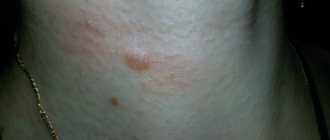
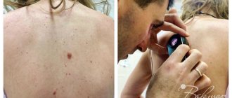
An example of preparation for laser dermabrasion surgery of a patient with an immature hypertrophic scar:
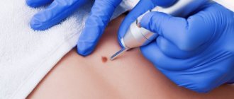
If there is a mature form of the scar, it is possible to perform laser dermabrasion surgery. This operation is performed under local application anesthesia using a high-energy CO2 laser, which allows for ablation (evaporation) of excess amounts of scar tissue, which subsequently leads to smoothing of the skin texture.
IV - stage of final scar transformation
4 months after the injury and up to about a year, the scar is finally formed and matures: the vessels die, the collagen fibers become stretched, the color fades, and the tissue becomes denser.
Speaking about the histological classification of physiological scarring, it consists of three stages:
- Fibroblastic. The fibroblastic stage occurs up to 30 days. It is characterized by the formation of young fibroblasts and the formation of a large number of vessels in this area.
- Fibrous. It is formed by the 33rd day of injury, mature fibroblasts are already present here, and collagen fibers accumulate.
- Hyaline. Formed by 42 days from the moment of damage. In the area of injury, the number of cells and blood vessels decreases.
Types of scars
Scars are divided into four types: normotrophic, atrophic, hypertrophic and keloid. Let's look at each of them below.
Normotrophic scars
Normotrophic scars are pale in color, close to the color of the skin, have good elasticity and are flush with the surrounding skin. Most often they are little noticeable and do not bother the patient. Normotrophic scars do not require radical correction; a course of chemical peels is sufficient.
Atrophic scars
Atrophic scars are often called “minus tissue” scars because they look like a depression. As a rule, atrophic scars are formed due to inflammation, trauma or chickenpox. They often have no pigment, so they appear white. These scars are formed due to insufficient formation of connective tissue. Examples of atrophic scars are stretch marks and post-acne.
Hypertrophic scars
They are excessive because they are formed due to excessive work of the cellular matrix. They rise above the surface of the surrounding skin and are capable of regressing within two years. They are a thick, dense tissue with a bumpy surface. As a rule, hypertrophic scars do not protrude beyond the boundaries of the injury. Most often it is formed as a result of burns, wounds, tattoos, and surgery. It can be easily excised and then corrected with chemical peels.
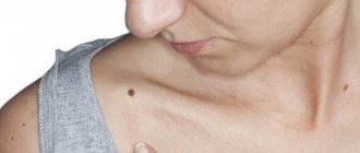
Keloid scars
They are pathological, as they are formed due to individual uncontrolled changes in the structure of scar tissue and too active work of the cellular matrix. They have a pronounced contour, purple color and protrude significantly above the surface of the skin. These scars can have different sizes that do not coincide with the boundaries of the injury. Scars of this nature bring a lot of discomfort to the patient, as they can be accompanied by itching, burning and pain. Keloid scars initially appear as small compactions, which then grow strongly and continue to grow for several years. This type of scarring must be monitored under medical supervision.
Scar correction methods
Let's consider the main modern methods of treating scar tissue:
Surgical excision of scars
This correction method helps to reduce the width of the scar, correct pathological phenomena in the relief of scar tissue, and excise foreign particles from the scar tissue. The scars become much neater and less pronounced. Most often, surgical excision is the only method to improve the appearance of the scar.
Chemical peels
They work best with normotrophic scars, but their use with atrophic and hypertrophic scars also shows good results. At the moment, the most popular method is the use of trichloroacetic acid in combination with ascorbic acid (using the nappage technique). This technique has a good stimulating effect.
Fractional photothermolysis
Photothermolysis is used with modified scar tissue. The focused beam creates a microcrack surrounded by intact tissue. Cells that are located in the area of damage begin to become active and promote regeneration. When using this method, the laser wavelength, which determines the depth and diameter of the microzone, is extremely important. Used to correct atrophic and hypertrophic scars.
Laser correction
The laser beam penetrates to a depth of 1 mm and activates regenerative processes with the synthesis of new collagen. Collagen fibers, displacing connective tissue, help improve the texture of the scar and smooth out the relief. The number of procedures is individually selected for each patient. After laser therapy, sun exposure is prohibited for three weeks.
What determines the risk of scarring?
The appearance of a scar and its nature depends on a number of factors:
- Depth and area of damage: if they are small, a scar may not appear at all.
- Tissue healing abilities.
- The presence of chronic diseases (for example, diabetes).
- Time for complete healing of the wound (it may increase if the wound has become infected).
The process of complete wound healing, which lasts for 21 days, ends with pathological scarring in only 33% of cases. If the process lasts more than 21 days, the probability increases to 78%.
Etiology and pathogenesis
It has been established that in 99% of cases, post-acne scars develop in the place of papules or pustules, and only in 1% of cases - in the place of comedones. It is difficult to remove acne scars; the healing of such defects is due to the complex interaction of tissue, humoral and cellular mechanisms. If the skin is damaged at the level of the papillary (upper) layer of the dermis, when fragments of the basement membrane between the epidermis and dermis are preserved, restoration can take place without gross defects. In such places, depigmented areas with focal atrophy remain on the skin.
However, when the basement membrane is completely destroyed, a scar is formed, which differs in structure and function from the surrounding skin. In its area, coarse fibrous collagen grows, blood and lymphatic vessels are deformed, and their number sharply decreases. At first the scar is immature , but gradually it becomes more natural in color and softer, forming a mature scar .
If catabolic processes (synthesis) exceed anabolic processes (destruction), excess collagen is formed - the scar begins to grow and becomes hypertrophic . It is distinguished by a pink tint and bulge above the skin level. With keloid, the scar area increases significantly due to the tumor-like proliferation of immature connective tissue due to the uncontrolled activity of fibroblasts. Features of keloid scars are their rapid growth with paresthesia (tingling), itching and pain.
The likelihood of the appearance and severity of scars after acne is influenced by the individual tendency to scar tissue, the time of onset and effectiveness of acne treatment, the activity of acne (mild, moderate, severe), the number of papules, pustules and comedones and other factors.
Clinical manifestations of acne scars
The main types of post-acne scars (Fig. 2 and 3):
- Hypertrophic.
- Keloids.
- Atrophic:
- chipped (icepick scars) - 60–70% of cases;
- rounded (rolling scars) - 20–30% of cases;
- rectangular (boxcar scars) - 15–25% of cases.
Atrophic scars after acne occur on average 3 times more often than hypertrophic and keloid scars.
Severity of post-acne scars:
- Macular - flat hyper- or hypopigmented (dark or light) lesions that do not change the skin texture.
- Mild - small atrophic or hypertrophic scars, invisible from a distance of more than 0.5 meters from a person. It is enough to simply hide them with decorative cosmetics or a beard (for men).
- Moderate - atrophic or hypertrophic scars of moderate severity, visible from a distance of more than 0.5 meters. They are not so easy to hide with makeup or natural facial hair, but you can straighten them with your fingers, stretching the skin in different directions.
- Severe - pronounced atrophic or hypertrophic scars, clearly visible from a distance of more than 0.5 meters. They cannot be straightened with your fingers when the skin is stretched.
In Russia, specialists from the Research Institute of Surgery named after. A.V. Vishnevsky developed a modified Vancouver scale . It can also be used in the analysis of post-acne scars ( Table 1 ).
Table 1. Modified Vancouver Scar Scale
| Points | Description |
| By color | |
| 0 | Normally pigmented (color of surrounding healthy skin) |
| 1 | Hyper- or hypopigmented (brighter or paler) |
| 2 | Various shades of red (immature scars) |
| In relation to the level of surrounding healthy skin | |
| 0 | At the level |
| 1 | Below level |
| 2 | Above level |
| By relief and surface quality | |
| 0 | Flat |
| 1 | Lumpy, uneven |
| 2 | With hyperkeratosis and ulcerations |
| By shape | |
| 0 | Scar cord or fold (the length of the scar is greater than its width) |
| 1 | Scar mass (the length and width of the scar correspond to each other) |
About half of all acne scars are of clinical significance, i.e. disrupt the structure and/or function of a specific area of the skin. By disfiguring the face, they negatively affect the emotional state of patients, reducing their self-esteem, causing psychological disorders and social maladjustment. Severe acne scars are considered a risk factor for suicide.
Rice. 1. Hypertrophic and keloid post-acne scars (www.acne.org, www.healthjade.com)
- Hypertrophic scars - appear within a month after healing, are located strictly at the site of the acne rash, are usually painless, lighten over time and can regress.
- Keloid scars - appear 3 or more months after healing, extend beyond the boundaries of the acne rash, cause pain and itching, darken over time and can continue to grow.
| Hypertrophic | Keloid |
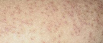
Rice. 2. Atrophic acne scars and their relative depth (Jacob CI, et al. J Am Acad Dermatol 2001; 45: 109–117. Hession MT, et al. JCAD 2015; 8: 50–58)
- Icepick scars - small in size (less than 2 mm), with clearly defined edges, cone-shaped (expand upward), deep (extend to the dermis and subcutaneous fat).
- Rolling scars - long (more than 4–5 mm), superficial, extending to the dermis, giving the surface of the skin a wavy appearance.
- Rectangular (boxcar scars) - medium in size (1.5–4.0 mm), the edges are smooth and steep (never tapering downwards), round or oval in shape, can be superficial or deep.
How to remove a scar
Scar management tactics depend on the type of scar and how fresh it is. The best treatment for scars is prevention—that is, if you are injured or have undergone surgery, spend adequate time and attention on the healing process of your wound. Carefully follow all the recommendations of the doctor who is observing you, regularly treat the suture before it heals.
If the scar begins to behave unusually - it hurts, acquires a new relief or a non-standard color, do not hesitate - show it to the surgeon. At an early stage, you can significantly improve the general condition of even a keloid scar. Using creams helps a lot.
If the intervention or injury took place a long time ago, and the scar has been on your body for more than a year, you can determine how to combat it after consulting a plastic surgeon. By contacting our doctor, you will receive a guarantee that you will be offered the most modern methods of transforming a scar into healthy skin.
Possible correction methods:
- Peeling . Chemical or abrasive peeling allows you to remove the unpleasant consequences of many effects on the skin. The top layer of skin peels off along with the scars, and the exposed healthy skin, as a rule, looks smooth. This method is recommended if you have scars on your face after chickenpox or acne.
- Injection technique . If you are concerned about a depressed scar at the site of injury, which is a “hole” against the background of healthy skin, you can restore the lost volume with the help of hyaluronic acid preparations. The “pit” will level out, however, after the hyaluronic acid is removed from the body, the procedure will have to be repeated.
- Laser correction . The most modern method used today. Allows you to get rid of scars completely, returning the skin to its normal texture and color. The technology is relatively simple - the laser either acts on the upper layers of the skin, “removing” the scar, or acts at a deep level, stimulating collagen production. The method is also good because it allows you to achieve results regardless of how pronounced the scar is - the condition will improve both if it is a tiny trace of acne, and if the reason for contacting a specialist is a pronounced, large scar.
Whatever method of getting rid of scars you choose, remember that after new interventions you need to take very good care of your skin: otherwise, a new one will simply appear in place of an ugly old scar. Contact our specialist if you have the slightest suspicion that the new scar is “manifesting itself” strangely.
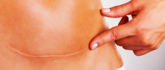
Where do scars come from?
A scar is a mark on the site of a healed wound. The human body fights any damage, including healing wounds and wounds. However, after healing has occurred, the skin at the site of injury becomes different. Especially if there was an inflammatory process during the healing process.
The connective tissue that allows us to regenerate differs in its properties from normal tissue - it has much lower functional properties. Sweat glands and hair follicles will not be restored at the site of the scar on the skin.
Scar tissue also differs in appearance from normal skin. Fresh scars are usually darker than the base skin tone, but over time, on the contrary, traces of damage become lighter.
Photos "before" and "after"
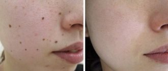
Subcision is the most effective way to treat acne scars. The result “before” and “after” of one procedure. Surgeon: Vasiliev Maxim.
Correction of deep scars. Before and after photos of one session. Completed by: Vasiliev Maxim.
Laser resurfacing of post-acne scars. Performed by dermatocosmetologist Elena Vlasova.