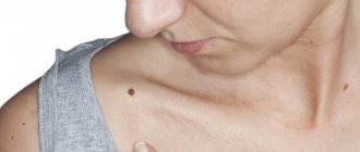
Dermatoscopy is an instrumental non-invasive study in which the doctor examines moles and other formations on the skin using a special device that creates multiple magnification. In oncology, dermatoscopy helps to distinguish harmless formations from skin cancer and identify melanoma in the early stages.
- Indications for dermatoscopy
- Why are moles dangerous?
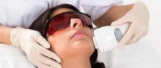
- How is dermatoscopy performed?
- Equipment for dermatoscopy at Euroonco
- How to assess the malignancy of a mole
- Advantages of dermatoscopy using the FotoFinder system
- Prices for dermatoscopy at Euroonko
"Pearls" on the Internet.
We will not analyze in detail the “pearls” that can be found on the Internet on the topic of dermatoscopy.
Just follow the first link for this request:
The author suggests that we urgently do dermatoscopy immediately after traumatization of a mole. In order not to waste precious time, suddenly the mole has already become malignant...
The article has a signature, but it is impossible to understand from it whether the doctor wrote it or not.
I am sure that any oncologist will agree with me - a person who gives such recommendations, at best, is far from diagnosing skin cancer. At worst, it has nothing to do with medicine at all.
Very often I have to deal with manifestations of depression in people who have read such publications.
But we only examined one of them...
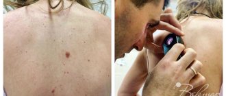
Equipment
A couple of decades ago, conventional dermatoscope devices were used for the procedure. Now doctors have digital devices in their arsenal. It is clear that they have many undeniable advantages over the devices of the previous generation.
In new models, the device's tube is connected to a computer monitor, where the image is displayed. Such devices are used for preventive research, primarily for people at risk of developing melanoma.
It is also important that, using a digital device, the doctor can create a map of moles over the entire area of the body within three minutes. This is necessary for further observation.
The research technique using digital devices is not only completely automated, but also simple and safe.
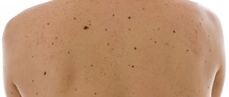
What kind of research on moles are there? How can I check my moles?
Let's try to figure out whether dermatoscopy is really “the best.” To begin with, we will briefly describe the options for examining skin formations.
Before deletion
We can examine moles in two ways:
- Clinical diagnostic method (examination, palpation, history of the birthmark). The effectiveness of this principle of diagnosis is not high - from 37 to 80%. The numbers speak for themselves - the method is not accurate.
- Dermatoscopy, scraping or puncture. The diagnostic accuracy of these methods does not exceed 95%. Of course, this is a very good indicator. However, out of 100 patients with melanoma or cancer, 5 will be misdiagnosed and people will not receive timely treatment.
- After removal,
there is only one research method: histology. It is this that allows one to determine the nature of a mole with almost 100% accuracy and carry out the necessary treatment if it turns out to be malignant.
Benefits of dermatoscopy
The advantages of the examination are undeniable:
- High accuracy (up to 97%);
- Simplicity;
- Ability to study the smallest moles;
- Delicacy towards the skin and the object of study;
- Painless;
- Safety;
- No contraindications;
- Quick results.
Another advantage is the fact that the use of the latest generation devices makes it possible to save an image of an object for further observation.
Histological examination
The Onco.Rehab Integrative Medicine Clinic will help you navigate and complete the study efficiently.
Why is a dermatoscope better than an oncologist's eye?
This device has 2 serious advantages:
- Tenfold (or more) magnification
- The deep structures of the mole are examined, not the superficial ones. When examining a mole, the human eye perceives only those rays of light that are reflected from its surface layers. In a dermatoscope, thanks to immersion oil, special conditions are created. With them, the rays of light from the built-in illuminator are reflected not from external, but from internal structures. Thanks to this, the doctor looks into the depths of the mole through a dermatoscope and can evaluate its structure.
I think comments are unnecessary here. The capabilities of human vision are limited.
What can be diagnosed
- diagnosis of warts and the ability to distinguish them from calluses, corns, injuries;
- diagnosing seborrheic keratosis, angioma, squamous cell carcinoma, basal cell carcinoma, dermatofibroma and other neoplasms;
- dynamic assessment of skin tumors;
- diagnosis of infectious and parasitic diseases: molluscum contagiosum, scabies, lice;
- diagnosing changes in the nail plates and assessing the condition of the vessels of the nail bed;
- diagnosis of hair and scalp lesions: cicatricial alopecia, alopecia areata, hair abnormalities;
- differential diagnosis of dermatoses: lichen planus, lupus erythematosus, scleroderma;
- assessment of age-related changes in facial skin.
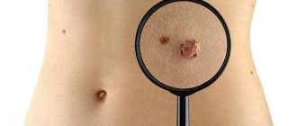
How is the malignancy of a mole assessed?
There are several methods by which the doctor decides - melanoma in front of him or a pigmented nevus (mole). I will describe only one, which I use most often in my practice. This technique is called the Argenziano scale or three-point rating system. According to it, there are three dermoscopic signs of melanoma. One point is added for each feature. If you score two or three points, a biopsy of the mole is indicated. These are the signs:
1) asymmetry of structure or color along two axes of symmetry
melanoma benign nevus
2) atypical pigment network
on the left side of the image there is an atypical pigment network, on the right there is a typical one
3) white-blue structures
in the middle and lower part of the formation there are characteristic light blue areas
Everything is quite simple, however, a lot of experience is required to correctly interpret the dermoscopic picture.
From blackhead and genetic mutations
The first descriptions of melanoma date back to the writings of Hippocrates in the 5th century BC and the work of the ancient Greek physician Rufus. The only thing that was known about the neoplasm was that it was dark and deadly, for which it later received the name “melas” (black) + “oma” (tumor).
Between 1650 and 1760 AD, there were many references to "deadly black tumors with metastases and dark fluid in the body" in the medical literature of Europe. The first surgical removal of melanoma in the Old World is attributed to the Scottish surgeon John Hunter of St. George's Hospital Medical School in London. Hunter himself called it a “cancerous fungal growth” on the jaw of a 35-year-old man - by the way, this melanoma has survived to this day and is now in the Hunter Museum of the Royal College of Surgeons of England in London's Lincoln's Inn Fields.
The famous inventor of the stethoscope, Rene Laennec, was the first to suggest that melanoma was a separate disease, unrelated to the black deposits that may be found in patients' lungs after autopsy.
In 1857, William Norris, a general practitioner from Stourbridge, developed general principles for the epidemiology and clinical management of melanoma. He was one of the first to identify the connection between nevi and melanoma, and also noted the possible dependence of melanoma on environmental factors, such as industrial pollution.
These and other key points are presented in Fig. 1 . And we will move on to the diagnostic criteria for melanoma.
Rice. 1. Main events in the modern history of the study of melanoma (adapted by Rebecca VW, et al. A brief history of melanoma: from mummies to mutations. Melanoma Res 2012; 22(2): 114-122)
Rule ABCD(EF)
Asymmetry, Border, Color, Diameter, Evolving, Funny looking
The famous Rule ABCD (initially without E and F) was proposed in 1985 by a group of American specialists. It was conceived as a simple tool that can be used not only by a doctor at an appointment, but also by an ordinary person in everyday life ( Fig. 2 ).
It is very easy to remember and pronounce the initial letters of the Latin alphabet:
- A – ( a symmetry) asymmetry of the neoplasm;
- B – ( b order) clarity and evenness of edges;
- C – ( color ) color;
- D – ( d iameter) diameter more than 6 mm;
- E – ( e volving) progression of malignant nevus over time (proposed in 2004);
- F – ( funny looking, formerly ugly duckling sign) the dissimilarity of the suspicious nevus to the surrounding ones (in 1998 the ugly duckling sign was proposed, and in 2015 it was reworked into the funny looking criterion).
Rice. 2. 10 steps of early self-diagnosis of melanoma according to the ABCD Rule (Friedman RJ, et al. Early detection of malignant melanoma: the role of physician examination and self-examination of the skin. CA Cancer J Clin 1985; 35: 130-151)
The ABCD rule is easy to apply in practice, although it has certain disadvantages. Thus, it is not suitable for the analysis of ulcerated lesions, since the B order criterion is not adequately assessed in them - it is obvious that the edge will be uneven. However, such tumors are relatively rare and so suspicious that they are usually missed.
The ABCD rule has also been criticized by some experts. For example, seborrheic keratosis and atypical nevi have many of the criteria for melanoma, which can mislead the diagnostician. But the authors sought to create a universal tool for the early detection of most typical melanomas, so they did not take into account individual exceptions.
It should be emphasized that not all melanomas meet the full set of ABCD(EF) criteria. It is the combination of signs (A+B+C, A+E+F, etc.) that makes skin lesions suspicious during screening. The sensitivity of the ABCD Rule (without E and F) for small melanomas (less than 3 mm) is 47.3%, specificity is 56%.
ABCD dermoscopic rule
Asymmetry, Border, Color, Different structural components
There is a dermoscopic version of the ABCD Rule proposed in 1994 by Wilhelm Stolz et al. Here, the asymmetry of the nevus in two planes, the clarity of its edges, the variety of colors and the presence or absence of 5 different structural components are assessed ( Table 1 ).
Table 1. The value of melanoma criteria according to the dermoscopic ABCD Rule
| Diagnostic criterion | Points | Factor |
| Asymmetry (measured in two planes) | 0-2 (1 point for asymmetry in each plane) | x 1.3 |
| edge (nevus is divided into 8 sectors, “like a cake”) | 0-8 (1 point for edge unevenness in each sector) | x 0.1 |
| Color (red, brown, dark brown, black, gray, blue) | 1-6 (1 point for each color) | x 0.5 |
| 5 different structural components (mesh, homogeneous areas, points, globules, stripes) | 1-5 (1 point for each component) | x 0.5 |
scoring more than 5.45 points are extremely suspicious for melanoma and should be studied histologically. The ABCD dermoscopic rule has a sensitivity for melanoma of 84.1% and a specificity of 83.5%.
CASH Algorithm
Color , Architectural disorder , Symmetry , Homogeneity
In 2006, Scott Henning and co-authors proposed the CASH algorithm. It was developed for specialists with little experience in dermatoscopy.
CASH examines 4 parameters ( Fig. 3 ):
- C – ( color ) color: light brown, dark brown, black, red, white, blue. 1 point for each color found.
- A – ( a rchitectural disorder) architectural disorder: scored from 0 (absent) to 2 points (maximum disorder).
- S – ( s ymmetry) symmetry: scored from 0 (absent) to 2 points (symmetry in two planes).
- H – ( h omogeneity) homogeneity: atypical network, dots/globules, pseudopodia, blue-white veil, regression structures, spots >10% of the lesion area, polymorphic blood vessels. 1 point for each structure found.
Any lesion scoring 8 or more on the CASH should lead the clinician to suspect malignancy. The CASH algorithm has a sensitivity for melanoma of 98% and a specificity of 68%.
Rice. 3. Dermoscopic picture of architectural disorder (Henning JS, et al. The CASH (color, architecture, symmetry, and homogeneity) algorithm for dermoscopy. J Am Acad Dermatol 2007; 56(1): 45-52)
- A – absence of disorder with a uniform arrangement of structures and colors ( dysplastic nevus );
- B – moderate disorder with heterogeneous asymmetrical globules ( macular nevus );
- C – marked disorder with a spotted network, many colors, multiple dots and globules ( melanoma, Breslow thickness 0.5 mm)
Mackey's 7-point list
7-point checklist by Rona McLeod MacKie
The 7-point checklist for early diagnosis of invasive melanomas was proposed in 1985 by Rona McLeod MacKie, MD.
Basic criteria for melanoma:
- Change in size over time
- Irregular pigmentation
- Fuzzy edge
- Presence of inflammation
- Itching or altered sensation
- Larger than ordinary nevi (diameter >7 mm)
- Weeping and/or crusting
In 1989, the 7-point checklist was refined:
- The first 3 points are called the main ( large ) criteria and received a value of 2 points
- The remaining 4 points are called additional ( small ) criteria and received a value of 1 point
Patients who score 3 or more points during screening should be immediately referred for a detailed examination ( Fig. 4 ).
In 2005, the UK National Institute for Health and Clinical Excellence (NICE) recommended the Updated 7-Point Checklist for use by all primary care professionals. It has a sensitivity for melanoma of 91.7% and a specificity of 53.5%.
Rice. 4. Early diagnosis of melanoma using the 7-point checklist: visible irregular pigmentation (2 points) and unclear margin (2 points) (Walter FM, et al. Using the 7-point checklist as a diagnostic aid for pigmented skin lesions in general practice : a diagnostic validation study. Br J Gen Pract 2013;63(610):e345-e353)
Argenziano's 7-point rule
7-point checklist by Giuseppe Argenziano
Developed in 1998 by former President of the International Society of Dermatoscopy (IDC) Giuseppe Argenziano and a group of co-authors:
- Atypical pigmentation is a network of unusual black, brown or gray spots and thick lines.
- A blue-white veil is a vague coating consisting of merging spots.
- Atypical vessels – not associated with areas of regression.
- Unusual strokes are linear structures that are not associated with a network of pigmentation lines.
- Unusual pigmentation – black, brown and/or gray areas that are irregularly distributed and of unusual shape.
- Atypical dots and granules are black, brown and/or gray round or oval structures of different sizes, randomly distributed within the neoplasm.
- Signs of regression are white and bluish areas, almost indistinguishable from a blue and white veil.
Melanomas do not necessarily have to have all seven of these features; often five or even three are sufficient ( Fig. 5 ). Argenziano's 7-point rule has a sensitivity for melanoma of 97% and a specificity of 71%.
Rice. 5. Melanoma with three criteria out of seven according to Argenziano’s Rule: above – unusual strokes, 1 point; bottom left – signs of regression, 1 point; lower right - atypical dots and granules, 1 point (Argenziano G., et al. Epiluminescence microscopy for the diagnosis of doubtful melanocytic skin lesions. Comparison of the ABCD rule of dermatoscopy and a new 7-point checklist based on pattern analysis. Arch Dermatol 1998;134(12):1563-1570)
Rule 4x4x6
4×4×6 Rule
Proposed in 2009 by the current President of the International Society of Dermatoscopy, Iris Zalaudek.
Dermoscopic criteria 4x4 :
- Color – black, brown, gray, blue ( Fig. 6 )
- Drawing (pattern) – globular, reticular, star-shaped, uniform blue ( Fig. 7 )
- Pigment distribution – multifocal, central, eccentric, uniform
- Localization – face, palms/toes, nail plate, mucous membranes
And 6 additional factors (... x6 ):
- Patient age
- Skin phototype
- Disease history
- UV exposure
- Presence of pregnancy
- Dynamics of nevus growth
Using the 4x4 dermoscopic criteria and six additional factors helps the clinician better identify melanomas and determine the management of multiple melanocytic nevi.
Rice. 6. The color of the neoplasm during dermatoscopy and its relationship with the depth of its occurrence in the skin: black – superficially in the epidermis, brown – in the dermoepidermal junction, gray – in the papillary dermis, blue – in the reticular dermis (Zalaudek I., et al. Using dermoscopic criteria and patient-related factors for the management of pigmented melanocytic nevi. Arch Dermatol 2009; 145: 816-826)
Rice. 7. Relationship between the dermoscopic pattern (pattern) of the neoplasm and its histological features (Zalaudek I., et al. Using dermoscopic criteria and patient-related factors for the management of pigmented melanocytic nevi. Arch Dermatol 2009; 145: 816-826)
Menzies method
Menzies Method
Proposed in 1996 by Scott Menzies, associate professor of dermatology at the Faculty of Medicine at the University of Sydney (Australia). The method is based on 11 signs of melanoma, which are “negative” and “positive”. For a preliminary diagnosis, both “negative” criteria must be absent and at least 1 of 9 “positive” criteria must be present.
“Negative” signs of melanoma (both must be absent):
- Symmetrical pigmentation is the symmetry of all patterns of the pattern, including color along any axis passing through the center of the lesion.
- This sign is often decisive in the diagnosis of benign pigmented lesions.
- Symmetry of the shape of the neoplasm is not required.
- Single color - meaning black, grey, blue, red, dark brown or tan.
- White doesn't count.
- The presence of a single color nevus excludes the diagnosis of melanoma. This is due to its invasion into the deep layers of the skin: melanin at the level of the stratum corneum appears black, in the middle of the epidermis - dark brown, in the dermoepidermal junction - yellow-brown, in the upper dermis - gray, in the deep dermis - blue.
- Thus, most melanomas have several colors, usually 5 or 6.
“Positive” signs of melanoma (at least one must be present, Fig. 8):
- Blue-white veil is an irregular merging blue pigmentation with a white coating of the “frosted glass” or “veil” type.
- Histologically, it presents with melanin in the middermis with compact orthokeratosis on the epidermal surface.
- The veil never covers the entire melanoma, unlike blue nevi.
- It is found in 51% of invasive melanomas (sensitivity of the sign is 51%) and has a specificity of 97%.
- Multiple brown dots – histologically represent intraepidermal melanoma cells.
- They should be small in size (precisely points, not globules) and accumulate in pockets, and not be scattered.
- The sign has a sensitivity of 30% and specificity of 97%.
- Pseudopodia are “stepped” projections along the edge of the tumor.
- May arise from a pigmented network or from the border of a solid tumor.
- In melanoma, they never occupy a uniform circular position, unlike Spitz nevus.
- They have the same histological substrate as radial veins (see below).
- Pseudopodia occur in 23% of invasive melanomas and have a specificity of 97%.
- Radial veins are finger-like projections along the edge of the lesion.
- Histologically, they are confluent foci of melanoma.
- Typically seen in superficial spreading melanomas.
- The sensitivity of radial veins is only 18%, but the specificity is 96%.
- Scar-like depigmentation is defined as white, scar-like areas within the tumor.
- It should be distinguished from the hypopigmented areas characteristic of nevi.
- Pure white color and clearly visible irregular borders help identify melanoma.
- The sensitivity of scar-like depigmentation is 36%, specificity is 93%.
- Peripheral black dots/globules – located at or near the edge of the lesion.
- Histologically, they are an accumulation of pigment in the stratum corneum of the epidermis.
- They are a deep black color, in contrast to the brown globules that are usually found in benign lesions.
- They should also be distinguished from the central black dots/globules of some dysplastic nevi.
- Peripheral blackheads/globules are found in 42% of invasive melanomas and have a specificity of 92%.
- Multiple colors (5 or 6) – in invasive melanoma, pigment is often detected at different levels of the epidermis and dermis, giving it different colors on dermatoscopy.
- There must be at least 5 colors out of 6 possible: red, brown, dark brown, black, gray and blue.
- The sensitivity of this sign is 53%, specificity is 92%.
- Multiple blue-gray dots - in areas of regression of melanocytic lesions, dermatoscopy may reveal macrophages (melanophages) that have absorbed the pigment.
- They resemble peppers scattered on the table, which is why they are called pepper-like dots.
- In large numbers, melanophages can form areas of a blue veil.
- They are a common symptom of malignant lentigo melanoma.
- Blue-gray dots are detected in 45% of cases and have a specificity of 91%.
- Expanded reticulum is an increase in the thickness of the pigmented reticulum lines found in melanocytic lesions.
- In melanoma, it is usually located focally rather than uniformly over the entire area of the lesion.
- An expanded mesh is also a sign of lentigo melanoma.
- Histologically, it represents an expansion of nests of nevus cells in the dermal-epidermal junction.
- The sensitivity of the sign is 35%, specificity is 86%.
When assessing the totality of signs, the overall sensitivity of the Menzies method for melanoma is 92%, specificity is 71%.
Rice. 8. “Positive” signs of melanoma (adapted from Dermoscopedia.org)
TADA method
Triage Amalgamated Dermoscopic Algorithm
In 2016, Tova Rogers and a group of co-authors proposed the Triage Amalgamated Dermoscopic Algorithm or TADA method.
Triage is the sorting of grapes with the selection of low-quality and unripe berries. The TADA method uses 3 sequential questions to “sort” malignant and benign skin lesions ( Figure 9 ):
- “ Does the lesion have obvious dermoscopic features of an angioma, dermatofibroma, or seborrheic keratosis? » – If “yes”, then no additional assessment is required; follow-up is recommended. If “no”, then the next question sounds.
- " Does defeat have architectural disorder?" » – Any architectural abnormality should be biopsied. If “no”, then the third question sounds.
- “ Does the lesion contain a star pattern, blue-black or gray color, shiny white structures, negative meshwork, ulcers/erosions, or vessels? » – If “yes”, a biopsy should be done. If “no”, then it is not melanoma.
The sensitivity of TADA for melanomas is 94%, specificity is 75.5%. The area under the curve (ROC AUC) for TADA is 0.8461.
Rice. 9. This is a schematic view of the signs studied in the TADA method (adapted from Rogers T., et al. Triage amalgamated dermoscopic algorithm (TADA) for skin cancer screening. Dermatol Pract Concept 2017; 7(2): 39-46)
conclusions
1. The quality of laboratory diagnostics of scabies in the Russian Federation does not meet the requirements of the Order of M.Z. RF No. 162 dated April 24, 2003 “On approval of the industry standard” Protocol for the management of patients. Scabies". It is used by 83.1% of doctors, constantly - by 57.8%, and the pathogen is detected only in 36.5% of cases. Lactic acid is rarely used (21.1%). 69% of specialists do not have dermatoscopes, 10.2% do not know the method, 20.8% ignore the method. Dermatoscopically, 27% more passages are detected than are visible to the naked eye. The frequency of detection of mites in intact scabies tracts (97%), follicular papules (21%) and vesicles (32%) varies. The diagnostic significance of the method increases in cases of reactive scabies associated with vesicles (36.4%) and pustules (66.1%).
2. For scabies complicated by secondary pyoderma, allergic dermatitis and microbial eczema, treatment should be given