A dermoid cyst or dermoid is a tumor that has the shape of a cyst with a wall of connective tissue. A dermoid cyst is an organoid teratoma consisting of connective tissue. A characteristic feature of such formations is that they are smooth on the outside (placed in an oval capsule) and rough on the inside. The inner layer is similar in structure to the skin and consists of periderm and stratified epithelium, with sweat and sebaceous glands, hair and fatty inclusions.
As a rule, dermoids occur in people of different ages and are located on the temporal region, the upper or inner edge of the orbit, on the eyelids, neck, scalp, in the oral cavity, in the area of the manubrium of the sternum, etc.
Localization
The development of a dermoid cyst is possible in almost any organ or tissue, as doctors note. Most often the presence of cysts is found in:
- the ovarian area in women of reproductive age, which can seriously affect the ability of the fair sex to conceive;
- the lower eyelid area (mostly children are affected, but the pathological neoplasm can be easily removed with the help of surgeons);
- the coccyx area, where the size of the ovarian dermoid cyst rarely reaches large sizes, and the formation can go unnoticed for a long time;
- the area of the anus, where without symptoms of inflammation, pathology is generally detected only by digital rectal examination, etc.
The discovery of dermoid cysts in any organ or tissue makes this pathology quite common. Moreover, as doctors note, the presence of dermoid cysts can either not make itself felt for a long time or cause severe discomfort.
Causes of cyst manifestation
- infections;
- parasites;
- injuries, burns;
- consequence of glaucoma;
- genetics;
- eye inflammation;
- reaction to medications;
- congenital iris dissection;
- allergic manifestations;
- dystrophic age-related changes;
- consequences of the operation.
A product of disruption of the activity of the membranes of the eye: iris, cornea, conjunctiva, caused by the above reasons, requires mandatory ophthalmological care and is an unpleasant cosmetic defect. Treatment or removal is necessary to avoid serious consequences.
Symptoms
Detection of cysts - if they are on the surface - rarely poses serious difficulties for the doctor. Dermoid cysts are not very large in size and are usually painless even on palpation. Depending on the contents, the neoplasm can be either dense or soft.
If a dermoid cyst is found in one of the internal organs, it may not make itself felt for a long period of time. Symptoms appear mainly when the tumor reaches a certain size. For example, if there is an ovarian cyst, a woman will complain of:
- pulling pain in the lower abdomen;
- more frequent urge to go to the toilet;
- the appearance of pain during sexual intercourse.
If inflammation of a cyst localized in the anal area develops, there will be pain during bowel movements and difficulty in taking a sitting position.
Symptoms that can characterize a dermoid cyst are very diverse. However, often due to the benign nature of the neoplasm, the pathology is an accidental finding.
Types of cysts
Depending on the source of occurrence, there are different types of eyeball cysts:
- congenital, associated with iris dissection;
- spontaneous ones affect people of different ages, the cause has not yet been identified; with serous, the ball is transparent, with pearl, its walls are dense;
- traumatic ones occur as a result of damage to the eyeball or after an unsuccessful operation;
- exudative are the result of glaucoma;
- due to infection, the ocular conjunctiva gives rise to a retention conjunctiva or, after intervention, an implantation conjunctiva grows;
- stromal is detected and disappeared unpredictably;
- dermoid is formed due to displacement of the epithelium.
The latter originates in utero due to the adverse effects on the mother of medications, radiation, and infection. Dermoid leads to edema, strabismus, amblyopia, astigmatism, and optic nerve atrophy. It cannot be cured, it must be eliminated.
How to get treatment:
- Make an appointment. By phone (89031830141), make an appointment from the website, by E-mail.
- Consultation. In person or remotely (E-mail, WhatsApp, Viber).
- Examination. Ultrasound of the kidneys and bladder. For proximal hypospadias or in combination with cryptorchidism - endocrinological examination.
- Hospitalization and collection of tests. Collection of necessary tests and preparation for surgery.
- Surgical treatment. Microsurgical reconstructive plastic surgery.
- Extract. Recommendations in the postoperative period.
Treatment price
The cost of removing atheroma, cysts, papillomas at MGK starts from 5,500 rubles. The figure is calculated individually and will depend on the volume of treatment and diagnostic procedures performed. You can find out the price of a particular procedure by calling a number in Moscow (the call is free for mobile phones and regions of the Russian Federation) or online, you must use the appropriate form on the website, you can also familiarize yourself with the “Prices” section.
Make an appointment
Diagnostics
Before the procedure to remove a pathological tumor, the doctor will have to carry out a number of diagnostic measures in order to ensure that the diagnosis is correct, as well as to exclude more dangerous pathologies. After all, dermoid cysts can be confused with a number of other diseases.
Dermoid cyst on ultrasound
Ultrasound examination is a simple, painless and non-invasive way to obtain information about the characteristics of the cyst before surgery. It is possible to find out its size, growth characteristics, determine the stalk of the neoplasm and draw conclusions about its blood supply.
MRI
MRI for dermoid cysts is used if the neoplasm has a complex location or there is reason to believe that it is of a low-quality nature. MRI helps to better plan removal of the cyst capsule.
Blood test for tumor markers
If the ovaries are affected in women, it is recommended to donate blood for tumor markers. A cyst on the ovaries can degenerate into a malignant neoplasm, and in order to choose the right therapy, the doctor sends the patient for a blood test for tumor markers. Based on the results, a conclusion is drawn about how dangerous the formation on the ovary is, and whether there is a need for additional treatment.
Histology
Histology when the ovaries or other internal organs are involved in the process is performed in order to exclude malignancy of the neoplasm. The tissue obtained during removal is examined under a microscope to look for atypical cells.
Contraindications
- Inflammation in the body;
- cold;
- diabetes;
- poor blood clotting;
- venous diseases;
- oncology;
- mental disorder in the acute stage;
- pathologies of the heart and blood vessels;
- pregnancy is considered on an individual basis.
Possible complications after surgery:
- hemorrhages due to tissue injury;
- infection due to exposure to microbes;
- seam instability;
- corneal erosion.
Relapses are rare and are possible when the capsule is not completely removed.
The following are suggested as preventive measures:
- apply makeup carefully;
- monitor the expiration date of products that come into contact with your face;
- do not use lenses;
- observe eye hygiene requirements;
- Healthy food;
- systematically undergo ophthalmological examinations.
The chance that the cyst will self-destruct is negligible, so you should not expose yourself to undue risk. Under certain circumstances, these formations can develop into malignant ones. If alarm bells appear, immediately make an appointment with a doctor at our clinic. We have all the technical and professional resources to remove an eye cyst and solve the problem in full.
Indications for surgical treatment
It all depends on the location of the formation, its size, development dynamics, the patient’s age and condition, the effect on the functions of internal organs or their aesthetic value.
For example, if it is located under the skin of the face, on the neck or other open areas of the body, is large, grows, or manifests certain symptoms, then it should be removed after a full examination.
In case of a small formation, which does not manifest itself in any way and was an accidental finding of the examination, it is not always worth resorting to surgery - you should consult with a surgeon and evaluate all the pros and cons. Patients are often advised to see a doctor and undergo regular examinations. Only if the pathological formation begins to grow or change in some way should it be removed surgically.
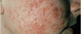
Manifestations of atheroma
Atheroma has characteristic manifestations in the form of a painless bulge, which is not fused with the surrounding tissues and does not differ in color. The diameter of the formation varies, it can reach 10 cm in diameter. The favorite localization of atheroma is skin areas rich in sebaceous glands (face, scalp, ears). Somewhat less frequently, sebaceous atheromas can form on the skin of the back and chest.
The addition of inflammation (it is provoked by the penetration of a bacterial infection) is characterized by redness of the atheroma and the appearance of pain. Further development of the infectious process causes the accumulation of pus in the cavity of the atheroma, which can come out through the fistula, or cause purulent melting of the surrounding healthy tissue.
Cyst removal methods
Removal of a dermoid cyst in most cases is not considered a complex procedure in surgical practice. Today, clinics use the laparoscopic method when it comes, for example, to ovarian cysts.
Laparoscopic surgery has become popular due to the fact that the intervention allows for a faster recovery and has a lower risk of complications.
However, if the pathological neoplasm is very large or localized in a hard-to-reach place, it is possible to remove the cyst through classical open surgery.
Women often worry that if the ovary is damaged, they will have to remove it entirely, but this is not always the case. If the surgeon can save the organ while ridding the patient of dermoid cysts, he will do so.
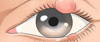
Treatment of eye cysts with laser
The first signs of an eyeball cyst may not be alarming, especially since the small lump may disappear. If it turns into a painful pea, the image becomes clouded and distorted, black flickering spots appear, the field of vision narrows, a visit to the hospital is necessary. The doctor will examine the eye, prescribe tests and make a diagnosis.
Afterwards, either drug treatment or surgical removal will be prescribed. Folk remedies can be used only after consultation with an ophthalmologist. When drawing up an action plan, the origin, size, location are taken into account. Treatment of eye cysts caused by infection occurs with the help of anti-inflammatory, antihistamine drops and ointments. The course usually lasts no more than two weeks.
If medications do not help, then the formation is removed using a scalpel, radio wave knife or laser beam. Previously, fluid was sucked out of the bubble, but after this procedure the formation grew again.
The equipment of modern ophthalmology clinics with advanced equipment and the level of training of specialists make it possible to carry out these difficult manipulations on an outpatient basis. It is very important to remove or destroy the capsule completely to prevent regrowth.
The use of a laser to eliminate a tumor helps to effectively cope with the task and has undoubted advantages:
- precisely directed action;
- destruction completely, without a trace;
- exclusion of infection;
- low likelihood of complications;
- fast recovery;
- absence of swelling and scars;
- healthy tissue is not affected.
Carrying out the operation
It is often performed under general anesthesia, but if it is small and located subcutaneously, local anesthesia may also be used.
As a rule, a skin incision is made and, if necessary, the cavity is opened, access is provided and complete excision is performed. If possible, laparoscopy is performed; if this is not possible, then open intervention is performed.
Surgical removal of the formation can be performed on part of an organ or the entire organ. We are, of course, talking about paired organs. Removal of the ovary is recommended for significant size pathological formations and for postmenopausal women. Removing one kidney with a cyst is only advisable if it is not possible to save the organ. The liver and pancreas can only be resected to the extent that the remaining part of the organ can cope with its function.
That is why, when pathology is localized in the liver, pancreas or other unpaired organs, it is recommended to eliminate them at the earliest stages, including in children, since an increase in the size of the formations can lead to damage to most of the organ and the impossibility of performing radical surgery.
Laser removal procedure
- Local anesthesia in the affected area;
- an incision measuring 2 mm;
- a specialist inserts a laser source through the hole;
- the beam directionally evaporates the affected area and seals the blood vessels.
If the eyeball cyst is large or an abscess has appeared, then surgical removal under anesthesia is recommended. It takes place in several stages:
- limiting the surgical area with sterile materials;
- introduction of a staining solution to clearly define the boundaries of the lesion;
- fixation of the capsule;
- removal with a scalpel;
- rinsing the cavity with an antibacterial solution;
- connecting the incision using a continuous absorbable suture.
Rehabilitation is long and relapse is possible, unpleasant consequences such as hemorrhage, infection, suture dehiscence. In the postoperative period, antibiotics are prescribed, it is advised to reduce the load, and avoid contact with water. The applied bandage is not removed from the eye for five days. It serves as a barrier to dirt and bacteria. There are a number of restrictions on operations.
Consequences of surgery for dermoid cysts
Surgery to eliminate dermoid cysts, if they are detected at an early stage of development, rarely ends in any complications. Basically, within a couple of days after the intervention, the patient can return to his normal rhythm of life.
Difficulties may arise if the removal of the dermoid formation is delayed too much. Possible problems include:
- increased risk of cyst recurrence;
- high risk of hormonal disruptions if the formation is localized on a hormone-producing organ;
- degeneration of cysts into malignant neoplasms.
Treatment of complications of dermoid cyst
Complications may require both medication and emergency surgical intervention. For example, during suppuration, the use of antibiotics and anti-inflammatory drugs is indicated, and removal of the dermoid formation is possible only after the inflammatory process has subsided.
In case of necrosis of the formation, its emergency elimination is indicated, since this can lead to serious complications in the form of suppuration, intoxication, sepsis and even death.
Increased or suspected malignancy is also an indication for removal. In this case, there is no need for emergency surgery, but it should be performed as soon as possible after full preparation (additional examination, anesthetic preparation).
Today, refusal from surgical intervention is considered advisable only in cases where the neoplasm is minimal in size, does not manifest itself in any way, and the likelihood of complications is extremely low. In all other cases, it is better to remove the formation at least in order to prevent the development of complications.
Epidermal cyst - causes
The mechanism of development of the pathological process in which an epidermal sebaceous cyst is the blockage of the duct of the sebaceous gland, while its secretion accumulates. Over time, the volume of accumulated sebum increases, and horny masses, which are exfoliated cells of the epidermis, are added. There are several reasons for the development of such education:
- A metabolic disorder that leads to a thicker consistency of sebum with blockage of the gland ducts.
- Inflammatory processes of the skin (including acne or acne, as well as inflammation of the hair follicles) lead to swelling, which reduces the diameter of the excretory ducts of the glands.
- Traumatization of the skin, followed by scarring, while the connective tissue compresses the ducts of the sebaceous glands.
- Insufficient hygiene leads to blockage of the sebaceous glands with horny skin scales.
- Excessive enthusiasm for various cosmetic products or their improper use.
- Hormonal imbalance, in which the regulation of the process of sebum formation, as well as its release to the surface, is disrupted.
- Genetic predisposition.
Simultaneous exposure to several causes significantly increases the risk of developing such a voluminous skin formation.