INTRODUCTION
Currently, dermatoscopy has become an integral part of the dermatologist's arsenal. The method serves as a useful non-invasive adjunct to the clinical examination of both pigmented and non-pigmented lesions. For melanoma, diagnostic accuracy was found to be 15.6 times higher using a dermatoscope than using visual inspection alone [1]. This led to the inclusion of dermatoscopy training in the residency program. Gradually, the scope of dermatoscopy expanded to include inflammatory and pigmentary dermatoses and is currently used to study diseases of the nails (onychoscopy) [2] and mucous membranes (mucoscopy) [3].
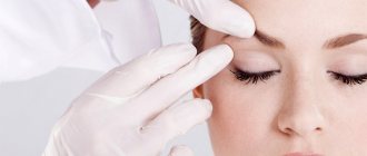
The dermatoscope provides ×10 magnification thanks to the lens. Both contact and non-contact devices can be used, and a cross-polarization filter helps to better visualize deeper structures [4]. Dermatologists have been reluctant to perform routine mucoscopies due to concerns about direct contact of the glass with the mucosa and the risk of transmission of infection. However, innovative measures such as the use of adhesive films and barrier pads have now circumvented these problems [Figure 1] [4, 5]. In this article, we briefly review the published literature on genital muscoscopy [Table 1].
Table 1
Characteristic dermoscopic features of several non-venereal genital dermatoses
| Genital dermatoses | Dermoscopic data | |
| Physiological state | Pearly penile papules (men) and vestibular papillomatosis (women) | Transparent papillary structures with a central vascular component containing irregularly shaped vessels. The bases of individual papillae remain isolated |
| Hyperplasia of the sebaceous glands | Yellowish papules with whitish ovoid structures | |
| Pigment disorders | Melanosis of the mucous membranes | Ring-shaped / parallel / structureless / reticular / globular pattern of uniform brownish pigmentation |
| Genital nevi | A homogeneous/globular structure pattern is usually observed. Atypical nevi have a mixed pattern of structure with a blue-white veil | |
| Melanoma | Multi-component structures with several colors. Blue, gray and white colors have high predictive value. White and blue veil. Amelanotic melanoma - polymorphic vessels | |
| Precancerous conditions | Extramammary Paget's disease and EC | Reddish areas with glomerular vessels |
| Vascular disorders | Angiokeratoma of Fordyce | Reddish-dark blue lacunae with a blue-white veil |
| Inflammatory dermatoses | Balanitis Zuna | Homogeneous orange areas with curved vessels |
EC=Erythroplasia Queyrat
NORMAL PHYSIOLOGICAL CONDITIONS OF THE GENITAL ORGANS
Vestibular papillomatosis
It is believed to be an anatomical variant of the vestibular mucosa, but the condition is often confused with genital warts, leading to unnecessary interventions. This is the female equivalent of pearly penile papules. Clinically, this manifests itself on the inner side of the labia minora in the form of thread-like projections. When dermatoscopy, it appears in the form of transparent papillae with a pronounced fibrovascular center containing randomly located vessels (Fig. 2a). The bases of individual papillae remain isolated. Dermoscopy of genital warts demonstrates multiple, randomly arranged papillae with tapering ends that are less transparent and wider than the structures found in vestibular papillomatosis [Figure 2b]. The papillae arise from a common base [6]. Hemorrhagic lesions in the form of black and red dots and stripes are also visible [7].
Pearly penile papules
They are benign physiological formations located in the area of the coronary sulcus. They are also mistaken for genital warts. On dermatoscopy, they appear as white, slightly translucent papillae arranged in a cobblestone pattern, with a central vascular component that is comma-shaped or may appear as a dot [Figure 3] [8].
Hyperplasia of the sebaceous glands
Modified sebaceous glands located above and distal to the prepuce are called Tyson's glands. Hyperplasia of the sebaceous glands or ectopic sebaceous glands are usually observed in the red border of the lips. On the genitals they appear as pink-yellowish, tiny papules with an umbilical depression. On dermoscopy, they appear as distinct yellowish structures with milky white ovoid structures that are slightly translucent [Figure 4]. They are surrounded by non-branching vessels. Similar lesions can be observed in the mucocutaneous fold of the labia minora in women [9].
Pigment formations
Pigmented formations located on the mucous membrane of the genital organs are observed in approximately 10% of women with fair skin. In men, these formations are located on the head of the penis, and in women in the vulva area. This includes melanotic macules or lentiginosis, nevi, melanoma, angiokeratomas, seborrheic keratosis, squamous cell carcinoma and basal cell carcinoma (BCC) [10].
Melanosis
Melanotic macules, or melanosis, are the most common pigmented lesions observed in the genital mucosa. It is believed that this is a violation of the migration of melanocytes into the epidermis [11]. They tend to appear in areas of the mucosa with non-keratinizing epithelium, rather than in areas of the external genitalia with areas of hair growth. They are benign pigmented macules that are characterized histopathologically by increased pigmentation of the basal layer. It usually appears in people with skin phototypes IV, V and VI. They are clinically present as single or multiple macules ranging in color from light brown to black and can range in size from 1 mm to 1 cm [Figure 5a]. They can often be mistaken for mucosal melanoma. However, standard algorithms such as ABCDE—asymmetry, margin, color, diameter, evolution—may not be suitable for the assessment of genital pigmented macules [10]. The various dermoscopic patterns are described as follows:
1. Ring-like pattern: multiple ovoid structures with hyperpigmented borders, which in some areas look like bunches of grapes [12].
2. Parallel structures: This can be seen in the typical small spots located on the lips, glans penis and vulva. The pigment bands are arranged in regular parallel lines [Figure 5b].
3. Structureless Areas: Structureless pigmented areas ranging in color from light brown to dark blue-gray. Clinically, these macules have a slightly larger diameter [13].
4. Reticular pattern: Similar to the reticular component of pigmented nevi located on the skin, where the pigment appears as a round to ovoid reticular component [Figure 5c].
5. Globular pattern: round-ovoid dark brown globular structures [14, 15].
Genital nevi
They are even more rare, with the prevalence of vulvar nevi being 2.3% among cases of melanocytic nevi [13]. A variety of vulvar nevi are atypical melanocytic nevi of the genital type (AMNGT), which have specific histopathological features. Vulvar nevi are usually present in childhood, in contrast to AMNHT, which is observed in women of the reproductive age group. Vulvar nevi are flat or dome-shaped papules that are pink to dark brown-black or blue in color. They have regular borders and are evenly pigmented. The most commonly described dermoscopy patterns are globular and homogeneous patterns. In addition, milia-like cysts can also be seen. Other patterns, such as parallel structures and cobblestone patterns, are also found and are sometimes difficult to distinguish from vulvar melanosis, which exhibits similar patterns. Sometimes a mixed pattern and radial radiation may raise suspicion for melanoma, and then histopathology can solve this problem [10, 13]. “Divided” or “kissing nevus” of the penis, which are a type of congenital melanocytic nevus, have also been described. It is a solitary nevus that initially divides after the epidermis invaginates to form the coronal sulcus. Dermoscopic features similar to those of congenital melanocytic nevi on the skin, in particular dots and globules, have been described [16]. In a study on genital melanocytic nevi, the authors found that most dermoscopic features were similar to melanocytic nevi on the skin. However, in some cases, differentiation between melanosis and melanocytic nevi, as well as between benign and malignant melanocytic tumors, was not possible using dermatoscopy alone [11]. The practice of routine prophylactic biopsy of melanocytic nevi should be abandoned, especially in children, in the absence of any alarming signs, both clinical and dermoscopic [17].
Atypical melanocytic nevi of the genital type
They make up 5% of all benign melanocytic formations of the genitals. The main histopathological features are as follows [18]:
1. Nested pattern: round or ovoid nests of melanocytes of various sizes that are oriented perpendicular or parallel to the dermoepidermal border.
2. Dyshesive nest pattern: adjacent scattered melanocytic nests, represented by epidermal and dermal melanocytes.
3. Crowded pattern: closely spaced, ill-defined nests of melanocytes located at the dermoepidermal border.
A mixed model of structure is most often observed during dermatoscopy. Parallel lines appear along with structureless areas or brown-black globules [14, 15]. A blue-white veil and irregular dots have also been described. All cases with a mixed pattern on dermoscopy should undergo biopsy to rule out melanoma [10].
REASONS FOR APPEARANCE
Men, especially after the age of thirty to thirty-five years, are most susceptible to the appearance of this neoplasm.
Hyperplasia of the sebaceous gland is a benign neoplasm and, according to general opinion, cannot degenerate into a malignant tumor.
However, especially with multiple manifestations, this skin defect can cause severe psychological discomfort due to its unaesthetic appearance. In addition, papules protruding above the surface of the skin can be damaged when shaving, bleed and become infected. In any case, sebaceous gland hyperplasia carries increased risks of inflammation as a result of mechanical stress. Therefore, people susceptible to this disease often resort to the procedure of removing tumors on the recommendations of dermatologists and cosmetologists.
The exact reliable cause of hyperplasia has not been established, although some experts link it to excessive sun exposure. But, since middle-aged and older men are more susceptible to this defect, that is, there is an age connection, the probable causes of the appearance of sebaceous gland hyperplasia, according to the general opinion of experts, lie in the field of physiology. They are associated with the rate of development and maturation of special cells formed in the gland that synthesize sebum - sebocytes - and the general hormonal status of a person.
At a young age, with a high hormonal status, the formation of sebocytes occurs faster, and their activity is higher. With age, hormonal status can decrease significantly, to which the sebaceous glands in the facial part are most sensitive. As a result, the development and maturation of sebocytes slows down, they accumulate in the duct and cause the growth of the sebaceous gland. Some researchers note the presence of a genetic predisposition to this skin defect. Also, this disorder is more often observed in people who naturally have enlarged pores and a generally oily skin type. In this case, it can manifest itself already in adolescence and after the appearance of single papules, their number can increase significantly.
BENIGN LOCATIONS
Seborrheic keratosis
Pigmented seborrheic keratosis, like its cutaneous counterpart, shows features such as polypoid structures, marrow-like structures, and finger-like structures on dermoscopy. Comedo-like structures, which are keratin-filled invaginations, are not observed in genital seborrheic keratosis due to constant friction. Milium-like cysts are usually visible because they are located intraepidermally [11,19].
Verruciform xanthoma
These are rare tumors composed of lipid macrophages that present clinically as yellowish plaques and nodules with a papillomatous surface in the anogenital areas. Dermoscopy reveals numerous yellowish papules with centrally located spiny vessels [20,21].
INDICATIONS FOR REMOVAL
In most cases, benign tumors are removed at the request of the patient.
- Khimichsky. Chemical treatment may include medication using ointments and tablets based on active ingredients that reduce the size of the sebaceous glands and reduce their activity. However, compared to more modern ones, this method is considered ineffective, since it takes a lot of time and does not always give the desired result, including subsequent relapses. It is also possible to remove papules using trichloroacetic acid, which destroys the sebaceous gland when applying lotions. This usually affects healthy areas of the skin.
- Mechanical. The mechanical method includes removal using a scalpel. But just as in the case of acid, excision is traumatic, accompanied by bleeding and, accordingly, the risk of wound infection, the healing of which requires additional care and can take a long time.
- Physical. Physical methods include electrocoagulation, that is, the destruction of papules due to the impact of high-frequency electric current on them. In this case, scars sometimes form, and healing of the treated skin can take two weeks or more. The method of cryo-destruction or freezing of papules can also be used. In this case, liquid nitrogen is used, which is used to treat the affected areas of the skin. Subsequently, a crust forms, under which healthy tissue is already formed.
- Laser removal. The most modern method of treating sebaceous gland hyperplasia is laser treatment of papules. And one of the main distinctive features and advantages of this method is accuracy. Thanks to this, only the affected areas are affected, and healthy tissues are not affected, as often happens when using other methods. Moreover, precise laser settings eliminate damage not only to areas adjacent to the surface of the skin, but also to those located in its depths, which, among other things, also contributes to accelerated healing. This method is the most painless and absolutely bloodless, since damaged blood vessels are immediately coagulated or sealed with a laser beam. The operation itself is performed under local anesthesia and takes about half an hour on average.
After the laser treatment procedure there are no scars left, that is, it is ideal from a cosmetic point of view. Thus, the highest accuracy, efficiency, accuracy of this method of treating sebaceous gland hyperplasia, as well as the speed of healing are important factors in favor of its choice. And it is better to make such a decision while the papules are still small and have not grown into the width and depth of the skin.
PRE-CANCEROR FORMATIONS
Extramammary Paget's disease
Extramammary Paget's disease is clinically characterized by erythematous plaques with oozing, ulcerated, and scaly nodules and focal hyperpigmentation. Areas of hypopigmentation can also be seen (Fig. 6a). Dermoscopy of extramammary Paget's disease reveals milky red areas, vascular structures, superficial scales, ulcers, pigment network, shiny white lines and white structureless areas [Fig. 6b]. Common vascular structures are stippled vessels and glomerular vessels [22]. Paget's disease is often mistaken for melanoma due to features such as irregular pigmented globules, white negative pigment network, and structureless blue-gray areas [23].
Erythroplasia Keira
It is the genital counterpart of Bowen's disease and has similar dermoscopic features. Glomeruloid vessels are characteristic, located in clusters or diffusely, which can be used to differentiate Queyre's erythroplasia from other causes of chronic balanitis. In addition, linear vessels and brown dots can be seen [24]. Dermoscopic features in pigmented Bowen's disease include pigmented streaks combined with other features such as comedo-like structures [25].
Vulvar intraepithelial neoplasia
This is a common cause of genital itching. About 15% of vulvar intraepithelial neoplasias are pigmented. Typical findings are milky white or pink areas associated with punctate or glomerular vessels and papillomatous structures. In the pigmented version, brownish dots with clearly defined boundaries and brain-like structures are visible [26].
MALIGNANT EDUCATIONS
Melanoma
Vulvar melanomas account for 1-3% of all melanomas observed in women. They are often found in older women and present as flat or raised pigmented lesions that are often larger than 7 mm in size. Non-pigmented variants have also been described. Multiple colors and a multicomponent structure pattern characterize the dermoscopic picture of mucosal melanoma [11, 27]. The presence of blue, gray and white colors with or without structureless areas had 100% sensitivity for diagnosing melanoma [28]. Reticular depigmentation and a network of fine whitish lines are valuable in the diagnosis of early vulvar melanoma [10]. In non-pigmented melanoma, dermoscopy reveals whitish, skin-colored papules with polymorphic vessels [29]. Asymmetry, multicomponent patterns, blue-white veil, and the presence of vessels should raise suspicion of malignancy and be confirmed by histopathology [30].
Basal cell carcinoma
Dermoscopy of genital BCC is similar to cutaneous BCC, consisting of bluish-gray ovoid globules, whitish homogeneous areas, and tree-like vessels. Non-pigmented lesions are difficult to diagnose because... they can mimic inflammatory diseases. In this case, clearly visible tree-like vessels help make the diagnosis [31].
Newspaper “News of Medicine and Pharmacy” 15(335) 2010
Seborrhea (seborrhea, Latin sebum - fat and Greek rhea - flow) Chronic recurrent disease. It occurs during puberty due to dysfunction of the sebaceous glands and increased secretion of sebum with its qualitative change.
Etiology, pathogenesis
Not known. An important role is played by genetic factors, pathological changes in the activity of the sebaceous glands, bacterial flora in the mouths of the hair follicles and sebaceous glands (corynebacterium acne, Staphylococcus alba, aureus), hormonal disorders (increased androgens, decreased estrogens), immunological abnormalities (decrease in T and B lymphocytes, nonspecific immunity), disorders of the central and autonomic nervous system, gastrointestinal tract, the presence of focal infection. Depending on the consistency and chemical composition of sebum, types of seborrhea are distinguished: oily (liquid and thick), dry and mixed forms of seborrhea.
Oily seborrhea
It is observed during puberty, when the secretory function of the sebaceous glands increases. Occurs in areas where there are many sebaceous glands (face, scalp, chest, interscapular area). The skin becomes oily, shiny, uneven, dirty-gray in color, and the mouths of the hair follicles are enlarged. Large yellow and white scales and crusts are observed on the scalp.
Liquid form of oily seborrhea. Women aged 10–14 years are most often affected. Chronic course with remissions in the summer. The occurrence is associated with vegetoneurosis. Sebum has a liquid consistency. Hair becomes oily, sticks together and becomes thinner. After washing they quickly become greasy. The pathological process may be complicated by secondary infection, which leads to a more severe course. There is an abundance of “dandruff” in the scalp area, and often at the age of 20–24 years, baldness develops first in the temporofrontal region and then in the parietal region.
Thick form of oily seborrhea. Men aged 16–20 years are affected. The skin is compacted, dull, brownish-gray in color. On the surface there are dilated openings of the sebaceous glands - “pores”, a thick secretion with an unpleasant odor. On the scalp, the hair is thick, hard, coarse, and scales appear. Complications may occur in the form of folliculitis, boils, and abscesses.
Dry seborrhea
It is mainly observed in children before puberty due to insufficient development of the pilosebaceous apparatus.
Seborrhea of the scalp (gneiss). In the first weeks of a child's life, dirty gray scales form on the scalp. After removing the scales, hyperemic skin appears, on the surface of which scales reappear after some time. On the scalp, upper and lower extremities, and lateral surfaces of the body, the skin is thin, dry, flaky, and follicular hyperkeratosis is noted. Hair is thinning and dry.
Mixed seborrhea
Most often, men suffer from symptoms of oily seborrhea on the face, and dry seborrhea in the scalp. There may be a mixed form of oily seborrhea, in which there are manifestations of liquid oily seborrhea on the face, and thick oily seborrhea on the scalp.
Differential diagnosis
Folliculitis of the head undermining. Folliculitis and ostiofolliculitis appear on the scalp. The process involves adjacent areas. Abscess formation occurs in the deep parts of the follicle and skin. Individual abscesses merge and are connected to each other by fistulous tracts. The skin is bluish-red, lumpy. In the center of the infiltrate, softening and fluctuation occurs. Multiple fistulas connecting to each other are formed, from which purulent masses are released. After healing, rough scars remain with no hair.
Seborrheic eczema. In areas of the skin where there are many sebaceous glands (scalp, face, eyebrows, nasolabial folds, chest, interscapular area), yellowish-pinkish erythematous spots and papules with yellow greasy scales and crusts appear on the surface. Weeping may occur. The folds of skin behind the ears are affected, where serous purulent exudate is released. Subjectively intense itching that precedes the appearance of a rash. Rashes can be localized for a long time on “favorite” areas of the skin, not change and not cause subjective sensations.
Acne
Etiology, pathogenesis
A number of predisposing factors are noted in the occurrence of acne: hypertrophy and hypersecretion of the sebaceous glands, anomaly of keratinization of the mouths of the follicles; the presence of microflora on the skin and in hair follicles, the inflammatory reaction of surrounding tissues. It is observed in persons with oily or mixed forms of seborrhea during puberty, more often in boys. Mainly found are Corynebacterium acne, Staphylococcus aureus, and Pityrosporum ovale. As a result of hypersecretion of sebum, partial emptying of the sebaceous glands, hyperkeratinization of hair follicles, the mouths of hair follicles are blocked with the formation of comedones (“black dots”), which appear with liquid and thick oily seborrhea.
Clinic
There are vulgar, or juvenile, papular, indurative confluent and phlegmonous acne. The most common are acne vulgaris in the form of hemispherical, pink, nodules ranging in size from 1–2 to 3–5 mm in diameter, which are localized in the face, chest and back. An abscess may appear in the central part of the nodule, which shrinks into yellow crusts. After regression, pigmentation or a superficial scar remains. More often observed with liquid oily seborrhea. With thick, oily seborrhea, a large number of comedones appear on the face, back, chest, and scalp with a pronounced inflammatory reaction around. The mouths of the hair follicles are dilated. The rashes are large in size and located deep in the dermis. The flow is sluggish. After a few weeks, an infiltrate forms, which softens and opens with the release of purulent fluid. After regression, deep scars remain. A severe form of thick, oily seborrhea is conglobate acne. In addition to comedones and atheromas, deep, dense, painful nodes are formed, ranging in size from beans to hazelnuts, which sometimes merge, forming conglomerates and abscesses. The skin over them is red-bluish in color. The nodes can open with the formation of long-term non-healing ulcers. After regression of the ulcers, rough scars with bridges and fistulas or often hypertrophic keloid scars remain. The rashes are located on the back, less often on the chest and face.
Differential diagnosis
Medicinal acne. Occurs in people who take various medications for a long time (bromine, iodine, glucocorticosteroid hormones, vitamins B6, B12), in the form of temporary papular and pustular rashes. There are no comedones or seborrhea. Goes away without treatment after stopping medication.
Acne syphilide. Follicular papules 2–3 mm in diameter appear, clearly demarcated from healthy areas of the skin. At the top of the nodule, a conical or spherical pustule with purulent exudate is determined, which shrinks into a yellowish-brown crust. After 1.5–2 weeks, the crusts fall off and depressed, pigmented scars remain. At the same time, roseola and papular syphilitic rashes can be observed. Serological reactions (RV, RIF, RIBT) are positive.
General treatment
A clinical examination is carried out to identify hormonal and neurovascular disorders and their correction. The diet is normalized. Constipation is being fought. Sanitation of focal infection. For neuroses and irritability, valerian, motherwort, and tranquilizers are prescribed. For psychosomatic disorders - adaptogens (pantocrine, tincture of Chinese lemongrass, ginseng, Rhodiola, Eleutherococcus). To improve the function of the sebaceous glands, it is recommended to take purified sulfur 0.5 g orally 3 times a day 30–40 minutes before meals for 2–3 months. To reduce the rate of sebum excretion, patients with acne vulgaris are recommended to take zinc sulfate 25–50 mg orally 3 times a day for 2 months. Vitamins A, group B (B1, B6, B12), and multivitamin preparations are prescribed. For severe forms of acne (globular, phlegmonous, cystic clinical forms), 13 cistretinoid is used in an initial dose of 40 mg, which is gradually increased to 60–80 mg. Improvement is noted after 4 weeks, and a more pronounced effect occurs after 6–12 weeks. Immunotherapy is carried out (staphylococcal antifagin: 0.2–0.4–0.6–0.8–1.0–1.2–1.4–1.6–1.8–2.0 ml in autologous blood every other day , 8–10 injections, or staphylococcal vaccine intradermally into the forearm, 0.1–0.2 ml once every 2–3 days, for a course of 1.0–2.0 ml). For disorders of the functional state of the gonads in women, ethyl estradiol 0.01–0.05 mg 3 times a day in the first 2 weeks of the intermenstrual period or 4 injections of estrone 0.05%, 1 ml daily. For men, octestrol 0.005 g for 10–15 days with a break between courses of 8–10 days. Externally for women, synestrol cream on an emulsion basis (0.01%, 0.1% and 0.15%). For men, synestrol cream 0.1% and 0.14% daily or every other day, 12–15 procedures (3–4 courses of treatment at intervals of 10–15 days). For abscessed, phlegmonous, indurative forms of acne, injections of trypsin or crystalline chymotrypsin are prescribed intramuscularly, 10 mg in 2 ml of isotonic sodium chloride solution every other day, 10–12 injections, to break down necrotic tissue and prevent hypertrophic scars.
External treatment
For oily seborrhea, the lesions are wiped with 1–2% aqueous solutions of sodium thiosulfate, tetraborate or bicarbonate, 1% tannin, 3–5% sulfur. As the inflammatory phenomena subside - 3-4% alcohol solutions of salicylic and boric acid, camphor. Powders with sulfur, boric acid, tannin or a water-shaken suspension with sulfur, ichthyol, and tar are prescribed. For mild forms of seborrhea, it is sufficient to wash the skin with warm water and soap and use alcohol and aqueous solutions or shaken suspensions, ointments (5-10% syntomycin emulsion, 2-3% heliomycin emulsion, 0.5-1% riodoxol). Physiotherapeutic methods include electrophoresis with zinc sulfate and ichthyol. Electrocoagulation of pustular elements and cryotherapy with liquid nitrogen are performed. In the absence of inflammatory phenomena, steam baths and mechanical facial cleansing, absorbable masks, and therapeutic massage are recommended. For acne, salicylic acid-based products are recommended as external therapy, for example the Clearasil ULTRA series, consisting of a cleansing gel, cleansing lotion, scrub and medicated cream. The active ingredients in the formula of these products deeply cleanse pores, have an anti-inflammatory and antibacterial effect, and also regulate sebum secretion. The composition of the products in this series includes hydrolyzed milk protein, which regulates the production of sebum, while simultaneously softening and moisturizing the skin. Clearasil ULTRA cleansing gel and cleansing lotion, in addition to hydrolyzed protein, contain 2% salicylic acid, menthol and coconut glycoside and have a strong cleansing effect, while moisturizing and mattifying the skin. The active ingredients of Clearasil ULTRA topical cream are 2% salicylic acid and 1.5% hydrogen peroxide. The cream effectively and quickly relieves inflammation, has an antiseptic and antibacterial effect.
Prevention
Elimination of factors contributing to the occurrence of the disease. Personal prevention measures (basic series Clearasil Stayclear for daily use based on 2% salicylic acid - gel for washing, cream gel for washing, lotion for deep skin cleansing, daily scrub). Sanitation of foci of infection. Treatment of concomitant diseases. Hardening the body (sports, swimming, sunbathing). Diet. Prevention of skin contamination. Identification and treatment of patients with initial manifestations. Clinical examination of patients with severe forms of acne. Early referral of patients to specialized cosmetic institutions.
Rosacea, rosacea, acne rosaua (lat. rosaceus - pink)
A chronic disease with periodic exacerbations, manifested by hyperemia of the facial skin. It occurs in all races, but predominantly in light-skinned people. More often it begins in the 3rd–4th decades of life and reaches its peak between the 4th–5th decades. English authors refer to rosacea as “Celtic hot flashes.” The high incidence among women is explained by the fact that they seek medical help at earlier stages of the disease more often than men.
Etiology, pathogenesis
An important point in the development of the disease is angioneurosis in the zone of innervation of the trigeminal nerve, caused by various factors: constitutional angiopathies, neurovegetative disorders, endocrine disorders (hormonal disorders, menopause, dysmenorrhea, taking hormonal contraceptives).
A common cause of rosacea is mites of the genus Demodex folliculorum, which is confirmed by their detection in infiltrates and the effectiveness of acaricidal drugs, metronidazole, and sulfur. Demodex folliculorum is a physiological representative of the skin microflora. With age, the population of follicles increases. It is not found in all patients. Often found in granulomas, as mites contribute to the development of a granulomatous, or lupoid, reaction. However, in most cases, the localization of rosacea elements is not associated with the follicular apparatus. Some patients also exhibit specific antibodies to Demodex. In all clinical forms of rosacea, mites and their larvae can be found at the mouths of the follicles and excretory ducts of the sebaceous glands. The highest density is observed during the papulopustular stage (the important role of the tick in the pathogenesis of this phase of the disease).
Many pathogenetic factors are important in the development of rosacea: unfavorable meteorological conditions, a sharp increase in environmental temperature (heat of stoves), exposure to emotional factors, irritating foods, alcohol, gastrointestinal disorders, impaired humoral and cellular immunity, focal infection, the presence of mites (“ ironmen"). Often there are concomitant diseases of a seborrheic nature, follicles, sebaceous glands, which often precede rosacea - from 20 to 83% of cases. Attention is paid to the allergic nature, which is sometimes confirmed by positive intradermal tests with various allergens, deterioration of the clinical picture after taking certain products.
Clinic
The rashes are localized on the face (forehead, nose, cheeks, less often the chin and other areas). At the onset of the disease, liquid erythema occurs, which persists from several minutes to several hours, and then disappears without a trace. Subjectively, patients note a feeling of heat and warmth. Under the influence of provoking factors, it appears again. There is a chronic course, relapses are replaced by remissions (months, years). Infiltration, telangiectasia, erythema with a bluish tint, bright red papules, pustules with sterile contents, papulopustular elements (rosacea) appear. Pustules merge, and inflammatory nodes, infiltrates, tumor-like growths, foci of hyperplasia of the sebaceous glands and connective tissue are formed. The sebaceous glands are enlarged. When pressed, a secretion with an unpleasant odor is released. The skin of the face thickens. Rhinophyma (thickening, knobby nose) forms. It is an independent form and occurs exclusively in men. There is constant venous congestion in the area of the tip and wings of the nose. Hypertrophy of the subcutaneous tissue and sebaceous glands develops. The shape of the nose becomes asymmetrical. Congestive cyanotic erythema, telangiectasia, dilated orifices of the excretory ducts of the sebaceous glands, clogged with comedones, are detected. There is an increase in the functional activity of the sebaceous glands. When pressed, a whitish, pasty-like secretion is released from the mouths of the follicles. Similar changes can be localized in other places: on the chin - gnathophyma; in the region of the glabella - metaphyma; on the earlobes - otophima; on the eyelids - blepharophyma. There are clinical forms of rosacea: erythematous, erythematous-papular, papulopustular and nodular. American and German authors note 3 stages of the disease: erythematotelangiectatic, papulopustular and pustular nodular.
There are special clinical forms of rosacea: - steroid rosacea, the phenomenon of “steroid skin” - mild subatrophy, extensive dark red erythema, telangiectasia, papulopustules; - lupoid (granulomatous) rosacea - disseminated brownish-red papules, small nodes in the periorbital and perioral areas. They fit tightly and form a bumpy surface. Diascopy reveals yellow-brown spots; - rosacea conglobata - large spherical abscess nodes, indurated fistulas. Often appear after taking medications containing halogens (iodine, bromine); - rosacea fulminans - observed in young women and is the most severe variant of rosacea conglobate. Acute onset. Inflammatory nodes merge into conglomerates. Fluctuation, sinuses and fistulas occur. The general condition suffers slightly; - Gram-negative rosacea - numerous follicles appear. Gram-negative bacteria are found in the contents of pustules. There are 2 types: 1) bacteria of the Enterobacteriaceae family, Pseudomonas aeruginosa - small pustular elements; 2) Proteus mirabilis - papules and nodes; - solid persistent swelling of the face (rosacea lymphoedema, Morbighan disease). Dense swelling forms on the forehead, chin, eyelids, nose, and cheeks. When you press on the surface, there is no hole left.
Eye lesions may occur: blepharitis, conjunctivitis, iritis, iridocyclitis, keratitis. Subjectively, patients feel a burning sensation, pain, photophobia, and complain of a foreign body sensation. It should be noted that in 20% of cases, eye damage precedes skin manifestations, in 27% it occurs simultaneously with them, and in 57% of cases, skin manifestations precede eye damage.
Forecast
With the development of rosacea keratitis (Kеratitis rosacea) is unfavorable. There is persistent clouding of the cornea and a significant decrease in visual acuity.
Demodicosis
A skin disease caused by worm-like mites of the genus Demodex folliculorum (Demodex). Mites are found in the sebum of comedones, in pustules, and scales (on the tip of the nose). The clinical picture is similar to that of rosacea.
Diagnostics
Comedones and pus are examined in a slightly warmed isotonic solution of sodium chloride or in alcohol with glycerin, scales are examined in alcohol with glycerin or in a 20% solution of caustic alkali (mites are found dead in alkali).
General treatment
Elimination of etiopathogenetic factors. Dairy-plant diet with the exclusion of extractive foods. Sedatives are prescribed. Antihistamines. Vasoactive agents. Immunomodulators. Biogenic stimulants. Vitamins (A, C, P, B1, B6, B12, folic acid). Sulfur preparations are recommended (purified sulfur 0.5 g 3-4 times a day, 15-20 days). For papular and papulopustular clinical forms, broad-spectrum antibiotics are prescribed. The use of protistocidal drugs often gives good results. Anti-demodectic treatment is important in complex therapy (the period of acaricidal treatment should be at least 25 days, taking into account the tick development cycle).
External treatment
20–30% sulfur ointment, Demyanovich’s method, 5% sulfuric acid, 10–15% aqueous solution of trichopolum with 20–30% dimexide are used. For acute inflammatory phenomena, creams are first prescribed: boric acid 1.0 g, naphthalan 1.5 g, lanolin 10.0 g, peach oil 10 ml, distilled water 10 ml or wheat starch, zinc oxide 2.5 g, lanolin 10 .0 g, peach oil 7.5 ml, distilled water 7.5 ml. After the acute phenomena have subsided, 5% ichthyol, 5% sulfur mash, sulfur-tar pastes, ointments with a gradual increase in the concentration of sulfur and tar to 15%, electrophoresis with a 1-10% solution of potassium iodide are recommended. Good results were noted with the use of “Yam” ointment (Ac. Salicylici 5.0; Sulf. praecir. 10.0; Ol. Terebinthinae restificate 2 ml; Picis liquidae Betule, Lysole aa 5.0 ml; Novosaini 1.0; Zinci cooli 10.0; Lanolini 25.0; Vaselini 35.0; Ol.Lavandulae 2 ml). It is a strong external irritant (possible exacerbation of the inflammatory process). Therefore, on the 1st day it is applied for 15 minutes (residues are removed with vegetable oil), on the 2nd day the exposure is extended to 30 minutes.
In severe cases, cryotherapy with liquid nitrogen (10–15 procedures), dermabrasion (grinding), and electrocoagulation are performed.
Prevention
Eliminate factors that cause dilation of skin blood vessels (insolation, low temperature, excitement, eating spicy food). Work involving exposure to the sun, wind, frost, or in hot workshops is not recommended. The use of photoprotective creams, ointments, powders. To prevent relapses, it is recommended to lubricate the skin of the face with hellebore solution 2-3 times a day (1 week).
Differential diagnosis
Acne vulgaris. They get sick in adolescence. Seborrheic areas are affected. Hemispherical pink nodules, ranging in size from 1–2 to 3–5 mm in diameter, are noted on the face, chest and back. An abscess may appear in the center of the nodule, which shrinks into yellow crusts. After regression, pigmentation or a superficial scar remains. Comedones with a pronounced inflammatory reaction around are detected. The mouths of the hair follicles are dilated. The presence of severe seborrhea, acute inflammation, pain.
Papulonecrotic tuberculosis of the skin. Superficial or deep, flat, dense nodules, ranging in size from 1–2 to 3–5 mm in diameter, appear. The skin over them is unchanged or pale red in color. In their center, a yellow pustule appears, shrinking into a dense brown or dirty-gray crust, after the rejection of which a bleeding, crater-shaped, limited ulcer is noted. After the ulcer heals, a depressed “stamped” scar remains, initially hyperemic, and then a white, round, smooth scar. Along with this, papular rashes are observed, which, when regressed, do not leave scars. The rashes can be single or multiple and are localized on the extensor surface of the extremities, less often on the torso, buttocks, face, genitals, and scalp. Tuberculin tests are positive.
VASCULAR FORMATIONS
Angiokeratoma
Angiokeratoma of Fordyce manifests as multiple, well-defined, bluish-black papules on the skin of the scrotum [Figure 7a]. They are often clinically mistaken for genital nevus, blue nevus and melanoma. Various sources indicate that 20% of angiokeratomas were misdiagnosed as melanoma [32]. Dermoscopy clearly shows pronounced reddish-dark blue lacunae with a blue-white veil [Figure 7B] [33].
Lymphangiectasia of the vulva
Acquired lymphedema and lymphangiectasia of the vulva usually occur due to damage to the lymph nodes that occur after trauma, infection, malignancy, surgery, radiation, etc. Clinically, they present as flesh-colored papules or transparent vesicles with a diffuse erythematous background. Piercing the lesion may release clear fluid. Dermoscopy reveals clearly defined, round-oval reddish lacunae with whitish septa and numerous small pinpoint lacunae. This is similar to the manifestations of lymphangioma. Dermatoscopy is relevant when lesions are sometimes confused with warts or molluscum contagiosum [34].
Skin defects of infectious nature
These formations are removed using electrocoagulation with the parallel use of immunomodulatory methods.
Papillomas
What do they look like? Like tiny “bags” or “droplets” the color of normal skin, hanging on a thin stalk or sitting like a rounded “button”. They can be either single or abundantly covering the skin. The appearance of papillomas indicates a weakening of local immunity to the human papillomavirus.
Where? On the skin of the neck, armpits and under the breasts, less often on other areas of the skin. Almost everyone has papillomas, “relatives” of warts.
How to get rid of it? It is advisable to combine electrocoagulation of papillomas with strengthening the local immunity of the skin. It is advisable to use oxygen-ozone therapy (daily application of ozonated oil and injection of an oxygen-ozone mixture twice a week 7-10 times), local use of ointments with an immunomodulatory effect (ointments with interferon).
Papillomas are superficial formations, so the skin heals quickly after their removal.
If there are a very large number of papillomas or their reappearance after their removal, systemic immunocorrection is prescribed: oral administration of immunomodulators or an injection course. For this, Lykopid, Immunomax, Panavir, Polyoxidonium, Kagocel and a number of other drugs are used (as prescribed by a doctor).
Warts
What do they look like? Formations of irregular shape with a rough surface. They can be quite large. The appearance of warts near the nail or on the sole of the foot is especially unpleasant - in these areas, their removal is most labor-intensive.
Where? Most often on the skin of the hands and feet, although they can be located anywhere.
How to get rid of it? Warts must be removed, as they are contagious - this is the most famous viral skin problem. Warts are removed by electrocoagulation or cauterization with liquid nitrogen. Electrocoagulation is preferable, as it allows you to “target” more accurately and not injure the skin around the wart, and the healing process is easier under the crust, and not under the bubble, as when using liquid nitrogen. Before removing a wart, it is advisable to inject an anesthetic, lidocaine or ultracaine, under it, which will eliminate pain.
For multiple warts, systemic immunocorrection is necessary, as well as examination to identify possible causes that contribute to decreased immunity.
"Calls"
What do they look like? Like calluses. Against the background of various “problems” with the feet (excessive sweating, orthopedic defects of the foot, wearing uncomfortable tight shoes, swelling of the feet), viruses and (or) bacteria can form painful inflamed thickenings of the skin - “corns”. They usually try to fight them with a pedicure, at home or professional, but, as a rule, this does not help.
Where? On the sole and sides of the toes.
How to get rid of it? Remove “corns” using electrocoagulation, remove background causes (sweating, swelling, etc.) and increase local immunity. An excellent effect is achieved by a combination of homeopathic injections that tonify blood circulation in the legs, eliminating excessive sweating of the feet with Dysport, and administering interferon preparations under the corn.
Treatment of “corns” is not a quick process, but it is very useful, since a painful “corns” that interferes with walking forces people to finally address the serious problems that contribute to its appearance.
The editors thank the specialists of the BioMi Vita clinic for their assistance in preparing the material.
INFLAMMATORY DISEASES
Balanitis Zuna
In this condition, well-defined reddish plaques are visible on the skin of the glans or the inner side of the foreskin [Figure 8a]. Dermoscopy reveals uniform reddish-orange areas with curved vessels [Figure 8b] [24]. The orange areas correspond to hemosiderin deposition, and the curved vessels represent vascular proliferation. Other types of vascular structures include crescentic vessels and convoluted, cupped, linear and stippled vessels [35].
Candidal balanitis
Candida balanitis is characterized by curdled-like structures, which are colonies of fungi, as well as linear vessels [24].
Psoriatic balanitis
Clinically, multiple erythematous papules and plaques are detected. Dermoscopy reveals uniformly distributed, dilated and dotted vessels on an erythematous background [36].
Lichen planus
Thick, linear and irregular hairpin-shaped vessels are distributed diffusely according to the localization of lichen planus plaques. The Wickham grid is clearly visible against an intense red background [5].
Lichen sclerosus
White, ivory-colored atrophic plaques are visible on the skin of the glans and labia minora, disrupting the normal architecture of the skin of the anogenital area [Figure 9a]. Dermoscopy reveals white structures that appear pinkish, consistent with sclerosis on histopathology [36]. The gray-blue dots are arranged in a pattern of scattered pepper, which corresponds to melanophages of the skin. Characteristic features are a marked decrease in the number of vessels and the presence of polymorphic vascular patterns [Figure 9b] [5]. Purple-red globules and spots are visible especially in women, consistent with blood stains due to excessive scratching.
SYMPTOMS OF THE DISEASE
Symptoms of the disorder are initially diagnosed visually by a specialist dermatologist.
The papules are soft to the touch and with light pressure on them the patient does not experience pain. Despite the fact that hyperplasia of the sebaceous glands itself is a benign neoplasm, it can be one of the symptoms accompanying more severe diseases, including those of internal organs. On the other hand, some skin diseases, including cancer, may have similar symptoms. Such diseases include lesions of the sebaceous glands and, in particular, sarcoidosis, nodular elastosis, mylin, rhinophyma and others. For this reason, to make an accurate diagnosis, especially if the intention is to remove papules, an examination by a qualified dermatologist is necessary. In addition, tests and biopsies are mandatory. Histological examination reveals an enlarged duct of the sebaceous gland as a sign of hyperplasia. And also the presence of a large number of immature sebacites in the lateral lobules of the gland, despite the fact that, on the contrary, less fat is secreted.
CONCLUSIONS
Dermatoscopy has become important in the diagnosis of non-pigmented dermatoses and for differentiating benign lesions from equivocal and malignant lesions [1, 28]. Many dermatologists in modern practice have included dermatoscopy in their routine work. Although this does not eliminate the need for biopsy, the technique avoids unnecessary biopsies of benign lesions. It also helps us carefully select the biopsy site for suspicious or atypical pigmented lesions.