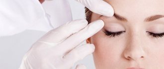
About the procedure
Sebaceous glands are located in the shallow layers of the dermis throughout the human body, except for the feet and palms.
However, their number is uneven: in some areas, for example on the face and scalp, there are more of them, and they all have ducts directed into the hair follicles. On skin devoid of vegetation (lips, genital mucosa and other areas) there are so-called free sebaceous glands that are not associated with hair follicles.
Sebum glands are produced throughout a person’s life; this process is most active in the first year of life and puberty.
Hyperplasia - thickening and enlargement of the sebaceous gland - usually develops in men after 30-35 years of age and the exact reasons that provoke this process have not been clarified at this time.
A number of experts believe that the culprit is hormonal imbalance in the body. This phenomenon is not life-threatening, but leads to pronounced cosmetic defects, since most often hyperplasia of the sebaceous glands occurs on the face: forehead, cheeks, nose, lower eyelids.
Externally, the disease manifests itself as papules on the skin - in the form of convex formations of a pale yellow color. Their size is up to 3 mm in diameter, dome-shaped, in the center there is a small recess from which, when squeezed, a small amount of sebum is released. In some cases, the defect is accompanied by the appearance of spider veins.
Read on topic:
Frenuloplasty
Structure and functions of the sebaceous glands
Sebaceous glands are derivatives or appendages of the skin. They were first described by the anatomist Malpighi in 1689. They are located on almost all areas of the skin, with the exception of the palms, soles and dorsum of the feet. In the overwhelming majority, the ducts of the sebaceous glands open into the mouths of the hair follicles of long, bristly or vellus hair. In this case, the sebaceous gland itself is located at the root of the hair on the border of the reticular and papillary layers of the dermis . In addition, the sebaceous glands are located isolated on the following areas of the skin: the area of the nose, forehead, chin, corners of the eyes, edges of the eyelids (glands of the cartilaginous edge of the eyelids - meibomian glands), the red border of the lips, the place where the skin meets the nasal mucosa, the lower third of the rectum intestines, skin of the nipples and periareolar region of the mammary glands, glans penis and inner layer of the foreskin (tysonian glands), as well as on the labia minora and clitoris.
The number of sebaceous glands on different parts of the body surface is not the same. For example, there are few of them in the area of the back of the hands, the skin of the legs and on the red border of the lips. On the contrary, in the area of the forehead, eyebrows, nasolabial triangle, chin (the so-called T-zone), on the scalp, as well as in the area of the ears, midline of the chest and in the interscapular area of the back, the number of sebaceous glands is large and reaches 400-900 per square centimeter. It is these places of large accumulation of sebaceous glands that are affected by seborrhea, a condition in which the secretion of sebum is increased. A large number of sebaceous glands are also present in the armpits, perianally and perigenitally. There they are anatomically connected not only with hair follicles, but also with apocrine sweat glands. Therefore, when they are blocked and inflamed, one of the severe forms of acne occurs - hidradenitis suppurativa, or inverse acne. These areas with a large number of sebaceous glands are usually called seborrheic.
The sebaceous glands are classified as simple alveolar glands with branched terminal sections and excretory ducts. The terminal sections are formed by several alveoli (sacs, lobules, acini), consisting of multilayered epithelium, which contains two types of cells: basal cells and sebocytes. Basal cells lie on the basement membrane and form the peripheral, or germinal layer of the terminal part of the gland. Sebocytes migrate from the basal layer and differentiate. These cells perform a secretory function; lipids accumulate in them in the form of large inclusions; Subsequently, the sebocytes are displaced in the direction of the gland duct, disintegrate and turn into a secretion - sebum. The type of secretion in which the lipid-producing cell completely dies and a gland secretion is formed is called holocrine, or holocrine. Consequently, the sebaceous glands are typical representatives of the holocrine type of secretion. Each terminal section has its own excretory duct, which are combined into a common one; it exits into the hair follicle. The wide and short common excretory duct is lined with stratified squamous keratinizing epithelium.
Sebaceous follicles and separately located sebaceous glands are abundantly supplied with blood from the superficial and deep dermal plexuses. This circumstance is important, since various metabolic products are released with the secretion of the sebaceous glands, as well as toxic and some medicinal substances (for example, preparations of iodine, chlorine, bromine, etc.) coming from the blood.
The innervation of the sebaceous glands is carried out by a complex nerve plexus surrounding the sebaceous glands, hair follicles, and sweat glands. These plexuses include fibers of the autonomic nervous system.
In different areas of the skin, the sebaceous glands are of different sizes. Typically, the smallest sebaceous glands are associated with long hair follicles, and the large and multilobed ones are associated with vellus follicles. Multilobular sebaceous glands in seborrheic areas are significant. They are quite large in size on the pubis, in the area of the labia majora, and also along the suture line on the skin of the penis. The skin of the legs, forearms and dorsum of the hands is equipped with small mono- or bilobed sebaceous glands. That is why the skin in these places is most often characterized by increased dryness.
During life, the sebaceous glands change their size. Thus, they are relatively large immediately after birth and in the first months of the child’s life, and then decrease. A sharp increase in the size of the sebaceous glands occurs with the onset of puberty. The sebaceous glands reach their greatest size by the age of 18-35. In old age, they atrophy to a large extent, so one of the signs of skin aging is its increasing dryness. It is also known that in girls, increased sebum secretion begins earlier than in boys. Subsequently, women experience a slowdown in the rate of sebum secretion throughout their lives, but this persists longer than in men.
The sebaceous glands secrete a complex secretion called sebum. On average, an adult produces about 20 grams of sebum per day. The secretion of the sebaceous glands onto the surface of the skin is facilitated by the contraction of the smooth muscle that lifts the hair. And the hair itself is also covered with a thin water-lipid film.
Sebum, secreted from the secretory section of the sebaceous glands, fills their excretory ducts, the mouths of hair follicles and is gradually distributed along the grooves of the skin, covering the entire surface of the skin with a layer 7-10 microns thick. At the same time, the secretion of the sweat glands reaches the surface of the skin; at the same time it mixes with sebum and emulsifies. Thus, a continuous, thin water-fat emulsion film, called the water-lipid mantle, is formed on the surface of the body. The composition of the water-lipid mantle also includes ceramides of the stratum corneum (see below). Emulsification of sebum occurs due to hydrophilic high molecular weight aliphatic alcohols and cholesterol included in its composition. Depending on the ratio of sebum and sweat on the skin, the resulting water-fat emulsion may contain more fat (water-in-oil type) or more water (oil-in-water type). So, for example, at high ambient temperatures and increased sweating, an “oil in water” emulsion is formed on the skin, and at low temperatures and slight sweating, an “water in oil” type emulsion is formed. In seborrheic areas, a water-in-oil emulsion is formed at higher temperatures.
In terms of its chemical composition, sebum is a mixture of lipids. It consists mainly of free and bound (esterified) fatty acids. In addition, small amounts of hydrocarbons, polyhydric alcohols, glycerol, cholesterol and its esters, wax esters, squalene, phospholipids, carotene, as well as metabolites of steroid hormones are found in sebum. Among free fatty acids, all fatty acids with a number of carbon atoms from 1 to 22 are found. These include higher and lower fatty acids, saturated and unsaturated, with a straight and branched chain of carbon atoms.
The main part of free fatty acids consists of higher fatty acids and their homologues with 14 (myristic), 16 (palmitic) and 18 (saturated - stearic and unsaturated - oleic) carbon atoms in the chain. Water-soluble lower fatty acids (formic, acetic, propionic, butyric, valeric, capronic, enanthic, pelargonic, capric and undecylenic) and their homologues are contained in sebum in small quantities. It is known that the content of free higher fatty acids is 25% relative to the weight of sebum, and free lower fatty acids are only 5.5%. It should also be emphasized that free lower fatty acids with the number of carbon atoms from 1 to 13 have fungicidal, bactericidal and virusostatic properties, thereby performing the function of a kind of “biological brake” in the protective system of the skin surface.
The skin as a unique structure is rightly compared by many authors to a brick wall, where the role of “bricks” is played by corneocytes (postcellular structures of the stratum corneum), and the “cement” is played by highly specialized and uniquely organized intercellular lipids. Such lipids include ceramides (from the English ceramides), cholesterol, fatty acids, as well as phospholipids, glycosylceramides, free sphingoid bases and cholesterol sulfate. These lipids form the main barrier to water, thereby preventing transepidermal water loss (TEWL). They also play the role of a special intercellular cementing substance, which provides adhesion strength to the postcellular structures of the stratum corneum and ensures the integrity of the skin. It is now known that in the stratum corneum of the epidermis there are six main classes of so-called “free”, not associated with corneocytes, ceramides and two main classes of ceramides covalently associated with the surface of corneocytes (classes A and B). The composition of ceramides in the stratum corneum of the skin in humans is highly variable and depends on race, concomitant medical diseases, age, environment and a number of other factors. Ceramides have a rather complex chemical structure. Most are long chains of sphingoid base with 16 to 22 carbon atoms; less commonly, they are represented by dihydrosphingosine, phytosphingosine and 6-hydroxysphingosine. Sphingoid bases are combined with various fatty acids (oleic, linoleic, etc.), including free lower fatty acids, which perform a number of important biological functions. The functions of ceramides include not only the retention of water in the skin, but also the regulation of the rate of desquamation, as well as the influence on the differentiation of keratinocytes. It has been shown that sphingosine is able to regulate the rate of renewal of the epithelial layer, preventing its rapid change without normal differentiation of keratinocytes.
Sebum secretion is regulated mainly by hormonal and to a lesser extent neurogenic mechanisms.
Hormonal regulation of sebum secretion is carried out through androgens, namely testosterone, which is the main hormone that enhances sebum secretion. It is for this sex hormone that there are receptors on the membrane of sebocyte cells. Interacting with the receptor on the surface of the cell that produces sebum, testosterone under the action of the enzyme 5-a-reductase is converted into its active metabolite - dehydrotestosterone, which directly increases secretion production. It has been proven that sebaceous glands of different locations have different numbers of testosterone receptors. This explains the fact that in a number of patients certain areas are often affected, for example, only the skin in the chin area or only the skin of the back, etc. The amount of biologically active androgen, as well as the sensitivity of sebocyte receptors to it, and the activity of 5-a-reductase, which determine the rate of secretion of the sebaceous glands, are genetically determined. In general, hormonal regulation of sebum secretion can be carried out at four levels: the hypothalamus, pituitary gland, adrenal cortex and gonads.
Hormones that suppress sebum secretion include estrogens, since their predominance leads to a decrease in the effect of androgens. However, the effect of estrogens that suppresses sebum secretion is much less pronounced than the effect of androgens, which stimulate sebum secretion.
As for the neurogenic regulation of secretion, it is mainly carried out by the autonomic nervous system. Increased sebum secretion is found in individuals with vagotonia; At the same time, other symptoms of increased vagal tone may appear - sharply increased sweating, persistent red dermographism, acrocyanosis. A change in the tone of the autonomic nervous system, which innervates the muscles that lift the hair, leads to a more rapid emptying of sebum from the sebaceous glands associated with the hair follicle. It is believed that not only the autonomic nervous system influences the secretion of sebum. It is also known that sebum secretion clearly increases with various lesions of the cerebral cortex (stroke) or with certain subcortical disorders in encephalitis, parkinsonism, diencephalic disorders, as well as with damage to peripheral nerves. Significant disturbances in sebum secretion also occur in patients with mental illnesses such as schizophrenia, manic-depressive and infectious psychoses, epilepsy, and various depressive states. Apparently, in all of these conditions, changes in the release of hormones from the pituitary gland and hypothalamus and an indirect effect on testosterone levels are more significant.
In conclusion, the biological role of the sebaceous glands should be emphasized. First of all, the sebaceous glands secrete sebum, which is part of the water-lipid mantle of the skin. The water-lipid emulsion formed on the surface of the skin has the following effects:
- Gives elasticity to the skin and prevents it from drying out.
- Regulates the rate of desquamation and differentiation of keratinocytes.
- Helps maintain a constant body temperature due to changes in the physical composition of the water-lipid mantle.
- Neutralizes alkalis that reach the skin surface with organic fatty acids and maintains a constant slightly acidic pH (4.5-5.5).
- Suppresses the proliferation of bacteria, fungi, viruses thanks to free lower fatty acids and some other substances that are part of sebum, sweat and the stratum corneum.
- It is one of the ways of excretion of various metabolic products, as well as medicinal and toxic substances.
Hyperplasia of the sebaceous glands: treatment
For an experienced dermatologist, diagnosing this disease will not cause any difficulties, although in some cases hyperplasia can be confused with skin diseases that have a similar clinical picture, for example, epithelioma of the sebaceous glands, xanthoma and others.
To confirm the initial diagnosis, a histological examination is prescribed, which shows the presence of many healthy lobules of the sebaceous gland located around the enlarged duct.
Drug treatment for this disease is ineffective, and no method has been developed to prevent the appearance of new papules. However, to get rid of existing ones, there are several effective cosmetic methods.
Laser removal of papules
The laser beam removes the enlarged gland duct and opens the papule. The procedure is performed using local anesthesia and lasts about 30 minutes. There are no scars left after the procedure. The appearance of bruises can be avoided due to the fact that the laser beam coagulates (solders together) the walls of blood vessels. Complete restoration of the skin takes about 10 days; during this period, the doctor prescribes the application of external agents that accelerate the regeneration process.
Electrocoagulation
The method is based on exposure to high-frequency electric current pulses. The enlarged gland is burned out using an electrode connected to an electric current generator apparatus. A crust remains in the affected area, which disappears after 10-14 days. Electrocoagulation is carried out under local anesthesia and does not leave bruises: blood vessels are also coagulated by the influence of current.
Phototherapy
The method requires a course (3-4 sessions) and is especially recommended for multiple papules. Their removal is carried out by exposure to light rays of a certain frequency. After a phototherapy session, slight redness of the skin and slight swelling may occur, although many patients undergo phototherapy correction without any adverse reactions.
Cryotherapy
Papules on the face are removed by cauterization with liquid nitrogen: an applicator soaked in it is applied to the papule for a few seconds, then wait until the nitrogen evaporates, after which the manipulation is repeated. The procedure is safe, does not cause significant pain and does not cause bleeding. A crust forms in the affected area, which disappears after a while. For multiple papules, several cryotherapy procedures are performed.
Newspaper "News of Medicine and Pharmacy" Dermatology (319) 2010 (thematic issue)
Seborrhea (seborrhea, Latin sebum - fat and Greek rhea - flow)
Chronic relapsing disease. It occurs during puberty due to dysfunction of the sebaceous glands and increased secretion of sebum with its qualitative change.
Etiology, pathogenesis
Not known. An important role is played by genetic factors, pathological changes in the activity of the sebaceous glands, bacterial flora in the mouths of the hair follicles and sebaceous glands (corynebacterium acne, Staphylococcus alba, aureus), hormonal disorders (increased androgens, decreased estrogens), immunological abnormalities (decreased T- and B- lymphocytes, nonspecific immunity), disorders of the central and autonomic nervous system, gastrointestinal tract, the presence of focal infection.
Depending on the consistency and chemical composition of sebum, types of seborrhea are distinguished: oily (liquid and thick), dry and mixed forms of seborrhea.
Oily seborrhea
It is observed during puberty, when the secretory function of the sebaceous glands increases. Occurs in areas where there are many sebaceous glands (face, scalp, chest, interscapular area). The skin becomes oily, shiny, uneven, dirty-gray in color, and the mouths of the hair follicles are enlarged. Large yellow and white scales and crusts are observed on the scalp.
Liquid form of oily seborrhea. Women aged 10–14 years are most often affected. Chronic course with remissions in the summer. The occurrence is associated with vegetoneurosis. Sebum has a liquid consistency. Hair becomes oily, sticks together and becomes thinner. After washing they quickly become greasy. The pathological process may be complicated by secondary infection, which leads to a more severe course. There is an abundance of “dandruff” in the scalp area, and often at the age of 20–24 years, baldness develops first in the temporo-frontal region and then in the parietal region.
Thick form of oily seborrhea. Men aged 16–20 years are affected. The skin is compacted, dull, brownish-gray in color. On the surface there are dilated openings of the sebaceous glands - “pores”, a thick secretion with an unpleasant odor. On the scalp, the hair is thick, hard, coarse, and scales appear. Complications may occur in the form of folliculitis, boils, and abscesses.
Dry seborrhea
It is mainly observed in children before puberty due to insufficient development of the pilosebaceous apparatus.
Seborrhea of the scalp (gneiss). In the first weeks of a child's life, dirty gray scales form on the scalp. After removing the scales, hyperemic skin appears, on the surface of which scales reappear after some time. On the scalp, upper and lower extremities, and lateral surfaces of the body, the skin is thin, dry, flaky, and follicular hyperkeratosis is noted. Hair is thinning and dry.
Mixed seborrhea
Most often, men suffer from symptoms of oily seborrhea on the face, and dry seborrhea in the scalp. There may be a mixed form of oily seborrhea, in which there are manifestations of liquid oily seborrhea on the face, and thick oily seborrhea on the scalp.
Differential diagnosis
Folliculitis of the head undermining. Folliculitis and ostiofolliculitis appear on the scalp. The process involves adjacent areas. Abscess formation occurs in the deep parts of the follicle and skin. Individual abscesses merge and are connected to each other by fistulous tracts. The skin is bluish-red, lumpy. In the center of the infiltrate, softening and fluctuation occurs. Multiple fistulas connecting to each other are formed, from which purulent masses are released. After healing, rough scars remain with no hair.
Seborrheic eczema. In areas of the skin where there are many sebaceous glands (scalp, face, eyebrows, nasolabial folds, chest, interscapular area), yellowish-pinkish erythematous spots and papules with yellow greasy scales and crusts appear on the surface. Weeping may occur. The folds of skin behind the ears are affected, where serous-purulent exudate is released. Subjectively intense itching that precedes the appearance of a rash. Rashes can be localized for a long time on “favorite” areas of the skin, not change and not cause subjective sensations.
Acne
Etiology, pathogenesis
A number of predisposing factors are noted in the occurrence of acne: hypertrophy and hypersecretion of the sebaceous glands, anomaly of keratinization of the mouths of the follicles; the presence of microflora on the skin and in hair follicles, the inflammatory reaction of surrounding tissues. It is observed in persons with oily or mixed forms of seborrhea during puberty, more often in boys. Mainly found are Corynebacterium acne, Staphylococcus aureus, and Pityrosporum ovale. As a result of hypersecretion of sebum, partial emptying of the sebaceous glands, hyperkeratinization of hair follicles, the mouths of hair follicles are blocked with the formation of comedones (“black dots”), which appear with liquid and thick oily seborrhea.
Clinic
There are vulgar, or juvenile, papular, indurative confluent and phlegmonous acne. The most common are acne vulgaris in the form of hemispherical, pink, nodules ranging in size from 1–2 to 3–5 mm in diameter, which are localized in the face, chest and back. An abscess may appear in the central part of the nodule, which shrinks into yellow crusts. After regression, pigmentation or a superficial scar remains. More often observed with liquid oily seborrhea. With thick, oily seborrhea, a large number of comedones appear on the face, back, chest, and scalp with a pronounced inflammatory reaction around. The mouths of the hair follicles are dilated. The rashes are large in size and located deep in the dermis. The flow is sluggish. After a few weeks, an infiltrate forms, which softens and opens with the release of purulent fluid. After regression, deep scars remain. A severe form of thick, oily seborrhea is conglobate acne. In addition to comedones and atheromas, deep, dense, painful nodes are formed, ranging in size from beans to hazelnuts, which sometimes merge, forming conglomerates and abscesses. The skin over them is red-bluish in color. The nodes can open with the formation of long-term non-healing ulcers. After regression of the ulcers, rough scars with bridges and fistulas or often hypertrophic keloid scars remain. The rashes are located on the back, less often on the chest and face.
Differential diagnosis
Medicinal acne. Occurs in people who take various medications for a long time (bromine, iodine, glucocorticosteroid hormones, vitamins B6, B12), in the form of temporary papular and pustular rashes. There are no comedones or seborrhea. Goes away without treatment after stopping medication.
Acne syphilide. Follicular papules 2–3 mm in diameter appear, clearly demarcated from healthy areas of the skin. At the top of the nodule, a conical or spherical pustule with purulent exudate is determined, which shrinks into a yellowish-brown crust. After 1.5–2 weeks, the crusts fall off and depressed, pigmented scars remain. At the same time, roseolous and papular syphilitic rashes can be observed. Serological reactions (RV, RIF, RIBT) are positive.
General treatment
A clinical examination is carried out to identify hormonal and neurovascular disorders and their correction. The diet is normalized. Constipation is being fought. Sanitation of focal infection. For neuroses and irritability, valerian, motherwort, and tranquilizers are prescribed. For psychosomatic disorders - adaptogens (pantocrine, tincture of Chinese lemongrass, ginseng, Rhodiola, Eleutherococcus). To improve the function of the sebaceous glands, it is recommended to take purified sulfur 0.5 g orally 3 times a day 30–40 minutes before meals for 2–3 months. To reduce the rate of sebum excretion, patients with acne vulgaris are recommended to take zinc sulfate 25–50 mg orally 3 times a day for 2 months. Vitamins A, group B (B1, B6, B12), and multivitamin preparations are prescribed. For severe forms of acne (globular, phlegmonous, cystic clinical forms), 13 cistretinoid is used in an initial dose of 40 mg, which is gradually increased to 60–80 mg. Improvement is noted after 4 weeks, and a more pronounced effect occurs after 6–12 weeks. Immunotherapy is carried out (staphylococcal antifagin: 0.2–0.4–0.6–0.8–1.0–1.2–1.4–1.6–1.8–2.0 ml in autologous blood every other day , 8–10 injections, or staphylococcal vaccine intradermally into the forearm, 0.1–0.2 ml once every 2–3 days, for a course of 1.0–2.0 ml). For disorders of the functional state of the gonads in women, ethyl estradiol 0.01–0.05 mg 3 times a day in the first 2 weeks of the intermenstrual period or 4 injections of estrone 0.05%, 1 ml daily. For men, octestrol 0.005 g for 10–15 days with a break between courses of 8–10 days. Externally for women, synestrol cream on an emulsion basis (0.01%, 0.1% and 0.15%). For men, synestrol cream 0.1% and 0.14% daily or every other day, 12–15 procedures (3–4 courses of treatment at intervals of 10–15 days). For abscessed, phlegmonous, indurative forms of acne, injections of trypsin or crystalline chymotrypsin are prescribed intramuscularly, 10 mg in 2 ml of isotonic sodium chloride solution every other day, 10–12 injections, to break down necrotic tissue and prevent hypertrophic scars.
External treatment
For oily seborrhea, the lesions are wiped with 1–2% aqueous solutions of sodium thiosulfate, tetraborate or bicarbonate, 1% tannin, 3–5% sulfur. As the inflammatory phenomena subside - 3-4% alcohol solutions of salicylic and boric acid, camphor. Powders with sulfur, boric acid, tannin or a water-shaken suspension with sulfur, ichthyol, and tar are prescribed. For mild forms of seborrhea, it is sufficient to wash the skin with warm water and soap and use alcohol and aqueous solutions or shaken suspensions, ointments (5-10% syntomycin emulsion, 2-3% heliomycin emulsion, 0.5-1% riodoxol). Physiotherapeutic methods include electrophoresis with zinc sulfate and ichthyol. Electrocoagulation of pustular elements and cryotherapy with liquid nitrogen are performed. In the absence of inflammatory phenomena, steam baths and mechanical facial cleansing, absorbable masks, and therapeutic massage are recommended.
For acne, salicylic acid-based products are recommended as external therapy, for example the Clearasil ULTRA series, consisting of a cleansing gel, cleansing lotion, scrub and medicated cream. The active ingredients in the formula of these products deeply cleanse pores, have an anti-inflammatory and antibacterial effect, and also regulate sebum secretion. The composition of the products in this series includes hydrolyzed milk protein, which regulates the production of sebum, while simultaneously softening and moisturizing the skin. Clearasil ULTRA cleansing gel and cleansing lotion, in addition to hydrolyzed protein, contain 2% salicylic acid, menthol and coconut glycoside and have a strong cleansing effect, while moisturizing and mattifying the skin. The active ingredients of Clearasil ULTRA topical cream are 2% salicylic acid and 1.5% hydrogen peroxide. The cream effectively and quickly relieves inflammation, has an antiseptic and antibacterial effect.
Prevention
Elimination of factors contributing to the occurrence of the disease. Personal prevention measures (basic series Clearasil Stayclear for daily use based on 2% salicylic acid - wash gel, cream-gel wash, lotion for deep skin cleansing, daily scrub). Sanitation of foci of infection. Treatment of concomitant diseases. Hardening the body (sports, swimming, sunbathing). Diet. Prevention of skin contamination. Identification and treatment of patients with initial manifestations. Clinical examination of patients with severe forms of acne. Early referral of patients to specialized cosmetic institutions.
Rosacea, rosacea, acne rosaua (lat. rosaceus - pink)
A chronic disease with periodic exacerbations, manifested by hyperemia of the facial skin. It occurs in all races, but predominantly in light-skinned people. Most often begins in the 3rd–4th decades of life and reaches its peak between the 4th–5th decades. English authors refer to rosacea as “Celtic hot flashes.” The high incidence among women is explained by the fact that they seek medical help at earlier stages of the disease more often than men.
Etiology, pathogenesis
An important point in the development of the disease is angioneurosis in the zone of innervation of the trigeminal nerve, caused by various factors: constitutional angiopathies, neurovegetative disorders, endocrine disorders (hormonal disorders, menopause, dysmenorrhea, taking hormonal contraceptives).
A common cause of rosacea is mites of the genus Demodex folliculorum, which is confirmed by their detection in infiltrates and the effectiveness of acaricidal drugs, metronidazole, and sulfur. Demodex folliculorum is a physiological representative of the skin microflora. With age, the population of follicles increases. It is not found in all patients. Often found in granulomas, as mites contribute to the development of a granulomatous, or lupoid, reaction. However, in most cases, the localization of rosacea elements is not associated with the follicular apparatus. Some patients also exhibit specific antibodies to Demodex. In all clinical forms of rosacea, mites and their larvae can be found at the mouths of the follicles and excretory ducts of the sebaceous glands. The highest density is observed during the papulopustular stage (the important role of the tick in the pathogenesis of this phase of the disease).
Many pathogenetic factors are important in the development of rosacea: unfavorable meteorological conditions, a sharp increase in external temperature (heat of stoves), exposure to emotional factors, irritating foods, alcohol, gastrointestinal disorders, impaired humoral and cellular immunity, focal infection, the presence of mites (“railroad cars”). Often there are concomitant diseases of a seborrheic nature, follicles, sebaceous glands, which often precede rosacea - from 20 to 83% of cases. Attention is paid to the allergic nature, which is sometimes confirmed by positive intradermal tests with various allergens, deterioration of the clinical picture after taking certain products.
Clinic
The rashes are localized on the face (forehead, nose, cheeks, less often the chin and other areas). At the onset of the disease, liquid erythema occurs, which persists from several minutes to several hours, and then disappears without a trace. Subjectively, patients note a feeling of heat and warmth. Under the influence of provoking factors, it appears again. There is a chronic course, relapses are replaced by remissions (months, years). Infiltration, telangiectasia, erythema with a bluish tint, bright red papules, pustules with sterile contents, papulopustular elements (rosacea) appear. Pustules merge, and inflammatory nodes, infiltrates, tumor-like growths, foci of hyperplasia of the sebaceous glands and connective tissue are formed. The sebaceous glands are enlarged. When pressed, a secretion with an unpleasant odor is released. The skin of the face thickens. Rhinophyma (thickening, knobby nose) forms. It is an independent form and occurs exclusively in men. There is constant venous congestion in the area of the tip and wings of the nose. Hypertrophy of the subcutaneous tissue and sebaceous glands develops. The shape of the nose becomes asymmetrical. Congestive cyanotic erythema, telangiectasias, dilated orifices of the excretory ducts of the sebaceous glands, clogged with comedones, are detected. There is an increase in the functional activity of the sebaceous glands. When pressed, a whitish, pasty-like secretion is released from the mouths of the follicles. Similar changes can be localized in other places: on the chin - gnathophyma; in the region of the glabella - metaphyma; on the earlobes - otophima; on the eyelids - blepharophyma. There are clinical forms of rosacea: erythematous, erythematous-papular, papulopustular and nodular.
American and German authors note 3 stages of the disease: erythematotelangiectatic, papulopustular and pustular nodular.
There are special clinical forms of rosacea:
- steroid rosacea, the phenomenon of “steroid skin” - mild subatrophy, extensive dark red erythema, telangiectasia, papulopustules; - lupoid (granulomatous) rosacea - disseminated brownish-red papules, small nodes in the periorbital and perioral areas. They fit tightly and form a bumpy surface. Diascopy reveals yellow-brown spots; - rosacea conglobata - large spherical abscess nodes, indurated fistulas. Often appear after taking medications containing halogens (iodine, bromine); - rosacea fulminans - observed in young women and is the most severe variant of rosacea conglobate. Acute onset. Inflammatory nodes merge into conglomerates. Fluctuation, sinuses and fistulas occur. The general condition suffers slightly; - Gram-negative rosacea - numerous follicles appear. Gram-negative bacteria are found in the contents of pustules. There are 2 types: 1) bacteria of the Enterobacteriaceae family, Pseudomonas aeruginosa - small pustular elements; 2) Proteus mirabilis - papules and nodes; - solid persistent swelling of the face (rosacea lymphoedema, Morbighan disease). Dense swelling forms on the forehead, chin, eyelids, nose, and cheeks. When you press on the surface, there is no hole left.
Eye lesions may occur: blepharitis, conjunctivitis, iritis, iridocyclitis, keratitis. Subjectively, patients feel a burning sensation, pain, photophobia, and complain of a foreign body sensation. It should be noted that in 20% of cases, eye damage precedes skin manifestations, in 27% it occurs simultaneously with them, and in 57% of cases, skin manifestations precede eye damage.
Forecast
With the development of rosacea keratitis (Kеratitis rosacea) is unfavorable. There is persistent clouding of the cornea and a significant decrease in visual acuity.
Demodicosis
A skin disease caused by worm-like mites of the genus Demodex folliculorum (Demodex). Mites are found in the sebum of comedones, in pustules, and scales (on the tip of the nose). The clinical picture is similar to that of rosacea.
Diagnostics
Comedones and pus are examined in a slightly warmed isotonic solution of sodium chloride or in alcohol with glycerin, scales are examined in alcohol with glycerin or in a 20% solution of caustic alkali (mites are found dead in alkali).
General treatment
Elimination of etiopathogenetic factors. A dairy-vegetable diet with the exclusion of extractive foods. Sedatives are prescribed. Antihistamines. Vasoactive agents. Immunomodulators. Biogenic stimulants. Vitamins (A, C, P, B1, B6, B12, folic acid). Sulfur preparations are recommended (purified sulfur 0.5 g 3-4 times a day, 15-20 days). For papular and papulopustular clinical forms, broad-spectrum antibiotics are prescribed. The use of protistocidal drugs often gives good results. Anti-demodectic treatment is important in complex therapy (the period of acaricidal treatment should be at least 25 days, taking into account the tick development cycle).
External treatment
20–30% sulfur ointment, Demyanovich’s method, 5% sulfur-tar alcohol, 10–15% aqueous solution of trichopolum with 20–30% dimexide are used. For acute inflammatory phenomena, creams are first prescribed: boric acid 1.0 g, naphthalan 1.5 g, lanolin 10.0 g, peach oil 10 ml, distilled water 10 ml or wheat starch, zinc oxide 2.5 g, lanolin 10 .0 g, peach oil 7.5 ml, distilled water 7.5 ml. After the acute phenomena have subsided, 5% ichthyol, 5% sulfur mash, sulfur-tar pastes, ointments with a gradual increase in the concentration of sulfur and tar to 15%, electrophoresis with 1–10% potassium iodide solution are recommended. Good results were noted with the use of “Yam” ointment (Ac. Salicylici 5.0; Sulf. praecir. 10.0; Ol. Terebinthinae restificate 2 ml; Picis liquidae Betule, Lysole aa 5.0 ml; Novosaini 1.0; Zinci cooli 10.0; Lanolini 25.0; Vaselini 35.0; Ol.Lavandulae 2 ml). It is a strong external irritant (possible exacerbation of the inflammatory process). Therefore, on the 1st day it is applied for 15 minutes (residues are removed with vegetable oil), on the 2nd day the exposure is extended to 30 minutes.
In severe cases, cryotherapy with liquid nitrogen (10–15 procedures), dermabrasion (grinding), and electrocoagulation are performed.
Prevention
Eliminate factors that cause dilation of skin blood vessels (insolation, low temperature, excitement, eating spicy food). Work involving exposure to the sun, wind, frost, or in hot workshops is not recommended. The use of photoprotective creams, ointments, powders. To prevent relapses, it is recommended to lubricate the skin of the face with hellebore solution 2-3 times a day (1 week).
Differential diagnosis
Acne vulgaris. They get sick in adolescence. Seborrheic areas are affected. Hemispherical pink nodules, ranging in size from 1–2 to 3–5 mm in diameter, are noted on the face, chest and back. An abscess may appear in the center of the nodule, which shrinks into yellow crusts. After regression, pigmentation or a superficial scar remains. Comedones with a pronounced inflammatory reaction around are detected. The mouths of the hair follicles are dilated. The presence of severe seborrhea, acute inflammation, pain.
Papulo-necrotic tuberculosis of the skin. Superficial or deep, flat, dense nodules, ranging in size from 1–2 to 3–5 mm in diameter, appear. The skin over them is unchanged or pale red in color. In their center, a yellow pustule appears, shrinking into a dense brown or dirty-gray crust, after the rejection of which a bleeding, crater-shaped, limited ulcer is noted. After the ulcer heals, a depressed “stamped” scar remains, initially hyperemic, and then a white, round, smooth scar. Along with this, papular rashes are observed, which, when regressed, do not leave scars. The rashes can be single or multiple and are localized on the extensor surface of the extremities, less often on the torso, buttocks, face, genitals, and scalp. Tuberculin tests are positive.
Preventive measures
As mentioned earlier, there are no preventive measures, since the exact causes of the development of the defect have not been identified.
As general measures, your doctor may recommend careful skin hygiene and a healthy diet, low in sugar and animal proteins, as they increase the activity of the sebaceous glands.
If the papules - as a cosmetic defect - do not bother the patient, then they are not removed: there are no medical indications for this, since the cells do not degenerate into malignant ones.
You can choose a clinic and make an appointment with a dermatologist at any convenient time using our portal.
Below are clinics and experienced doctors who provide treatment for sebaceous gland hyperplasia in Moscow and Russian cities. You can find out the cost of treatment for sebaceous gland hyperplasia, contraindications, ratings and reviews, and also make an appointment online.