In this article we will tell you what papillomavirus is and how it is dangerous. Do papillomas need to be removed? And also about the features of the laser removal method and contraindications to its implementation.
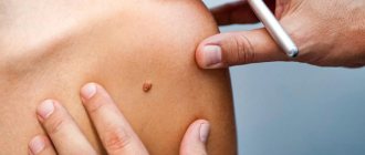
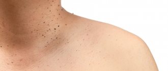
Papillomas are benign formations on the skin and mucous membranes caused by the human papillomavirus (HPV). In their appearance, the formations resemble a pedunculated papilla (rapula) - translated as a nodule. This type of formation occurs most often and its distinctive feature is the ability to appear on any part of the body: face, neck, foot, head, chest, back, genital area and even mucous clouds. Such growths are most often flesh-colored or cinnamon in color. The formations can be either single or multiple; they can connect with each other and create growths that occupy large areas of the body.
Causes of papillomas
According to statistics, more than 90% of the population are carriers of HPV, but it does not affect everyone. This is due to the state of the immune system. The lower the immunity, the higher the likelihood of papillomas. If you have growths characteristic of papillomavirus, this indicates a decrease in immunity.
You can become infected with HPV in different ways:
- Physically, even with a handshake
- Sexually
- When using common household and hygiene items
- When visiting baths, saunas, nail salons
- Perinatal route - infection can pass from mother to child.
There are many varieties of papillomavirus, conditionally they are divided into 3 groups according to risk level:
- Short
- Average
- High
Low-level papillomas can disappear on their own over a period of 6 months to 2 years, while high-risk ones can degenerate into cancerous formations. There is a high risk of degeneration into a malignant formation in papillomas located on the genitals. In women, HPV can cause uterine cancer, the inability to conceive a child, the cause of infertility, failure to carry a pregnancy to term, and postpartum complications.
Danger of laser radiation
Depending on a number of conditions, laser radiation can be both beneficial and harmful to the human body. The positive effect is determined by the ability of the laser beam to precisely “burn out” damaged tissue areas. The potential danger is varied and is determined by several factors:
- Source power;
- Duration of exposure;
- Wavelength;
- Concentration on one point;
- Reflection of radiation;
- Environment.
In response to irradiation, the body undergoes organic (directly at the site of exposure) and nonspecific changes occurring in human organ systems.
Most often, laser radiation affects the eyes (retina and choroid), as well as the skin. If we are talking about damage to the body by visible spectrum rays, then it is worth mentioning the observed disruptions in the functioning of the endocrine, metabolic and immune systems.
However, all of the above side effects are typical for industrial lasers. Cosmetology uses the latest generation of installations that do not cause serious damage to the human body.
Do papillomas need to be removed?
Changes in color and size are one of the possible signs of the onset of malignant processes in the body. Experts recommend contacting a medical facility if the papilloma is bleeding, it is inflamed and bothers you, it is a cosmetic defect, and also if there are signs of its degeneration. If papillomas are not removed in time, then the risk of its growth and degeneration is very high. Formations detected in the early stages, including malignant ones, are almost always curable.
Diagnostics
If the above symptoms appear, it is recommended to promptly seek help from a gynecologist. To detect papillomas inside the vagina, the doctor will collect anamnesis and send the patient for further diagnostics (laboratory and instrumental). The medical history includes the collection of information about possible unprotected sexual intercourse, the approximate time of formation of growths, and other disturbing symptoms.
Further diagnostics consists of the following activities:
- linked immunosorbent assay. It is used to determine the presence of antibodies to the virus in the patient. This allows you to determine the degree of progression of HPV;
- PCR (polymerase chain reaction) analysis. It allows you to establish not only the presence, but also the quantity and type of pathogen present in the body;
- histological analysis. This diagnostic method is aimed at determining changes in the structure of vaginal tissue;
- colposcopy. Using a special device, the doctor examines the vagina and cervix to determine the affected areas;
- anoscopy. This method involves examining the patient’s anal area for the presence of papillomas;
- oncocytology. The specialist takes scrapings from the cervix and cervical canal and examines them in the laboratory.
Based on the diagnostic examination data, the doctor determines or denies the presence of papillomavirus infection. If a virus is detected, the patient is immediately prescribed treatment. During the growth of condylomas in the vagina, the virus is not contagious. The risk of infection appears when the pathogen reaches the epidermis.
Laser removal of papillomas: features of the method
There are several ways to remove papillomas:
- Surgical removal
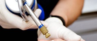
- Cryotherapy – exposure to low-temperature nitrogen
- Electrocoagulation
- Use of chemical reagents.
All these methods are of course practiced in modern medicine, but these procedures are painful and require a long period of rehabilitation.
Today, modern medicine offers an effective and painful method for removing papillomas and other formations using laser surgery. Laser removal is the most optimal method, which is used by doctors around the world, allowing you to get rid of problems in a short time without the risk of relapse.
Impact of laser radiation on the human body
Despite the apparent simplicity of the procedure, there are a number of contraindications to its implementation:
- Oncological diseases (except melanoma and basal cell carcinoma);
- The period of bearing a child;
- Problems with blood clotting;
- Epilepsy;
- Diabetes mellitus and insulin resistance;
- Individual intolerance;
- Diseases of the heart and blood vessels.
Side effects of laser tumor removal may occur after surgery:
- Moderate pain and swelling in the injured area is normal, but severe pain and extensive inflammation are alarming symptoms;
- Capillary bleeding or a consequence in the form of hyperpigmentation may occur;
- If the rules of postoperative rehabilitation are violated, there is a possibility of wound infection.
How does laser removal work?
A distinctive feature of this method is the targeted effect on the papilloma itself, while healthy areas of the skin are not affected. The formation is evaporated layer by layer. The laser is capable of treating tissue areas of any depth, while this method is bloodless, the laser seals the capillaries and the blood instantly stops. Most often, just one procedure is enough to completely get rid of the problem and get an excellent cosmetic effect.
A huge advantage is that it can be used on any part of the body and even on the mucous membranes. The doctor independently regulates the frequency of pulses and the dosage of laser radiation - this avoids overheating and eliminates the risk of burns during the procedure. After the procedure, a crust appears at the site of laser exposure, which cannot be removed independently. It will remove itself in about a week, and healthy skin will form in its place.
Preparation for the procedure
Two weeks before laser therapy you must:
- limit exposure to the sun, use products to protect against UV rays, and do not visit the solarium;
- do not carry out cosmetic procedures in the affected area (peelings, hardware cosmetology sessions, etc.);
- if the neoplasm is located in an area where the risk of irritation is increased (for example, it constantly clings to the collar), the papilloma must be covered with a plaster
Before the sessions, the doctor will definitely tell you about contraindications and identify their presence.
Hardware diagnostic methods
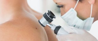
Dermatoscope HEINE DELTA 20 T
Designed for dermoscopic examination for early diagnosis of malignant melanomas, for the study of non-melanocytic formations, basal cell carcinoma and dermatofibroma.
Dermatoscopy is a non-invasive method of visual diagnosis of the skin, which, thanks to the use of high technology, allows us to move from a subjective assessment of the condition of the skin to an objective one and document identified skin changes.
The essence of the method is that the surface layers of the skin are examined using a special device - a dermatoscope at 10x magnification. This allows us to more thoroughly study the symmetry of the neoplasm, its boundaries, and structure.
CLEAR VISION 3D
The most phenomenal and new development in the field of diagnostics today, absorbing all the advantages of its older brother and multiplying them thousands of times.
The evolution of cinema from 2D to 3D did not pass by other areas of activity, because until recently the Vision system was famous for its so-called two-dimensional or 2D model for diagnostics.
It is not difficult to guess that covering the treated area from several sides will give a much greater effect than “observation from one angle.”
Diagnosis of 3D in our clinic is carried out free of charge.
“3D” in our case is not a tribute to fashion, but a visual confirmation of technological progress. The installation allows for three-dimensional scanning of the client’s face using a single short image. There is no need to worry about the quality, since the device is equipped with six high-resolution cameras located at three angles, which gives us images from both sides (profile and frontal) simultaneously in one shot. If time is money, then Clear Vision 3D understands this like no other!
DUB-TPM
Ultrasound diagnostics of skin.
Ultrasound diagnostic scanning is a well-known and proven technique, which currently accounts for more than 1/3 of all diagnostic procedures in medical practice. Modern devices are already quite easy to use and are available to many clinics.
However, these studies have not previously been used in dermatology, which was due to the difficulty of technically solving this problem. In conventional devices, the sensors have a frequency of 3-10 MHz, at which it was impossible to image the structures of the epidermis, dermis and hypodermis.
The German company TPM has created unique devices with sensor frequencies of 20-100 MHz. This technique is called high-resolution digital ultrasound imaging with the ability to study the most superficial layers of the skin.
Based on research materials conducted using the DUB TPM apparatus (the list is attached separately), many articles and 2 monographs have been published.
Until now, the main method for studying skin morphology has been histological and pathomorphological examination. This technique is quite labor-intensive and expensive; in addition, a biopsy sample is examined that has already been treated with various chemical reagents.
Ultrasound diagnostics of the skin fills the gap that previously existed between external research methods and histology, since this non-invasive method allows the study of the skin in vivo.
The importance of ultrasound scanning for skin diagnosis cannot be overestimated. This method has a number of undeniable advantages - non-invasiveness, painlessness, safety and high measurement accuracy. All studies are carried out without damaging tissue and can be repeated on the same area of skin many times.
The new tool allows you to see a section of the skin and subcutaneous fat down to the muscle fascia. We can examine the skin at various time intervals, documenting all the features. The data is digitized and placed in a database. It is easy to carry out a comparative analysis of images obtained over time, the images are saved on any digital media and the data is transmitted in publicly accessible formats via the Internet.
Ultrasound examination of the skin should become the “gold standard” in skin diagnostics, as in obstetrics, gynecology and cardiology.
Distinctive features of the DUB system
- TPM was the first to develop and commercialize skin ultrasound and set the standard for skin ultrasound throughout the world.
- Only DUB devices are equipped with sensors with a maximum frequency of up to 100 MHz and a resolution of up to 8-10 microns.
- Scan modes A,B,C.
- Three-dimensional scanning.
- Cinema loop without limitation of shooting duration.
- Digitization of the signal means the image is more detailed and clear.
- Digital data processing.
- View multiple images taken at different times.
- Innovative image processing algorithms.
- Saving raw data.
- Advanced software package.
- The use of an open system with water allows you to obtain 10-20% more information than using a system with film.
- At the moment, the device has no analogues throughout the world.
Device capabilities:
- 1) Study of the condition, structure and size of all layers of skin and skin formations.
- 2) In-depth diagnosis of morphological and functional changes in acute and chronic skin diseases, including scar changes and lipodystrophies.
- 3) Assessment of the dynamics of skin condition in normal and pathological conditions.
- 4) Facilitation of diagnosis of small skin rashes.
- 5) Timely early diagnosis, since with the help of ultrasound scanning it is possible not only to identify characteristic signs of skin manifestations at the earliest stages, but also to carry out preclinical diagnosis, prevention or timely treatment.
- 6) Diagnosis of skin conditions in the case of any manifestations difficult to detect with the naked eye.
- 7) Visualization, determination of size, volume and depth of invasion, as well as assessment of skin tumors and skin metastases, selection of treatment methods, setting parameters and monitoring effectiveness.
 Preoperative measurement of the depth of spread and volume of tumors during surgical interventions, including electrosurgery, cryosurgery, laser or radiation therapy.
- 9) Study of age-related skin changes.
- 10) Determination of the depth, intensity and duration of the therapeutic effect, choice of method.
- 11) Evaluating the effectiveness and monitoring of therapeutic, physiotherapeutic and surgical treatment methods, including cosmetic procedures (for example, mesotherapy, peelings, plastic surgery, tattoo removal, hardware procedures, etc.).
- 12) Preliminary diagnosis and evaluation of the results of the introduction of fillers, hyaluronic acid, collagen, synthetic or semi-synthetic gels, etc.
- 13) Early diagnosis of osteoporosis.
- 14) Study of skin elasticity.
- 15) Study of mucous membranes.
Advantages of using this method:
- 1) A non-invasive technique for visualizing the internal structures of the skin in vivo, which allows you to obtain important information that is not available with other research methods.
- 2) The method is indispensable for assessing the dynamics of skin condition in dermatology, cosmetology and dermato-oncology. Allows you to monitor the condition of skin manifestations and use the data in the primary diagnosis, prevention and treatment of most skin diseases.
- 3) The ability to save data in computer memory and on any electronic media, print out photographs for medical records, send via the Internet for consultations with colleagues.
- 4) An objective assessment of the dynamics of the patient’s skin condition is an important legal aspect in resolving conflict situations.
- 5) Visual visualization of the state of the internal structures of the skin and its relief is a strong psychological factor in explaining to patients the need for therapeutic measures.
- 6) With the help of DUB, it is easy to prove the effectiveness of treatment in a form accessible to the client. This is a powerful marketing tool to attract new customers.
- 7) The presence of this method increases the rating of the institution and indicates high equipment and the use of advanced technologies.
- In addition to the histological picture, it increases the accuracy of the pathological diagnosis.
- 9) Conducting scientific and educational programs.
- 10) Conducting consultations and consulting and diagnostic activities.
A new aspect of using this method for manufacturers is to assess the effects on the skin of various products, including cosmetics, medications, and devices.
VIVASCOPE® 1500 MULTILASER
The most accurate diagnostic device in dermatology, dermato-oncology and cosmetology.
Confocal laser scanning microscope for in vivo histological examination of skin by fluorescence using lasers of 3 wavelengths (785 nm, 658 nm, 445 nm)
- Combining reflected laser and fluorescence detection technology
- Optical non-invasive biopsy
- Real time display
The VivaScope 1500 Multilaser system combines reflected laser acquisition technology with fluorescence confocal laser scanning microscopy. Similar to the standard VivaScope 1500, skin areas can be viewed in vivo using infrared light. The wavelengths used are 785 nm (near infrared), 658 nm (red) or 445 nm (blue). All three lasers are integrated in one device.
Before using VivaScope, a fluorescent dye (non-toxic to the body) is applied to the area of tissue that needs to be examined. The corresponding laser radiation excites the fluorophore and the resulting fluorescence allows a contrast image to be obtained, which helps to display the histological structure due to the distribution of the dye.
With the VivaScope 1500 Multilaser it is possible to image various functional aspects of tissue changes in vivo. Living tissue can be visually examined sequentially using all available laser wavelengths.
What is needed to undergo treatment?
Today, there are several ways to remove papillomas and other formations on the skin and mucous membranes, but the laser method is superior to all these methods in terms of efficiency. The specialists of Dr. Novikov’s center have been successfully working in the field of laser medicine and cosmetology for many years. The procedure for removing papillomas in Moscow in our center is carried out on an outpatient basis. We remove papillomas in both men and women on any part of the body as effectively as possible. You can make an appointment with our specialists by calling +7, or use the convenient feedback form.
Are there any special recommendations for skin care after the removal procedure?
We issue the following recommendations in the form of a reminder to our patients:
- During the first 24 hours, do not wet the removal site.
- Treat the wound with Fukortsin solution 2 times a day for 3-5 days or Baneocin powder.
- Men: do not shave in the area of removal for 3-4 days.
- Women: do not apply cream or decorative cosmetics to the removal area for 3-4 days.
- You cannot pick or remove the crust yourself!!! in order to reduce the likelihood of atrophic/keloid scars, hypo- and hyperpigmentation.
- In cases of removal of large tumors, after the crust is rejected, apply Levomekol ointment locally 1-2 times a day for a month.
- Before going outside, be sure to use sunscreen on the face and open areas of the body with an SPF of at least 50, especially on sunny days (to prevent pigmentation of the treated areas).
Prevention of papillomavirus
Doctors at Dr. Novikov’s clinic take a comprehensive approach to the treatment of papillomas, therefore, after removal, in order to avoid the reappearance of formations, they recommend undergoing antiviral and immunostimulating therapy.
In order to avoid relapse, as well as to prevent infection, carriers of this virus are recommended to:
- Get tested periodically for the presence of HPV in your blood.
- Control the growth of existing formations
- Have only protected sex
- Use only your own hygiene products
- Eat properly
- Strengthen the immune system
- When visiting saunas and baths, use your own replacement shoes
Why do vaginal papillomas occur?
The main cause of papilloma in the vagina is the papilloma virus. It enters the body through unprotected sexual contact with a carrier. However, there are a number of factors that increase the likelihood of infection. These include sexually transmitted diseases (for example, gonorrhea and chlamydia).
Also, the reasons that contribute to the development of papillomas are:
- frequent change of sexual partners;
- disruption of the normal vaginal microflora (dysbiosis);
- low immunity;
- inflammation of the ovaries;
- severe stressful situations;
- pregnancy;
- deviations from the endocrine system;
- lack of vitamins in the body.
If several of the reasons described above are present, the risk of infection increases significantly. The incubation period is no more than 3 months, but under the influence of unfavorable circumstances for the immune system, this time can be reduced to 14-20 days.
Contraindications for laser papillomas removal
The procedure for removing papillomas with a laser is safe, but there are still reasons when you need to refrain from this method of removal. Now we will look at them:
- Oncology or suspected cancer
- Pregnancy and breastfeeding period
- Diabetes
- Herpes during an exacerbation
- Acute viral infections
- Endocrine system diseases
- Epilepsy
- Exacerbation of chronic diseases
Important! Health issues should be dealt with by professionals. If you have papillomas and you notice signs of their formation or they cause discomfort. The only correct solution to the problem is to contact specialists at a medical institution.
In no case should you remove the formation on your own, as you can aggravate the condition and delay the healing process. Timely elimination of papillomas and other skin problems will avoid the development of serious pathologies. At Dr. Novikov's laser medicine center you can undergo a consultation and receive qualified medical care. Experienced specialists will help you quickly and painlessly get rid of papillomas and other formations.
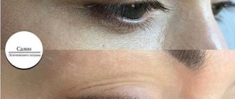
Advantages of treatment of papillomas on the eyelids and prices at MGK
Removal of papilloma on the eyelids should only be performed by a specialist who has undergone professional training and has the appropriate skills. To avoid possible complications, this procedure should be entrusted to an experienced doctor. At the Moscow Eye Clinic you will be able to undergo all the necessary tests, based on the results of which the attending physician will recommend the most effective treatment methods in your case.
Removal of papillomas on the eyelids is performed on an outpatient basis under local anesthesia. During the procedure, sterile instruments and disposable consumables are used, which eliminates the risk of infectious complications.
Minimally invasive surgical interventions on the eyelids at the Moscow Eye Clinic are performed by ophthalmologist Zargaryan Asmik Erdzhanikovna, who performs all types of minor operations on the eyelids using a minimally invasive surgical technique using the SURGITRON radiosurgical device (USA). The surgical treatment of eyelid neoplasms at the clinic is also carried out by ophthalmologists Yulia Valerievna Yakovleva and.
Author:
Dagaev Adam Huseinovich 5/5 (1 rating)
Honey. portal: