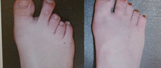
Valgus deformity is a common disease also known by several other names. Doctors talk about hallux valgus and hallux valgus. People not associated with medicine simply call it “ossicle” or hallux valgus.
The pathology is easy to determine. A thickening in the form of a callus appears on the inside of the foot. It hurts periodically and gradually increases in size. At the same time, the big toe begins to deviate towards the second toe and, in advanced cases, crosses with it. Along with aesthetic imperfections, a serious problem arises: the inability to use shoes, even everyday ones. The next stages in the development of the disease are arthritis and bursitis, the inability to move independently.
Most often, the first toe is deformed in women who prefer to wear high-heeled shoes (above 4 cm). In addition, this is an occupational disease of ballerinas. Excessive loads do not make themselves felt immediately, but after a while, when it is already difficult to correct the situation.
Surgical methods of treatment
Globally, all operations to remove hallux valgus are divided into minimally invasive and open. Minimally invasive ones are performed through small incisions in the skin. In most cases, there is no need for stitches. Recovery is quick and easy.
There are three categories of open surgical interventions:
Manipulation of soft tissues.
An example is the McBride operation. The muscles and ligaments of the foot are cut. Bones are not affected. The intervention is effective for minor deformities and can be performed in case of contraindications from the osteoarticular system.
Osteotomy.
An artificial fracture of the metatarsal bone is performed. The doctor selects the location of the fracture. Due to this, the position of the metatarsus is corrected
Arthrodesis.
The operation is used in advanced stages of hallux valgus deformity, when it is necessary to return the physiological shape of the joint.
The doctor, from all the methods of surgical intervention available to him, selects the one that best solves the problems of a particular patient.
The operation requires preliminary preparation. We need to get some test results. These include clinical and biochemical blood and urine tests, a blood test with a detailed coagulogram, and tests for blood-borne infections. Electrocardiography is prescribed, and x-rays of the foot are performed (necessarily in two projections).
If no contraindications to surgery to remove hallux valgus are identified, the method of anesthesia is agreed upon. Most often, local anesthesia is sufficient.
Sign up for a free consultation:
The operation requires standard preparation: consultation with a surgeon, tests for hospitalization and consultation with an anesthesiologist based on the test results. In addition, it is necessary to take an x-ray to clarify the type of syndactyly and when examining the patient, pay attention to possible concomitant congenital diseases.
How long does the patient spend in the hospital after surgery? When are stitches removed?
As a rule, spending one or two days in a hospital is enough. Some patients can leave the same day after surgery, for example, if surgery was performed on the fingers. If the separation of syndactyly was on the fingers of the lower extremities, then you can be discharged no earlier than the next day after the operation.
Suture removal usually occurs in two stages. At the first stage, we remove the fixing bandage. This usually happens 7-9 days after surgery. The second stage is to remove the stitches completely; this occurs within 10 to 16 days. As a rule, removable, that is, non-absorbable threads are used. We remove them approximately 14 days after the operation and can also remove them in 2 stages. At the first stage, after one, after two stitches, and then finally.
If the patient is from out of town, does he need to stay in Moscow for all 14 days or can he leave?
This is a standard problem for all out-of-town patients. You can come and go, but this involves a lot of movement, which will create an additional burden.
Can patients from other cities use self-absorbing sutures?
Self-absorbing sutures are not recommended here because more severe scarring may occur. It is possible to remove sutures at your place of residence, but we also do not encourage this, because it is necessary to control the edges of the wound and the condition of the skin flap. For out-of-town patients, we sometimes offer to send photographs. From the photo you can also understand what condition the wound is in, but it is still advisable for the patient to come for dressings and follow-up examinations.
How often should I come for dressing changes?
Of necessity. The first dressing is done the next morning after the operation, after which we send the patient home. If the dressing is dry and clean, then the next one will be 7-9 days after the operation to remove the fixing bandage.
If a child is born with this problem, then at what age is it better to correct syndactyly?
If we are talking about legs, then there is no need to rush from operations. If we are talking about hands, then it is advisable to separate syndactyly before school. There are currently no standard ideas about the optimal age. But they try to operate syndactyly on the hands at the age of 12-24 months: if the operation is performed later, it will be more difficult for the child in social and everyday life.
Does syndactyly occur on both arms and legs and how often does this occur?
It happens, but not often. Unilateral syndactyly is approximately twice as common as bilateral syndactyly.
Syndactyly involves the fusion of only two fingers or maybe three or four?
In different ways, there are many types of syndactyly, ranging from two fingers to total syndactyly. The frequency of occurrence also varies quite widely: in half of the cases syndactyly of the ring and middle fingers occurs, least often - in 4-5% of cases, the thumb and index fingers are fused - this is the first interdigital space.
How often does underdevelopment of the fingers themselves occur?
With simple cutaneous syndactyly of the middle and ring fingers, both of them usually grow normally; it is simply a violation of the formation of the interdigital spaces. The middle and ring fingers are not very different in length, so as a rule, there are no violations here. The situation is more complicated with the 1st and 4th interdigital spaces, when one of the two fingers is much shorter than the other. In this case, the longer finger may become deformed to the point of flexion contracture. That is why the division of syndactyly of these fingers must be performed in early childhood.
If a patient comes in with three or four fused fingers, do you correct everything at once?
You need to look at how pronounced the fusion is, what can be done in terms of plastic surgery. But most likely, two or three stages will be required for complex syndactylies. Here the question concerns the blood supply to each of the fingers, which can be significantly damaged during a one-stage operation, which can lead to partial or even complete necrosis.
What is the approximate cost of surgery in Moscow to separate syndactyly for the arms and legs?
We make no difference in separating hand and foot syndactyly. The approximate cost of dividing syndactyly of one interosseous space is from 35,000 rubles.
Can this operation be performed under compulsory medical insurance?
As far as I know, in early childhood such operations are performed under compulsory medical insurance or high-tech medical care programs. This pathology is not included in voluntary health insurance programs. People often ask on forums: if my son has severe syndactyly, will he be accepted into the army?
If syndactyly does not lead to functional impairment, then this is not a reason for deferment from the army. The basis for deferment is an operation performed on the eve of conscription. Then a delay is given for 2-6 months, depending on the complexity of the operation and the characteristics of the rehabilitation period. Of course, each case needs to be examined individually.
Distal osteotomy
The most common surgical method for correcting hallux valgus deformity is distal osteotomy. The operation takes place in several stages:
Get an online consultation
right now.
Get
First stage
Exostoexotomy or removal of the “bone”. In untreated cases, this stage may be the only one. Through a small skin incision or even without an incision, endoscopically, the exostosis itself (bone) and the inflamed joint capsule are removed.
Second phase.
Remote osteotomy. Using an artificial fracture, the position of the first metatarsal bone is leveled. The fracture site is secured with titanium screws. They are removed a month later, after the bone has completely healed.
Third stage.
Dissection of the muscle responsible for abducting the thumb.
Fourth and final stage.
Securing the thumb in a physiological position.
Surgical treatment of hallux valgus: indications
It is possible to correct the position of the thumb using conservative methods in childhood, when the bone skeleton is still developing. In all other cases, only surgery will help. The main indications are:
- discomfort when walking due to unbearable pain;
- deviation of the joint by several tens of degrees;
- a significantly protruding bone, making it impossible to wear shoes;
- violation of the position of the foot, making it difficult to move;
- swelling of the thumb;
- inflammatory manifestations and other complications of hallux valgus.
The doctor determines the indications individually for each patient. Typically, surgical methods are used when conservative methods (conventional methods, without surgery) become ineffective.
The correction provides patients with:
- beautiful feet without protruding “bones”;
- absence of excruciating pain;
- lifelong effect without relapse with properly selected shoes and favorable rehabilitation;
- rapid restoration of motor activity and performance.
It is important to understand that a bunion is not only an aesthetic defect, but also a serious problem that increases the risk of disability. Timely correction protects physical and psychological health.
Treatment of deformity of the joint of the big toe with surgery is 98% effective.
If conservative methods do not have the desired effect for a long time, you should consult an orthopedist and think about surgical intervention.
Myths about surgery to remove hallux valgus
There are many myths among people regarding surgical removal of a bunion. Let's dispel some of them.
You cannot have surgery if you have arthritis or arthrosis. In fact, these diseases are considered relative contraindications. The operation is possible, the doctor will just treat you with more attention.
It is useless to operate: the bone will soon grow anyway. With a high-quality intervention, the probability of re-development of hallux valgus deformity tends to zero. Relapses occur, but are extremely rare.
You will have to sit for a long time without moving and wear a cast. Modern technologies make it possible to move independently within a few hours after the procedure. The bones are held together by special structures, so there is no need for plaster.
Team of Doctors
First and second day. Bed rest. You can move around, but it’s better not to get active. It is recommended to keep your leg elevated and occasionally, if there is no pain, move your toes.
The third day. You can start moving slowly. It is important to choose a comfortable orthosis and shoes, which after surgery should be special, removing the load from the operated part of the foot. In most cases, Baruk shoes (the French surgeon who invented them) are used, which relieve the load on the forefoot when walking. At first, to get used to it, you can provide yourself with additional support in the form of a cane or crutches. After three to five days they will no longer be needed.
10-14 days. Period of doctoral supervision. In the case of a serious operation, the patient may spend all this time in the clinic. Physical therapy and physiotherapy sessions are recommended. It is not advisable to stand on your feet for a long time.
After 1-1.5 months, there is no need for an orthosis, but after surgery it is better to use shoes with suitable orthopedic insoles.
After 2 months, you can exercise on exercise bikes and swim in the pool.
Swelling of the foot and ankle may persist for up to three months. At this time, it is recommended to use cool compresses and wear compression stockings.
After 4-6 months, it is possible to move around without an orthosis, resume sports, and wear high-heeled shoes. High-quality recovery and quick rehabilitation require choosing comfortable wide shoes. Don’t forget to create comfortable conditions for your feet so that the unpleasant situation does not happen again.
Sign up for a free consultation:
Does this autograft always survive?
On the hands, almost one hundred percent engraftment usually occurs; on the legs, the probability of complete engraftment is less, since immobilization of the feet is often difficult: the patient needs to move around and come for dressings. This area is constantly in motion and inevitably gets injured, even though we apply special bandages that press the flap to the surface of the wound. There may even be a situation where, due to sweating under this bandage, the flap begins to macerate. Maceration of this flap leads to its partial and sometimes complete necrosis. In the literature, there is data on the use of materials of artificial origin or allografts, but we traditionally work with autoskin - this material is always available, does not require additional costs, and the percentage of engraftment of one’s own tissues is very high
What are artificial materials?
Artificial leather, Alloderm and others. But, I repeat, one’s own tissues – free autograft, transferred flaps from the back of the hand – are the “gold standard” of hand surgery. All the publications that I came across on the use of synthetic or allomaterials were of a research and not practical nature.
Will the patient walk with this artificial material all his life?
If we are talking about a classic transplant, it is always an autotransplantation of an epidermal skin flap. Therefore, surgery for cutaneous syndactyly involves a lot of nuances and is not as simple a correction as it seems at first glance.
As for bone syndactyly, this is work for orthopedists and traumatologists. Plastic surgeons, as a rule, do not do this. There are also many correction options here. These could be reconstructive bone surgeries; the fused bone can be split, creating two bone blocks... But orthopedists and traumatologists will tell you better about this.
Will the function of finger flexion after surgery be good or conditional?
With cutaneous syndactyly, the function of flexing the fingers does not suffer at all, the only difference is that the fused fingers bend together, but the tendons of the flexor and extensor muscles of such fingers are their own. After surgery, they will also bend. But since we create a scar deformation, and the scar tissue stretches worse, the flexion may be a little worse at first. The fingers need to be developed, first passively, and then actively, then the scar will not pull. There are isolated cases when hypertrophic scars form, but in most cases everything ends well.
How long after correction of cutaneous syndactyly should one begin to develop the operated fingers?
Development can begin 2-4 weeks after surgery. At first, passively, to prevent the scar from shrinking during the ripening process and to prevent . The scar takes up to six months to form, and all this time you need to work with a brush. After about another 2 weeks, you can already actively develop, for example, using rubber balls or hand expanders. We also quite often use special silicone gels or dressings to prevent severe scarring. How does bone syndactyly behave in terms of flexion?
Bone form of syndactyly
– this is the most surgically complex type of this pathology. The flexion-extension function can be significantly hampered, since the finger bones, joints, and tendons can be significantly deformed. When dividing bone syndactyly, speaking about the results of the operation, one must take into account, first of all, functional aspects. It is not always possible to obtain a satisfactory result.
Syndactyly also occurs in stars. Ashton Kutcher, for example, has zygodactyly - fusion of the second and third toes. This, by the way, is the most common variant of the anomaly. The actor did not undergo surgery, as he considers it not a flaw, but a highlight.
Let's talk more about the operation. What are the nuances of surgical correction? What anesthesia is used?
Correction of cutaneous syndactyly can be done under local and regional anesthesia, but it is not worth it, because in this case it is often necessary to do anesthesia in two places: on the fingers and where we will take the flap. These are additional negative sensations for the patient. It is better to perform this operation under conduction anesthesia, under spinal or epidural anesthesia with an additional intravenous component, that is, with sedation, or under general anesthesia with an inhalation component. This is ideal because the operation can last 2-2.5 hours.
The difficulty of the operation is not in dividing the syndactyly and removing the fibrous cord, but in not damaging the vascular and nervous structures, in transplanting the skin flap, and in the correct pattern of the Z-shaped flap. A straight cut should not be made on the fingers to avoid scar contracture, which will not allow the fingers to move fully. The incision lines must be planned in a certain way so that the edges of the skin do not become necrotic and at the same time are mobile enough for suturing the wound defect.
A Z-shaped scar stretches better than a straight scar. And autodermal flaps are also cut out to fit the resulting z-shaped wound surface. We sew these flaps with special thin threads, and then the flap is additionally fixed with a pressure bandage, which presses it to the entire wound surface. Without this, the flap cannot take root completely. At first, the nutrition of the flap occurs diffusely, then a capillary network begins to form.
Cost of surgery to remove hallux valgus
The doctor calculates the exact cost of the operation. It is very difficult to name a specific figure - it varies from twenty to eighty thousand rubles. This depends on the type of surgical intervention chosen by the attending physician (in accordance with the stage of the disease and physiological characteristics of the patient), the type of anesthesia, outpatient or inpatient stay, materials and instruments used. To keep the price low, come get rid of hallux valgus in a timely manner, on an outpatient basis, using minimally invasive intervention.