Congenital chest deformities in children
Congenital deformity of the chest in children may be associated with genetic characteristics and changes in the formation of the sternocostal complex, which can lead to a gradual increase in deformity until the growth of the child’s or adolescent’s skeleton is completed.
Congenital pathology is associated with improper development of the skeleton (vertebral column, ribs) due to an imbalance of mineral and endocrine metabolism. The consequence may be a specific development of the body:
- painful thinness;
- narrow shoulders;
- high growth;
- protrusion of the shoulder blades and collarbone;
- sunken chest when inhaling;
- long limbs;
- curvature of the spine (scoliosis or kyphosis).
Hereditary deformity is determined in 20-65% of cases of chest deformity. There are diseases and specific syndromes where this type of deformation is one of the symptoms. For example, pathology often develops against the background of Marfan syndrome.
Marfan syndrome
This disease is characterized by funnel-shaped and keeled deformities of the chest.
Marfan syndrome has the following symptoms:
- asthenic physique;
- arachnodactyly;
- dissecting aortic aneurysm;
- dislocation or subluxation of the lenses of the eyes (or other vision pathology);
- biochemical changes in the metabolism of collagen and glycosaminoglycans.
The development of chest deformity can be facilitated by dysplasia of connective and cartilaginous tissues, which is caused by enzymatic disorders.
Sporadic (non-hereditary) forms of deformity
Non-hereditary deformity of the chest develops due to teratogenic factors that affect the fetus during its development. Most often, abnormal development is caused by asynchronous, inharmonious growth of the sternum and costal cartilages.
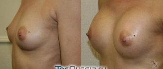
What is breast cancer?
Breast cancer occurs when breast cells, due to mutations, begin to divide uncontrollably and spread into surrounding tissue. These cells form a tumor, which can be detected by palpation of the breast or mammography (x-ray of the breast).
A tumor can form in different parts of the breast. Most breast tumors develop from the epithelium of the ducts that deliver milk to the nipple (ductal carcinoma). Others develop from glandular cells (lobular carcinoma). There are also less common forms, including nonspecific cancer, sarcomas, and breast lymphoma.
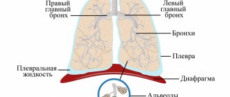
The tumor can grow locally, spread through the lymphatic system to the lymph nodes (regional metastasis), and spread throughout the body (distant metastasis).
Regional lymph nodes include axillary, intrathoracic, supra- and subclavian lymph nodes - on the same side as the detected tumor in the mammary gland.
The stage, prognosis and extent of treatment of breast cancer depend on the size of the tumor, the involvement of regional lymph nodes and the absence or presence of distant metastases.
Acquired chest deformities
Acquired chest deformity in a child develops against the background of diseases of the lungs and ribs (including tumor-like formations). This pathology can lead to other disorders of the body, for example, improper functioning of the respiratory system or psychological problems.
The acquired deformity is characterized by weakened immunity; the child often suffers from acute respiratory viral infections.
Physiological development is inhibited, fatigue appears after weak physical exertion. There are sharp changes in blood pressure.
Acquired curvature of the chest in a child can develop after suffering musculoskeletal diseases:
- Tuberculosis;
- Rickets;
- Scoliosis (Taking into account the fact that the spine and the sternocostal complex are an interconnected system, with severe deformities of the spine, a pronounced deformation of the chest is sometimes observed. More often, the posterior chest wall is deformed in the form of a rib hump, but there are also accompanying deformities of the anterior chest wall);
- Costal osteomyelitis;
- Rib tumors.
Pathology can be provoked by purulent-inflammatory processes in the soft tissues of the chest walls and pleura, injuries and burns of the chest. In some cases, the deformity is a consequence of cardiac surgery after median sternotomy, which can change the growth of the sternum in a child.
Normosthenic physique
Normosthenic girls (mesomorphs) have a proportional figure. Their main parameters:
- average height;
- thin waist;
- long and slender legs.
Normosthenic (mesomorphic) type is considered the most harmonious. Its owners are not prone to any special diseases; they are distinguished by good health and endurance. It is believed that these athletic girls were lucky to be born with innate coordination, speed and agility. The best sports are volleyball, basketball, and tennis. As women of this sporty and athletic type age, they should pay more attention to proper nutrition and healthy physical activity, eat more apples, pears and other fruits to easily lose weight if necessary.
It is useful to walk a lot, breathe fresh air, and take vitamins. Otherwise, there is a risk of gaining excess weight and developing unpleasant diseases.
The diet of normosthenics should include less fat than the menu of asthenics. Their percentage should be 10-20%. The share of proteins is 30-40%, carbohydrates – 40-50%.
The wrists of normosthenics have a circumference of up to 18.5 cm.
Types of chest deformation
Ryzhikov Dmitry Vladimirovich
Ryzhikov Dmitry Vladimirovich (Head of the Department of General Bone Pathology of the Federal State Budgetary Institution "National Medical Research Center for Pediatric Traumatology and Orthopedics named after G.I. Turner, Candidate of Medical Sciences, doctor of the highest qualification category, traumatologist-orthopedist)By type, we most often see the corpo-costal type, this is a deformation of the sternum in the lower part with the involvement of the ribs.
The manubrial type (manubrio-costal) is much less common, this type includes deformation of the manubrium of the sternum (this is the upper part of this bone).
Orthopedists also differentiate between asymmetric forms of deformation and its elasticity.
At what age and by what symptoms can chest deformation be detected in a child?
Among the patients of our Center there are children of any age. Most patients are admitted with a congenital form of the pathology. Sometimes a child is born with an already noticeable chest deformity, but most often we see situations where the deformation becomes noticeable for the first time at the age of 6-8 years and progresses significantly at 10-13 years. Chest deformities can increase while there is potential for skeletal growth, that is, on average until 15-17 years of age. And the higher the height of the parents and the more active the growth of the children, the higher the risk of developing a very pronounced deformity. In contrast to limb deformities, chest deformities often include in their list of symptoms and disturbances in the functioning of the chest organs.
Diagnosis of breast cancer
Diagnosing breast cancer in most cases does not cause difficulties, since this disease belongs to the so-called. "cancer of external localizations."
Examination and palpation of the mammary glands are important techniques for understanding the size, structure, location and a number of other signs of tumor formations in the mammary gland.
The doctor may order additional tests.
Ultrasound of the mammary glands . A study using an ultrasound sensor, which allows you to evaluate the structure of breast tissue and identify pathological formations. Ultrasound is also used during biopsy to obtain more accurate results when collecting cells or tissues.
Mammography . X-ray examination of the mammary glands. It is especially important from the point of view of screening - examination of healthy women without symptoms of breast cancer, but is also the main diagnostic method. Using mammography, you can identify pathological formations in the mammary glands and indirectly judge their nature. When describing mammograms, the standardized Bi-RADS algorithm is used, which, with a greater or lesser probability, allows the identified formations to be classified as potentially malignant. A targeted biopsy is also performed using a mammographic examination.
MRI of the mammary glands . It is prescribed to clarify the nature of a previously detected formation. It has an advantage in assessing the breasts of women with a high risk of hereditary cancer (for example, in the presence of identified mutations in the BRCA 1 and 2 genes).
Core biopsy . Taking a sample of the identified tumor using a special needle, which is inserted into the tumor under ultrasound and/or mammography guidance. Based on the obtained material, a histological examination is performed to confirm the diagnosis, as well as a special immunohistochemical study, which makes it possible to determine a number of important parameters - for example, the sensitivity of the tumor to hormone therapy and targeted therapy.
Fine needle aspiration biopsy . Performed if lymph node damage is suspected. After receiving the material, it is subjected to cytological examination to identify malignant cells.
Osteoscintigraphy . The study allows you to assess the condition of the skeleton, since breast cancer often metastasizes to the bones.
Additional tests may be ordered to determine the extent of the disease. For example, computed tomography (CT) and magnetic resonance imaging (MRI) can help rule out distant metastasis. Determining the stage of cancer development is necessary for the doctor to choose the most appropriate treatment.
Histological examination. Analysis of tissue obtained during biopsy. It is mandatory for diagnosis. It is also carried out after surgical treatment, which makes it possible to more accurately determine the stage of the process. If chemotherapy was administered before surgery, histological examination allows us to determine the response of the tumor to the treatment, which is an important prognostic sign.
Immunohistochemical study (IHC). A type of morphological diagnosis that allows you to evaluate the expression of various receptors on the surface of tumor cells. In breast cancer, a number of parameters are necessarily determined: estrogen receptors, progesterone receptors, Her2\neu overexpression and the Ki-67 proliferative activity index.
Molecular genetic research. Allows you to more accurately determine the presence of overexpression of the Her2\neu gene in cases where, according to IHC data, it is not possible to accurately determine the status of this important marker.
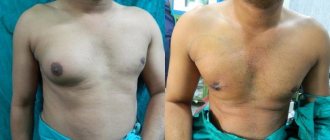
Unique treatment methods at the G.I. Turner National Medical Research Center
Vissarionov Sergey Valentinovich
Vissarionov Sergey Valentinovich (Director of the G.I. Turner National Medical Research Center for Pediatric Traumatology and Orthopedics of the Ministry of Health of Russia, Doctor of Medical Sciences, Professor, Corresponding Member of the Russian Academy of Sciences, laureate of the Government of the Russian Federation)Today, our Center is one of the few that performs unique operations to eliminate pectus excavatum deformity in children (PCD) through the use of low-traumatic methods of surgical correction. In some situations, we can treat keeled chest deformities in children conservatively by using special braces developed in our Center to correct this pathological condition.
The treatment of chest deformities in children and adolescents is carried out by the General Bone Pathology Clinic of our center.
- Conservative methods and surgical treatment of chest pathology
How to get treatment at the Turner Center for Pediatric Traumatology and Orthopedics (formerly the G.I. Turner Research Children's Orthopedic Institute)
The decision on the possibility and necessity of hospitalization at the National Medical Research Center clinic is made after consultation with a specialist from the specialized department and consideration by the Subcommittee of the Medical Commission of the Center for Patient Selection.
For those patients who cannot come for an in-person consultation, there are 2 options:
- Contact a doctor at your place of residence and request a telemedicine consultation in the doctor/doctor mode.
- Send a request for an absentee consultation, attaching all necessary documents. Including a scan of the attending physician's report. It is important for the doctor to obtain information on the form of the deformity (photos of the chest on the right and left are required), it is also important to send data on the FUNCTION OF THE HEART AND LUNG (ECG, echo-kg, spirography)
Signs and symptoms of breast cancer
The presence of a dense tumor in the mammary gland, which does not decrease in different phases of the menstrual cycle, increases over time.
- Nipple retraction
- Nipple discharge
- Pain in the breast or nipple area
- Skin infiltration (orange peel-like skin) in the breast area
- The appearance of dense nodes in the axillary area
- Ulceration of the skin in the breast area
- Swelling of all or part of the breast (even if the lump is not clearly felt)
- Unexplained weight loss
- Weakness
- Redness, peeling, or thickening of the skin of the nipple or breast