Types of skin cancer
There are 3 types of common malignant skin tumors. They differ both in incidence (i.e., the chance of getting sick) and in the degree of danger to life - basal cell carcinoma, squamous cell carcinoma and melanoma.
Melanoma is one of the rare and dangerous skin tumors. It accounts for only 4% of the total number of malignant skin tumors, but is the cause of almost 80% of deaths in this localization. You can read more about melanoma here.
Sign up for the webinar “Carcinogens in cosmetics: truth, lies and... marketing”
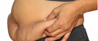
What is the structure of benign neoplasms
The growths consist of cells that have partially retained their original functions and are capable of growing slowly. They are similar in structure to the tissues from which they originated. They can put pressure on nearby tissues, but do not penetrate them, since they have a capsule in their structure. They respond well to hardware and surgical treatment and, as a rule, do not cause relapses.
There are always congenital formations on the skin - moles or warts, as well as acquired ones. The latter are formed on the surface or in the subcutaneous layer as a result of metabolic disorders, decreased immunity, or under the influence of a virus.
Basal cell skin cancer
Basalioma is the most common, but at the same time the safest type of skin cancer. Death from basal cell carcinoma is possible only in very advanced cases or with aggressive forms (basosquamous) tumor. The favorable course of basal cell carcinoma is due to the fact that it almost never metastasizes (only 0.5% of cases).
Symptoms and signs
Most often, basal cell carcinoma occurs on the skin of the nose, a little less often on the face and much less often on other parts of the body.
The peak incidence occurs over the age of 40 years. The youngest patient diagnosed with basal cell carcinoma by histology was 39 years old.
What basal cell skin cancer looks like depends on the form:
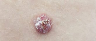
- Nodular form (synonymous with nodular). The tumor is presented in the form of a nodule. It can be distinguished from other skin formations by an increased number of vessels on the surface, a waxy sheen and small gray-blue inclusions. All these signs are visible in the photo.
Nodular form of basalioma
In addition, on the surface of nodular basalioma there may be another characteristic sign - ulceration.
Nodular basal cell carcinoma with ulceration
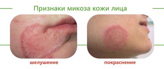
- The superficial form of basal cell carcinoma in most cases is presented as an area of redness on the skin. Elements of peeling and the waxy sheen already mentioned above are also possible.
Superficial form of basalioma
- Scleroderma-like form of basalioma is very rare and often presents difficulties in diagnosis. It is characterized by a lighter and harder seal compared to the surrounding skin.
Scleroderma-like form of basalioma
- The pigmented form of basal cell carcinoma makes up a very small part of the total number of these tumors. It is distinguished by a large amount of pigment. In this regard, basal cell carcinoma is often mistaken for melanoma when examined without a dermatoscope.
Pigmented form of basalioma
- The ulcerative form of basalioma can reach very large sizes and in advanced cases is practically untreatable.
Ulcerative form of basalioma
Photos in the initial stage
Unfortunately, basal cell skin cancer is extremely difficult to diagnose in the early stages, i.e. when it is minimal in size. Here are some photos:
Basalioma of the skin of the nose, nodular form, size 5 mm
Basalioma, nodular form, 3 mm in diameter
Nodular basalioma of the temporal region, diameter 2 mm
Diagnosing basal cell carcinoma in the early stages, when the tumor is small, can present significant difficulties. Only a combination of a comprehensive examination of the entire skin, a thorough determination of the history of the existence of the formation and dermatoscopy will help in establishing the diagnosis of basal cell carcinoma at an early stage.
Basaliomas with high and low risk of recurrence (NCCN, 2018)
Area H: Facial mask (including eyelids, eyebrows, skin around eyes, nose, lips [skin and red border of lips], chin, lower jaw, skin/grooves in front and behind the auricle, temples, ears), genitals, palms and feet .
Area M: cheeks, forehead, scalp, neck and legs
Region L: trunk and limbs (excluding shins, palms, feet, nails and ankles)
Notes
- Localization, regardless of size, may be a sign of high risk
- Histological forms of low risk: nodular (nodular), superficial, keratotic, piloid, with differentiation towards skin appendages, Pincus fibroepithelioma
- Area H means high risk regardless of size
- Morphea-like, basosquamous (metatypical), sclerosing, mixed infiltrative, micronodular in any part of the tumor
To assign a tumor the status of “high risk of recurrence”, only one of the factors from the right or left column is sufficient.
Treatment of basal cell carcinoma
The main goal of treatment for basal cell carcinoma is complete removal of the tumor while maximally preserving the cosmetic properties and functions of those parts of the body where this tumor has developed.
As a rule, the best results are achieved by surgical methods. However, the desire to maintain functionality and cosmetic properties may lead to the choice of radiation therapy as the primary treatment modality.
Depending on the degree of risk of relapse (see above), the approach to treating basal cell carcinoma may vary.
In patients with superficial basal cell carcinoma and a low risk of recurrence, when surgery or radiation therapy is contraindicated or inappropriate, the following treatments may be used:
- 5-fluorouracil ointment;
- Imiquimod ointment (Aldara, Keravort);
- photodynamic therapy;
- cryodestruction.
Mohs micrographic surgery may be recommended for patients at high risk of recurrence.
Chemotherapy for basal cell carcinoma includes drugs that are inhibitors of the hedgehog signaling pathway – vismodegib (Erivedge) and sonidegib (Odomzo). These drugs can help in cases where surgical methods, like radiation therapy, are not applicable or contraindicated.
What you need to know about basal cell carcinoma?
- In the vast majority of cases, basal cell carcinoma does not pose a threat to life.
- If a histological examination of a distant formation results in basal cell carcinoma, there is nothing to worry about. It is important to make sure that the formation is completely - be sure to consult with an oncologist.
- If, after removal of a basal cell carcinoma, the histological examination contains the phrase “tumor cells in the resection margin” or something similar, further treatment in order to completely remove the tumor.
- strongly do not recommend removing basal cell carcinoma without histological examination, since even a very typical-looking formation may not be what it seems at first glance.
- Basalioma needs to be treated . Observation is a bad option for a diagnosis like this. Treatment of advanced forms (see photo of ulcerative form) is extremely difficult and expensive.
- If you have already had a basal cell carcinoma removed, you should regularly have your entire skin examined by an oncologist in order to possibly identify another such tumor.
- The likelihood of metastasis in the metatypical (basosquamous) histological type is higher than in other types.
Methods for removing skin tags
There are several known ways to remove skin growths:
- Simple cutting with scissors or a scalpel;
- Removal with radio knife;
- Removal with a coagulator;
- Removal with liquid nitrogen;
- Laser removal;
- Plasma removal.
Rice. 5A. Stages of removing skin growths with a laser. Anesthesia
Rice. 5 B. Stages of removing skin growths with a laser. Laser vaporization
Rice. 5V. Stages of removing skin growths with a laser. Laser vaporization.
Rice. 5G. Stages of removing skin growths with a laser. Laser vaporization
Rice. 5D. Stages of removing skin growths with a laser. Final result
Each of them has the right to exist. However, since skin growths in most cases are removed solely for cosmetic reasons, removal methods should be chosen that will not subsequently leave a visible mark on the skin. These methods include: laser and plasma vaporization.
Squamous cell carcinoma
It is less common than basal cell carcinoma, the second most common type of skin cancer, and has a slightly less favorable prognosis. However, it should be noted that the course of the disease much less malignant than that of melanoma.
Metastases occur relatively rarely - on average in 16% of cases [1]. In patients with squamous cell skin cancer less than 2 cm in size, the 5-year survival rate is about 90%; for larger sizes and tumor invasion into the underlying tissue, it is less than 50% [1].
It can occur on any part of the body, including the genitals and mucous membranes, but most often in places exposed to sunlight.
Symptoms and signs
What squamous cell skin cancer looks like depends largely on the clinical form of the disease.
The keratinizing form is a raised or flat surface covered with horny scales that can grow and fall off. If damaged, it may bleed.
Keratinizing form of squamous cell skin cancer
It must be remembered that it is the keratinizing form of squamous cell carcinoma that may be hiding under the mask of the cutaneous horn . In this regard, such formations should always be removed only with histological examination:
The cutaneous horn should be removed with histology - a keratinizing form of squamous cell carcinoma may be hidden under its mask
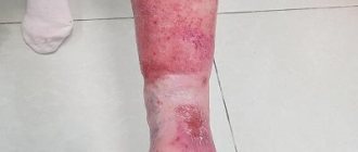
Non-keratinizing endophytic form (growing towards surrounding tissues). Most often it looks like a long-term non-healing wound or ulcer, which can deepen and expand over time.
Non-keratinizing endophytic form of squamous cell skin cancer
The exophytic non-keratinizing form of squamous cell skin cancer appears as a nodule that rises above the level of the skin. The surface of the node may be eroded or wet.
Exophytic nonkeratinizing form of squamous cell skin cancer
Photos in the initial stage
The initial stage of squamous cell carcinoma refers to a condition when the malignant process is limited to the epidermis - the outermost layer of the skin. It is referred to in the diagnosis as in situ or intraepidermal squamous cell carcinoma. This disease is not life-threatening if completely removed.
There are 2 forms of this phase of the disease:
Bowen's disease
Most often it is represented by single flat plaques, with clear boundaries, an asymmetrical shape, and uneven edges. The size reaches 7–8 mm. The formation may gradually increase, and peeling or crusting is often observed on the surface.
The color is red or brown, located on any part of the body. [3]
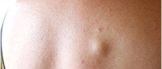
On my own behalf, I will add that in my practice, histologically confirmed Bowen’s disease occurred only once. It looked like a small (3 x 4 x 3 mm) flesh-colored lump with a smooth surface on the skin of the shaft of the penis in a 43-year-old man.
Bowen's disease
Erythroplasia Keira
The second form of early-stage skin cancer, which develops most often on the skin of the foreskin of the penis or the glans. Much less commonly, the disease affects the female external genitalia.
The most common appearance of Queyre's erythroplasia is a bright red spot with clear boundaries and a moist, shiny surface [3].
Erythroplasia Keira
Treatment of squamous cell skin cancer (NCCN, 2018)
As in the case of basal cell carcinoma, squamous cell carcinoma is divided into groups of high and low risks of recurrence and metastasis.
Area H: Facial mask (including eyelids, eyebrows, skin around eyes, nose, lips [skin and red border of lips], chin, lower jaw, skin/grooves in front and behind the auricle, temples, ears), genitals, palms and feet .
Area M: cheeks, forehead, scalp, neck and legs
Region L: trunk and limbs (excluding shins, palms, feet, nails and ankles)
Notes
- The rim of hyperemia should be taken into account when measuring size.
- Excisional biopsy is preferred over incisional biopsy.
- The modified Breslow thickness measurement should exclude parakeratosis and crusting and should be taken from the base of the ulcer, if present.
- Localization, regardless of size, may be a sign of high risk.
- Area H implies high risk regardless of size.
The basic principles and methods of treatment for squamous cell carcinoma are the same as for basal cell carcinoma.
The main goal is to maintain functionality and cosmetic qualities. method is considered to be removal of the tumor, including 4–6 mm of healthy tissue with a low risk of recurrence and metastasis. For high-risk tumors, Mohs micrographic surgery or wider excision is recommended than for low-risk tumors.
Radiation therapy is useful in cases where other methods cannot be used. Platinum drugs (cisplatin, carboplatin) as well as EGFR inhibitors (cetuximab) can be used in chemotherapy for squamous cell carcinoma.
Table of contents
- Etiology and pathogenesis
- Clinical manifestations
- Principles of treatment
Furrows on the face (deep wrinkles, creases, folds) are visible changes in the relief of the facial skin in the form of local depressions of more than 3 mm. Usually combined with other signs of chrono- and photoaging of the skin, underlying structures and the whole organism.
In our company you can purchase the following equipment for treating furrows:
- AcuPulse (Lumenis)
- UltraPulse (Lumenis)
How to avoid getting skin cancer? What to avoid?
Sunlight. The most proven cause of both types of skin cancer, as well as melanoma, is exposure to sunlight. If you like to travel to hot countries, have fair hair and skin, or your work involves prolonged exposure to the sun, you should seriously consider UV protection.
Precancerous skin diseases are the next factor that may precede the development of the squamous cell form: actinic (solar) keratoses and cheilitis, leukoplakia, human papillomavirus infection of the mucous membranes and genitals. This type of tumor can also develop against the background of scar changes after burns or radiation therapy.
Contact with carcinogens
Various chemicals can lead to the development of skin cancer: arsenic and petroleum products.
Weakened immune system. People taking immunosuppressive drugs after an organ transplant or people living with HIV have an increased risk of developing squamous cell skin cancer.
Chemotherapy
It is carried out both as an independent method of treatment and in combination with surgical intervention. Prescribing chemotherapy before surgery is intended to reduce the pathological focus. The purpose of this method of treating skin cancer after surgery is designed to completely destroy cancer cells.
The disadvantage of this method is the inability to exclude the effects of drugs on healthy cells. The question of the need for chemotherapy is decided by the attending physician based on the individual characteristics of the development of the tumor process.
Which doctor treats papillomas and where is the best place to remove papillomas on the body in St. Petersburg?
Many patients are interested in which doctor treats papillomas. This is done by specialists - dermatologists, gynecologists, urologists and oncologists. It all depends on the location of the tumor.
Specialists at the Diana Clinic in St. Petersburg conduct an initial examination and confirm the diagnosis. Then the papillomas are removed with a laser and radio knife, using the latest devices. By contacting our medical center, the patient receives a detailed consultation and can undergo all tests, including oncology. Removal of papillomas is carried out without pain or complications.
How dangerous are papillomas on the body: why they need to be removed
Papillomas are harmless only in appearance. These neoplasms, in the absence of timely and adequate treatment, quickly spread throughout the body to healthy tissue. This process is called autoinaculation. The result of the proliferation of neoplasms is at least multiple warts.
Of course, warts - papillomas on the body - are very ugly, but this is not the only problem. A good dermatologist will tell you why papillomas on the body are dangerous - such neoplasms are susceptible to melanization - malignant degeneration of the tumor. How quickly the process will proceed and whether it will begin at all depends on the location of the papilloma and the type of HPV. Genital warts growing on the cervix are especially dangerous. The degeneration of such formations into cancer occurs in every second case.
The structures that form on the skin as a result of damage by the papilloma virus are usually not accompanied by pain, itching or burning, which often causes negligence on the part of the patient. But since papillomas almost always come into contact with clothing, there is a high risk of damage. The result will be severe bleeding, infection and the formation of unsightly scars on the skin. From all this follows the answer to the question of whether it is necessary to remove papillomas on the body - of course it is necessary!
4.Treatment
The following treatment methods are used:
- Taking heavy doses of vitamin C. This does not destroy existing keratomas, but is a good prevention of the appearance of new ones.
- External hormonal agents. Local hormone therapy in the form of ointments reduces inflammation and disrupts the blood supply to the keratoma, slowing its growth.
- Intensive protection against tanning, as the main factor in the appearance and enlargement of keratomas.
- Diet. Replacing animal fats with vegetable fats in the diet.
- Sometimes it is recommended to pay attention to constant stress factors (quitting a job, normalizing family relationships).
Based on the results of the examination, keratoma removal is most often prescribed, especially if the keratoma is constantly peeling off, injured or exposed to solar radiation. Modern medicine offers several ways for this:
- laser removal;
- cryodestruction;
- radiosurgery;
- surgical excision.
Sometimes folk remedies are used to treat keratoma, but they are not very effective and, at best, lead to a slowdown in growth, but not to the disappearance of the keratoma. At the same time, some aggressive folk methods of self-medication contribute to the transformation of education into malignant.
Prevention of keratomas includes moderation in sun tanning and its complete exclusion during the most dangerous hours of the day, proper nutrition, adequate sleep, giving up bad habits and a sedentary lifestyle, reducing and avoiding stressful situations, a balanced diet rich in vitamins.
Clinical manifestations of the tear trough
The main places where deep furrows appear are in the eye area (in the corner, above and below the eyelids), on the cheeks, near the ears, between the eyebrows, in the corners of the mouth, and on the chin. It is also worth noting the nasolabial folds and horizontal lines on the forehead and neck, which gradually deepen with age.
Classification of wrinkles according to the Modified Fitzpatrick Wrinkle Scale (MFWS):
- Class 0 (no wrinkles) - no visible wrinkles, skin uniformly smooth.
- Class 0.5 (very small) - wrinkles barely visible to the eye.
- Class 1 (minor) - slight indentations in the skin.
- Class 1.5 (pronounced) - depressions less than 1 mm.
- Class 2 (medium) - clearly visible relief bends from 1 to 2 mm.
- Class 2.5 (medium deep) - visible wrinkles more than 2 mm deep, but less than 3 mm.
- Class 3 (deep) - grooves on the skin more than 3 mm.
Despite the general similarity of localization, wrinkles are located differently in each person, forming an individual pattern - the so-called “wrinkleprint”, similar to fingerprints. In experiments, it was found that the future localization of wrinkles and furrows on the face can be determined approximately 8 years before their appearance. Moreover, for this you do not need to perform complex tests - just smile ( Fig. 2 ).
Rice. 2. Diagnosis of wrinkles at rest ( Baseline neutral ), when smiling ( Baseline expression ) and after 8 years at rest ( 8-year neutral ). In the third row of each column you can see the features of the patterns of periorbital wrinkles at rest, when smiling and after 8 years at rest (Farage MA, Miller KW, Maibach HI (Eds.) Textbook of Aging Skin. Springer 2010: 916)