- Preparation
- Indications
- Types of surgical interventions
- Organ-conserving surgery for breast cancer
- Lumpectomy
- Oncoplastic resections
- Assessment of oncological risk of operations including primary reconstruction
- After breast cancer surgery: nutrition, prognosis, risk of recurrence
- How long do you live after surgery for breast cancer?
- Proper nutrition after breast cancer surgery
Preparation
In preparation for surgical treatment of breast cancer, patients undergo a comprehensive examination, which allows them to determine the extent of the operation and the possibility of using alternative methods of antitumor treatment. Standard laboratory tests, tumor biopsy, CT, MRI, ultrasound are performed, and consultations are held with specialists to resolve the issue of preoperative preparation and choice of anesthesia.
Directly at the planning stage of the operation, it is necessary to inform the doctor about all medications taken, including over-the-counter medications, medicinal patches, ointments, dietary supplements, etc. The doctor may adjust the prescriptions.
The evening before the operation, you must take a hygienic shower. The last meal should be no later than midnight. Then you can only drink liquid, in a volume of no more than 350 ml. You should also take a shower the morning of surgery. If the patient is taking oral medications, they should be taken with a minimal amount of water, ideally in one sip. This must be done at least 2 hours before surgery.
Immediately before the operation, you need to remove contact lenses, dentures, and all jewelry, including religious items or piercings, if any.
Types of surgical interventions
Surgical treatment of breast cancer is undoubtedly the main method of complex treatment. Its effectiveness increases significantly when combined with chemotherapy, hormonal therapy and radiation therapy.
One of the main principles of breast cancer treatment at Euroonko is to carry out mainly organ-preserving operations and operations for complete removal of the mammary gland (mastectomy), taking into account individual indications.
The essence of organ-conserving surgery for breast cancer is to remove only the focus of the breast tumor with a small amount of surrounding healthy tissue (lumpectomy and quadrantectomy ). This operation is usually followed by a course of radiation therapy to the area of remaining breast tissue and regional areas.
It is important to know that for invasive cancer, both of these operations are combined with the mandatory removal of axillary lymph nodes - lymphadenectomy. For non-invasive forms of cancer, a complete three-level removal of lymph nodes is currently not performed, since this sharply worsens the quality of life of patients - swelling of the upper limb (lymphedema), impaired mobility in the shoulder joint, and chronic pain develop.
Therefore, at Euroonko, as part of the first comprehensive examination, a biopsy of the “sentinel” lymph node is mandatory. The essence of this technique is to determine whether the axillary lymph node is affected by cancer. This technique makes it possible to carry out organ-preserving treatment and preserve axillary lymph nodes if they are not affected by metastases. This certainly has a positive effect on the patient’s future quality of life. The presence of cancer cells in the sentinel lymph node indicates a high risk of detecting these cells in distant organs and tissues of the body, that is, the risk of developing metastases. In this case, MRI and scintigraphy are performed. We mandatory carry out histological and immunohistochemical studies of surgical material (removed breast tissue and lymph nodes).
Sectoral breast resection
This operation is performed for nodular mastopathy (a combined diagnosis that includes situations with a lump in the mammary gland of unknown origin). A skin incision is made either above the seal, or along the edge of the areola, or along the submammary fold. The seal is removed, the resulting defect in the gland tissue is sutured, and an intradermal suture is applied.
A special sectoral resection technique is used for intraductal papilloma (usually a small tumor located in the duct and manifested by discharge from the nipple). A dye is injected into the duct. A skin incision is made along the edge of the areola, a stained duct is found behind the nipple, it is crossed at this point, and isolated to the periphery of the nipple so that the papilloma is removed. The gland tissue and skin are sutured with an intradermal suture.
Central breast resection
It is used for intraductal papilloma, when it cannot be localized, for multiple intraductal papillomas located in the central sections of the ducts. The operation is acceptable in cases where breastfeeding is not expected. After a skin incision made along the edge of the areola, all ducts are crossed behind the nipple. The gland tissue with the central sections of the ducts is isolated to 2–3 cm and removed. The gland tissue defect is sutured and an intradermal suture is applied.
Nipple resection
It is used for nipple adenoma, a rare benign tumor, or as a diagnostic step for the morphological diagnosis of Paget's cancer. The nipple is resected wedge-shaped and interrupted sutures are applied with thin suture material. Some of the ducts intersect, which can complicate subsequent lactation.
Mastectomy
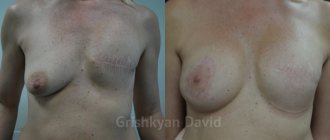
Mastectomy - removal of the breast (without lymph nodes). Performed for non-invasive forms of cancer (ductal carcinoma in situ, lobular carcinoma in situ), hereditary breast cancer syndrome, as a preventive operation. If simultaneous breast reconstruction is not planned, a thin linear scar remains on the breast. In cases where the operation is combined with simultaneous reconstruction of the mammary gland, mastectomy is performed using the technique of skin-sparing mastectomy (the nipple-areolar complex is removed, all other skin of the gland is preserved) or subcutaneous mastectomy (all skin of the gland is preserved). After such operations, a “skin bag” remains, which must be filled by a plastic surgeon. The aesthetic result of such operations is usually very good.
Radical mastectomies
Radical mastectomy involves removing the entire mammary gland, along with the underlying fatty tissue, fascia, and lymph nodes. The pectoral muscles are also supposed to be removed, but modifications now exist to preserve them.
Radical mastectomy according to Halsted
Radical mastectomy, that is, an operation that includes removal of the mammary gland with pectoral muscles and fatty tissue of levels 1–3, began to be performed by William Stewart Halsted since 1882 at the John Hopkins Hospital (John Hopkins Hospital, Baltimore, Maryland, USA). The first description of an operation performed on 13 patients dates back to 1891, this description was part of an article on wound healing (WS Halsted “The treatment of wounds with especial reference to the value of the blood clot in the management of dead spaces.” John Hopkins Hospital Rep., 1890–1891. 2:255.). The removal of fatty tissue is due to the presence of lymph nodes here, which were often affected by cancer metastases and consisted of multiple dense nodes of various sizes. The anatomical division of fiber is made relative to the pectoralis minor muscle: fiber outward from the pectoralis minor muscle is level 1 fiber, anterior and posterior from the pectoralis minor muscle - level 2, inward from the pectoralis minor muscle - level 3. The removal of muscles was explained by the fact that in advanced forms of the disease (which were the majority), the lymphatic vessels passing through the muscles and fascia covering the muscles were affected by the metastatic process.
The disadvantages of the operation include deformation of the chest wall. Currently, the indications for radical mastectomy according to WS Halsted are invasion of the pectoralis major muscle by the primary tumor and involvement of the Rotter lymph nodes, as well as palliative operations.
Patey & Dyson modified radical mastectomy
DH Patey and WH Dyson in 1948 proposed a modified method of radical mastectomy, which differs from the WS Halsted operation by preserving the pectoralis major muscle. The block of tissue removed includes the mammary gland, pectoralis minor muscle and lymph nodes of levels 1–3. In most cases, the operation is not inferior in effectiveness to the Halstead operation; its advantage is less trauma and less deformation of the chest wall. At the same time, not everything is simple with the remaining pectoralis major muscle. When removing the pectoralis minor muscle, 1-2 small nerve branches (lateral pectoral nerve and a branch of the medial pectoral nerve) innervating the outer part of the pectoralis major muscle are inevitably transected. Subsequently, of course, this leads to atrophy of the outer part of the pectoralis major muscle.
Madden modified radical mastectomy
Modification of radical mastectomy according to JL Madden involves preserving both pectoral muscles and removing fiber from levels I and II.
Radical mastectomy with pectoral muscle sparing
It is a variant of modified radical mastectomy, developed at the Federal State Budgetary Institution Russian Cancer Research Center named after. N. N. Blokhin RAMS. It involves removal of the mammary gland, removal of levels I–III fiber without removal of the pectoralis minor muscle, in contrast to the Patey & Dyson operation. The advantage of the operation is the complete removal of fiber and preservation of muscles and their innervation.
Palliative mastectomy
All previous operations performed for cancer were called “radical”. This meant that the disease was removed with the smallest “roots” and should not return; the operation was aimed at preventing the development of metastases. In other cases, when the tumor has already metastasized or when the local spread of the disease is so great that the development of metastases is most likely to follow after the operation, the operation cannot claim to be called radical. In such cases, it can be performed for palliative purposes, that is, in order to eliminate the immediate troubles associated with the presence of a tumor - tumor decay, bleeding; or in order to reduce the volume of tumor tissue and create conditions for more effective drug treatment.
Interview with Sarukhanov Georgy Mikhailovich
Sarukhanov Georgy Mikhailovich is a plastic surgeon with extensive experience in performing plastic surgery on the chest.
More than 3,000 different types of mammoplasty have been performed. Proficient in many techniques for breast augmentation, reduction, and lifting. He is actively involved in scientific activities, has many publications in specialized journals, and regularly makes presentations at Russian and international conferences and congresses. Georgy Mikhailovich, we would like to ask you a few questions, as a plastic surgeon who has absolute authority in the field of mammoplasty, who has performed a huge number of similar operations and, in our opinion, understands the psychology of patients who have decided to undergo breast plastic surgery. The popularity of mammoplasty has not subsided over the years. Why do you think women undergo such operations?
Ideal female breasts
From time immemorial, beautiful female breasts have been considered one of the main advantages and a source of pride for the fair half of humanity. The female body has always been an inexhaustible source of inspiration and an eternal muse for all artists, poets and musicians. Of course, the canons of the beauty of female breasts have changed over time and large, lush breasts have not always been in “fashion”. Nevertheless, the shape of the breast itself, regardless of its size, is, in my opinion, the main criterion for assessing women and the motivating factor for contacting us, plastic surgeons.
Do you, as a man, and not a plastic surgeon, support this desire of women? Have you ever had a person come to you with a request to enlarge your breasts, for example, to size 6 or reduce them to size 1? How do you feel about such wishes of patients?
Probably every woman wants to please not only men, but also herself, first of all. After all, the desire for harmony and perfection is a natural desire. I don’t see anything “reprehensible” in the fact that you want to be beautiful and loved, to live in “harmony” with yourself, as they say. Of course, we are all different in our tastes, views and desires. Therefore, in our practice, we sometimes encounter “non-standard” requests. And if the proposals, as you say, “increase to size 6 or reduce to size 1,” are quite adequate, justified, and most importantly, will not harm health or lead to serious complications, then we will meet you halfway and agree to perform the operation .
Which type of breast surgery do you do best?
The operation that succeeds best is the one that you approach thoughtfully, having weighed everything and choosing the right tactics and methods. The main success is obtaining a result that not only pleases you as a performer (and most surgeons are quite self-critical of the work done), but also brings great joy and satisfaction to the patient. Then you feel that it was not in vain that you chose such an interesting and unique specialty...
Okay, then let's look at each type of breast surgery separately.
Organ-conserving surgery for breast cancer
The history of the development of organ-sparing operations for breast cancer is relatively short. Such operations became possible due to a combination of three main factors: 1) earlier detection of the disease; 2) the realization that expanding the scope of surgery for early forms of cancer does not lead to improved survival of patients; 3) the use of radiation exposure to the preserved mammary gland as a powerful means of reducing the likelihood of local relapse.
G.Crile Jr. in 1975 presented 10-year results of a randomized trial comparing breast-conserving partial mastectomy with total mastectomy. In the comparison groups there were 42 patients with primary operable cancer. Cancer mortality rates over 10 years were 34% and 38%, respectively.
Patient reviews
Irina Kashevarova: I have been fighting breast cancer for 9 years now. I started this fight in Israel, where I was operated on and had my first chemotherapy. Then there were several other medications. In my now “professional” opinion, everything was done efficiently and I was treated by great doctors, and Professor Inbar is still treating me, but now I don’t leave Moscow. The world turned out to be small, when I asked on the phone almost 9 years ago my doctor friend from the USA, who had gone there a long time ago, where I should do procedures with medications, he advised me of his friend Dr. Aronov, and when, having found out, he gave the address, it turned out that it was in my yard. Incredible. Yes. Now the relapse is again, I’m already tired of fighting, but I’m only 44 years old and I’ll still do it. Everyone at the Medis clinic and Dr. Sasha himself help me immensely in my life.
Zhanna Asherova: We immediately began to treat my mother to the maximum, having studied the entire “market” of medical services. We sold the apartment that we were renting out and invested everything in treatment. I literally became almost a professor at the Russian Medical Journal. And now, looking back, I understand that I didn’t do everything right. It was wrong to follow the Soviet path, and the oncology center of the Soviet spill is a Soviet one. Yes, we found the best surgeon there, we organized everything so that the operation could be performed. But after hanging around there, they went to a private clinic, first to one, but from there they immediately moved to Medis. And I didn’t regret it for a second.
Lumpectomy
The minimal surgical procedure in terms of the volume of breast tissue removed, lumpectomy (lump, lump, piece, lump), was developed during research by the National Breast and Bowel Supplementation Project (USA, NSABBP). The study included patients with a tumor size of no more than 4 cm. Groups of patients with different types of treatment were compared: lumpectomy (group 1), lumpectomy with radiation therapy (group 2), modified radical mastectomy (group 3).
During a 12-year follow-up, local relapse in the mammary gland developed in patients of the 1st group in 35%, of the 2nd group - in 10%. There were no significant differences in overall survival and survival without distant metastases between the compared groups. The general conclusion about the equal effectiveness of breast-conserving treatment and radical mastectomy was also confirmed at 20-year follow-up. The rate of local recurrence after lumpectomy was 39.2%, after lumpectomy with irradiation - 14.3%.
From the point of view of surgical techniques, organ-sparing operations of the first stage (partial mastectomy, quadrantectomy, radical resection) in their original form (wide wedge-shaped resections without additional restoration of the shape of the gland) are a thing of the past. Modern organ-saving operations are lumpectomy and oncoplastic resection. A detailed analysis of world experience on the oncological risk of organ-conserving operations is presented below.
Having gained our own experience in performing lumpectomy, we came to the need to modify it. The modification concerns two points: the tumor must be removed with a supply of healthy tissue around it, and the gland tissue must be sutured. For small tumors (up to 1–2 cm), lumpectomy remains the best operation: non-traumatic and elegant. If the tumor is large or centrally localized, in order to preserve the shape of the gland, there is a need for additional efforts to move tissue, and/or intervention on the contralateral gland to maintain symmetry, that is, the need to perform oncoplastic resections.
Innovative diagnostic tests
You can order from us separately:
- Oncotype DX test - to predict cancer metastasis (about $6 thousand)
- Oncofocus test - targeted (individual) selection of chemotherapy from Cambridge (costs only about 3 thousand $)
- Prosigna test is a new product approved by the FDA as the most effective means of combating relapse of breast cancer (test cost more than $5 thousand)
- PET CT with an expert opinion from two experts at once!
During the examination, it is extremely important what type of biopsy is performed. Today in the Russian Federation, stereotactic or aspiration fine-needle biopsy or core-needle biopsy is performed. If breast cancer is suspected, a core-needle biopsy : by taking a larger area of breast tissue, such a biopsy makes it possible to obtain enough histological material for IHC and making an accurate diagnosis of breast cancer. A fine-needle biopsy will not be indicative; it can only be used for analysis of lymph nodes. A thick-needle biopsy is also necessary when the formation is not palpable or is located deep in the tissues of the gland. This uses x-rays or ultrasound to determine where the needle will be inserted.
Oncoplastic resections
The term “oncoplastic resection” is generally accepted in the world literature and implies the performance of breast resection for cancer using plastic surgery methods to restore the shape of the gland, and it is also possible to combine it with simultaneous surgical intervention on the opposite gland to restore symmetry.
One of the first operations that can be classified as oncoplastic resection (the term “oncoplastic resection” was proposed later) was breast reconstruction according to A. Grisotti - the most successful method of restoring the shape of the gland after removal of its central part. After resection of the central part of the gland along with the nipple and areola, a skin incision is made vertically down from the medial edge of the resulting wound, which is then extended laterally along the inframammary fold. Below the wound defect, part of the skin is de-epidermised, leaving an island of skin corresponding in size to the areola. In the projection of the vertical part of the skin incision, the gland tissue is dissected throughout its entire thickness to the submammary space, and the entire lower-outer quadrant of the gland is mobilized. The mobilized gland tissue is rotated, its part located under the skin island is moved to the central section and sutured. Subsequently, tattooing of the newly created “areola” and nipple plastic surgery can be performed.
In Russia, the period of oncoplastic resections started in the early 90s, when an operation using the inverted “T” reduction plastic technique was proposed. The operation was performed for lower tumor localizations; reduction plastic surgery of the opposite gland was mandatory.
Currently, there are a lot of options for oncoplastic resections; we can say that there are as many of them as there are patients. The technique and course of the operation is dictated by the oncological situation, the shape of the mammary glands, the characteristics of the tissue condition, and the surgeon’s favorite techniques.
Organ-conserving operations are not automatically an adequate type of treatment. A thorough examination of patients who are planning such an operation is necessary. It is better to have a mastectomy than breast-conserving surgery that does not meet the oncological criteria.
The “cleanliness” of the resection margins is the main indicator of the adequacy of organ-saving surgery. Organ-conserving surgery is recognized as a radical option for local treatment only in combination with radiation therapy.
Assessment of oncological risk of operations including primary reconstruction
Our clinic performs a wide range of oncological surgeries, including simultaneous breast reconstruction. The question arises: is it safe? Does additional surgery provoke the rapid development of metastases?
To answer these questions, we analyzed information about 503 patients with stage I–III breast cancer who received treatment at the Federal State Budgetary Institution Russian Cancer Research Center named after. N. N. Blokhin RAMS in 1992-2002. The main group included 124 patients, average age 41.5 years (24–67). Women were operated on with radical mastectomy with preservation of the pectoral muscles in combination with primary breast reconstruction: an expander (n=14) or a skin-fat flap on the latissimus dorsi muscle using an endoprosthesis (n=18), or a transverse rectoabdominal flap on a muscular pedicle (n=92). The control group consisted of 379 patients, mean age 40.1 years (26–79). 145 patients underwent breast-sparing surgery, 234 underwent radical mastectomy with preservation of the pectoral muscles. The groups were comparable in terms of the main factors influencing prognosis (stage, age, treatment). Drug and radiation treatment was carried out according to general principles. Median follow-up duration was 63.7 (20.4–140.5) months.
The local recurrence rate was:
- in the mammary gland after organ-preserving operations - 4.1%;
- after modified radical mastectomy - 1.7%;
- after modified radical mastectomy with primary reconstruction - 1.6% (p>0.05).
Over the entire period of observation, relapse of the disease (that is, not only locally, but in any organs and tissues) was observed in 18.6±3.5% in the group with breast reconstruction (in 23 patients) and in 18.2±2.0 % in the control group (in 69 patients, p>0.05). The disease-free survival and overall survival curves in the compared groups were not statistically different.
According to multivariate analysis, the fact that primary breast reconstruction was performed does not affect the development of disease recurrence. Analysis of the factors influencing the relapse of the disease shows the predominant influence of such known factors as T, N criteria, age, and chemotherapy. The fact of primary reconstruction did not have a statistically significant effect on the process of tumor recurrence. Thus, primary breast reconstruction can be safely performed in patients with breast cancer.
However, the greater the volume of operations, the more likely complications of their healing are, especially in patients with concomitant diabetes mellitus, obesity and long-term smoking. In them, prolonged wound healing may delay adjuvant radiation therapy and chemotherapy. Therefore, for patients with planned adjuvant chemotherapy or radiation therapy who have the listed factors that impair wound healing, it is preferable to refuse primary reconstruction.
| More information about breast cancer treatment at Euroonco: | |
| Surgeons-oncologists-mammologists | from 5,100 rub. |
| Removal of a breast tumor | from 101,200 rub. |
| Emergency oncology care | from 12,100 rub. |
After breast cancer surgery: nutrition, prognosis, risk of recurrence
Surgery is the most radical method of treating cancer. But, even if the tumor is completely removed and the doctor has declared remission, there is still a risk of relapse in the future. Every woman who has successfully completed treatment should be under the supervision of a doctor.
You will have to visit a mammologist every few months. Over time, the doctor will invite you for examinations less and less often, after 5 years - approximately once a year (if no relapses have occurred during this time). Your doctor will order a mammogram 6–12 months after surgery, which will then need to be done annually. For certain indications, regular examinations by a gynecologist, determination of bone density and other studies are prescribed.
Progress of the operation
The intervention can last from 1 to 3 hours, depending on the extent of the cancer. General anesthesia is used.
Radical mastectomy for breast cancer occurs in several stages:
- Disinfection of the surgical field.
- Operative access – making incisions.
- Removal of glandular tissue.
- Removal of axillary tissue with lymph nodes, as well as the subclavian vein located along the course, in the subscapular and subclavian regions, in the space between the chest muscles.
- Installation of drainages.
- Suturing and dressing.
How long do you live after surgery for breast cancer?
The average life expectancy after breast cancer surgery is estimated by the five-year survival rate. It refers to the percentage of patients who remain alive five years after diagnosis. Five-year survival rate for breast cancer primarily depends on the stage at which treatment is started:
- Stage I - almost 100%.
- Stage II - 93%.
- Stage III - 72%.
- Stage IV - 22%.
In addition to the stage, factors such as age, the general health of the woman, the type of tumor, and lifestyle also play a role. There are no specific recommendations that would help to significantly improve the prognosis of survival after surgery for breast cancer. You need to lead a healthy lifestyle in general: eat right, maintain physical activity, monitor your body weight, avoid smoking and alcohol.
Indications:
- tumor size is more than 5 cm (1 quadrant);
- lack of effect from other methods of therapy or contraindications to them;
- multiple cysts (recurrent polycystic disease) or nodes;
- incomplete removal of cancer cells during a previous operation;
- large-scale inflammation of the mammary gland, accompanied by extensive suppuration;
- non-invasive form of cancer;
- large tumor with signs of decay (palliative measure);
- prevention of oncology of the second breast, if a diagnosis has already been made and BRCA mutations have been detected;
- phyllodes tumor;
- Stage II-III cancer and pain in both mammary glands;
- a tumor with a diameter of 2 cm, located near the nipple, accompanied by chest pain.
Proper nutrition after breast cancer surgery
After treatment, the body recovers, so it must receive enough protein. In the immediate postoperative period, you should not worry about excess calories, even if you are overweight. Now it's important to recover. You can lose weight later.
Some substances found in plant foods help improve health and reduce the risk of relapse:
- Phytoestrogens found in soy have been shown in some studies to help reduce the risk of recurrence of estrogen-positive cancer. Other studies have not found this effect.
- Antioxidants are found in many fruits and vegetables, especially broccoli, blueberries, carrots, and mangoes. They help protect cells from damage.
- Lycopene is one of the antioxidants that gives tomatoes their red color and grapefruit their pink color.
- Beta-carotene gives orange color to carrots and apricots. There is some evidence that it helps prevent cancer.
Should you take dietary supplements? Nutritionists believe that a diet rich in a variety of fresh foods is much better than dietary supplements.
Bibliography:
- S. M. Portnoy, S. N. Blokhin, Kh. S. Arslanov, et al. Assessment of the oncological risk of simultaneous reconstructive operations for breast cancer. Questions of Oncology, 2008, No. 6, 720–723.
- Effect of radiotherapy after breast-conserving surgery on 10-year recurrence and 15-year breast cancer death: meta-analysis of individual patient data for 10,801 women in 17 randomized trials Early Breast Cancer Trialists' Collaborative Group (EBCTCG)* Lancet 2011; 378:1707–16.
- Bolotina L.V., Zakiryakhodzhaev A.D., Malygin S.E., Medvedev S.V., Pak D.D., Petrovsky A.V., Portnoy S.M., Rozhkova N.I., Rasskazova E. .A., Saribekyan E.K., Stenina M.B., Trofimova O.P., Tyulyandin S.A., Frolova M.A., Khmelevsky E.V. Clinical guidelines for the prevention, diagnosis and treatment of patients with breast cancer glands. Medical alphabet. 2014. T. 1–2. No. 8. pp. 32–49.
- Portnoy S.M., A. Yu. Serbny. Surgical method for treating breast cancer. In: “Current aspects of clinical mammology.” Ed. E. B. Polevoy and S. M. Portnoy. "Author's Academy", Moscow, 2014. pp. 74–115.
Book a consultation 24 hours a day
+7+7+78
Advantages of the Melanoma Unit clinic
- Russian specialists of the clinic completed an internship in Israel and were approved as professors.
- All doctors regularly study in Europe and the USA, many are engaged in teaching.
- Once a month, the best Israeli doctors consult in Moscow (you can find out the price of a consultation regarding a mastectomy by phone).
- The treatment plan for each patient is coordinated with the Israeli center, where all documents are sent.
- Treatment protocols comply with the recommendations of the NCCN (National Comprehensive Cancer Network), a national comprehensive cancer control network that brings together 28 American cancer centers.
- The cost of mastectomy, announced to the patient at the reception, is final; it will not increase during the treatment process.