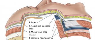
Atheroma (sebaceous gland cyst) is a benign neoplasm located immediately subcutaneously, resulting from blockage of the sebaceous gland duct. Atheroma occurs on any areas of the skin of the body where hair grows, but the favorite place is the face, scalp, back, neck, and groin area. Atheroma has a dense capsule of connective tissue, with secretion components.
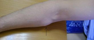
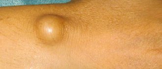
Visually, it looks like a subcutaneous ball, ranging in size from several millimeters to tens of centimeters. On palpation, this formation is round in shape, soft, elastic, freely moving under the fingers, there is no pain.
When atheroma suppurates, most often as a result of injury, the symptoms change: the formation increases many times in size, swelling and hyperemia increases around it. On palpation, the formation is denser, painful, and there is hyperthermia around it. When the process is neglected, the body temperature rises and weakness sets in. Into the flesh until an abscess forms.
To diagnose atheroma, in addition to visual examination, ultrasound diagnostics are often used to differentiate from lipoma. On ultrasound, atheroma looks like a hypoechoic neoplasm of soft tissue, located immediately subcutaneously, at a depth of 1–10 mm. Round in shape, with smooth and clear contours, the capsule can be traced throughout. The formation is most often homogeneous, without inclusions, with distal pseudo-enhancement. With CDK there is no blood flow.
Causes of atheroma
A sebaceous cyst can be acquired or congenital. Congenital atheroma is formed during fetal development when the layers of the epithelium are displaced. It detaches from the covering layer and deepens, and by the time the child is born, cysts appear in these foci. The sebaceous glands begin to work approximately from the 6th month of intrauterine development. Cysts are the result of the natural desquamation of epithelial cells and the production of vernix lubrication. Such atheromas are often hereditary.
The causes of atheroma in adults are varied. The sebaceous glands produce sebum, or sebum. They have a complex branched structure and a holocrine type of secretion. By secreting sebum, the cells of the sebaceous glands are completely destroyed, and the defect is replaced by the division of new cells.
The excretory ducts of the sebaceous glands most often open at the base of the hair follicle, less often in the skin. Therefore, atheromas develop on areas of the body covered with hair and never occur on the palms or soles. The work of the sebaceous glands is regulated by hormones. In men and women, their activity increases testosterone.
For the development of atheroma, the influence of several factors is necessary:
- high activity of the sebaceous glands - during puberty, as well as in women with hyperandrogenism, the sebaceousness of the skin increases. The glands produce a large amount of sebum, which can accumulate and form a cyst under the influence of additional factors;
- changes in the composition of sebum - the chemical composition of sebum includes triglycerides, vitamin E, squalene, fatty acids, and the remains of destroyed cells. If its consistency changes, the secretion from the gland slows down;
- blockage of the duct - skin injuries, constant friction leads to disruption of the integrity of the epithelium or its microtraumas. In this case, the duct may become clogged. And when combined with other factors, this creates conditions for the development of atheroma;
- poor hygiene – insufficient or irregular cleansing of the skin when it is too greasy leads to the accumulation of fat, creating conditions for the proliferation of bacteria. In some cases, inflammation occurs.
Sometimes atheroma develops after mechanical hair removal. Pulling it out leads to the development of local inflammation, blockage of the duct and the formation of atheroma.
Atheroma may develop after treatment of the boil. Trauma to the skin and subsequent recovery can lead to the formation of scar tissue. If it blocks the lumen of the gland duct, the accumulation of sebum forms atheroma.
Classification
The cellular structure of atheromas is different. Since this does not manifest itself clinically, the histological classification is practically not used and is of interest only to researchers. The more common classification is based on the mechanism of formation of sebaceous gland cysts:
- True or primary. These are congenital formations that are formed at the stage of intrauterine development of the fetus. It is usually diagnosed in children from the moment of birth. Often the formations are multiple in nature and rarely exceed 5 mm in diameter. Mostly located in the perineal area.
- False or secondary. They occur mainly in adults as a result of blockage of the duct of unchanged sebaceous glands. Over time, the cyst gradually increases and can reach 10 cm in diameter. Localization of false atheromas is most often observed on the head, neck and upper back.
What does atheroma look like?
A cyst formed in the sebaceous gland can be recognized by characteristic changes in the skin. Atheroma looks like an elastic subcutaneous node, usually it has a soft or moderately dense consistency. The diameter of the atheroma depends on the duration of its existence, so it can vary from 1 cm or more. But atheromas are never gigantic in size, exceeding a few centimeters.
Atheroma has a pseudocapsule of connective tissue, which forms as the cyst enlarges. It is mobile and not fused to the surrounding tissues, but is fixed to the skin at one point. It corresponds to the exit of the gland duct.
The skin over the atheroma is of normal color, consistent with the surrounding tissues. There are no vascular changes around the atheroma.
Atheroma on the head
The first signs of atheroma may appear at the age of 20-30 years. But young people do not always go to the doctor when they find a small painless nodule at the base of the hair on the scalp. It slowly increases in size, sometimes the patient does not seek help for several months or years.
Atheroma on the head occurs more often than on other parts of the body. Sometimes several tumors appear. At the point where the duct exits onto the skin, which coincides with the hair shaft, the duct sometimes opens slightly and a cheesy content with an unpleasant odor comes out of the cyst.
But you cannot count on self-healing of atheroma; after emptying, sebum and dead cells will begin to accumulate in it again. The cyst will increase to its previous size.
With the long-term existence of atheroma on the head, the nutrition of the hair is disrupted. This leads to their loss and the development of baldness in the affected area, or alopecia. After treatment, hair restoration does not occur.
Atheroma on the back
Increased sweating and oily skin, poor hygiene leads to blockage of the sebaceous glands located on the back and the formation of atheroma. More often it occurs on the back of the neck, along the spine - in these areas there are more hair follicles with sebaceous glands.
Initially, atheroma on the back can be felt with your fingers while taking a shower. It doesn't hurt and feels like a small pimple. The cyst grows very slowly and does not cause concern.
But if the formation is located in an area of high friction, for example, on the neck, it can be injured. When a bacterial infection penetrates, the atheroma becomes inflamed and suppurates. It becomes swollen and painful. The skin becomes red.
Spontaneous opening of a suppurating atheroma is possible. Curd-like contents with an unpleasant odor are released. It contains cholesterol, cellular debris, lipids and fatty acids. The color of the discharge is white with a yellowish tint.
The danger of atheroma on the back is low. But after inflammation it can lead to the formation of an abscess. The spread of infection to adjacent tissues causes widespread inflammation.
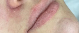
Atheroma on the face
In areas of increased greasiness on the face, under certain circumstances, atheromas may appear. In men, sometimes this is the area of the chin and mustache. A small nodule that does not differ from the color of the skin is mistaken for acne or a pimple.
Atheroma on the face grows at a slow pace. It becomes a cosmetic defect, so patients see a doctor earlier than in other cases. If you try to squeeze it out yourself, there is a risk of introducing an infection into the gland.
Inflammation of the sebaceous gland on the face is especially dangerous in the area of the nasolabial triangle and the wings of the nose. Venous outflow is directed towards the cranial fossa and brain. Therefore, there is a risk of infection and the development of meningitis.
Regardless of the location, atheroma almost never degenerates into a malignant tumor. Literary sources describe isolated cases where skin cancer was diagnosed at the site of atheroma, but there is no evidence that it was a consequence of a sebaceous gland cyst.
Festering atheroma
A large number of opportunistic microorganisms live on the surface of the skin. Usually these are bacteria from the groups:
- staphylococci;
- streptococci;
- peptococci;
- coli.
But with sufficiently active immunity, the proliferation of microorganisms is inhibited. When the immune defense is weakened, bacteria penetrate into the cavity of the cyst, and its contents become a breeding ground for them.
Suppurating atheroma is a complication that requires urgent surgical care. An abscess forms at the site of the cyst. This is accompanied by a deterioration in the general condition, malaise, pain in the area of the atheroma, its swelling and hyperemia. Inflamed atheroma can lead to the formation of an abscess, and in advanced cases, when pus breaks through into the subcutaneous tissue, phlegmon forms.
Diagnosis of atheroma
If you find a lump or small knot at the base of the hair follicle, you should contact a dermatologist or surgeon. Diagnosis of atheroma begins with examination and palpation. The criteria for atheroma are as follows:
- dense elastic formation;
- the surface is smooth, rising above the rest of the skin in the form of a round knot;
- usually located at the base of the hair follicle;
- fused to the skin in the area of the excretory duct, the rest of the part is movable;
- color does not differ from healthy;
- the size is often no more than 1-2 cm, sometimes increases to several centimeters.
When the cyst suppurates, visible swelling and pain appear. In some cases, the general condition suffers, but more often the body temperature remains within normal limits.
A biopsy of atheroma is not performed, because it extremely rarely turns into a malignant tumor. But differential diagnosis with other benign skin formations is necessary. More often it is a lipoma. It differs from atheroma in the following respects:
- located in subcutaneous fat;
- not fused with surrounding tissues, mobile;
- painless, soft, doughy consistency when pressed;
- very rarely suppurates.
After removal of the atheroma, histological examination of the material is not necessary. But upon morphological study of the tissues, it turns out that the structure of the skin layers is preserved. But the closer the cyst was to the surface of the skin, the fewer hair follicles in this area. In the dermis, a decrease in the number of fibrous structures and degenerative changes in collagen and elastic fibers can be observed. The upper layers are sclerotic. The walls of the vessels are thickened, and the sweat glands are atrophied. In the basal layer of the epidermis, the content of melanin may increase.
The cyst wall is a capsule of connective tissue, the thickness of which can vary greatly from patient to patient. Sometimes it is mild, but with a history of inflammation of the atheroma or its long-term existence, the capsule is thickened. Inside the capsule there is an epithelial lining of 5-6 layers of cells. The flat epithelium is located in the form of a palisade, but there are no signs of keratinization in it. If there was trauma to the cyst or its suppuration, then the thickness of the epithelial layer increases.
Atheroma on ultrasound
Ultrasound examination is not mandatory when diagnosing atheroma. But it is worth resorting to if differential diagnosis with other soft tissue neoplasms, including malignant ones, is necessary.
Ultrasound of atheroma allows you to assess the condition of the cyst, its structure, thanks to the use of modern linear sensors with a frequency of 30 MHz. The study is complemented by Dopplerography, elastometry and echohistography.
There is no need to prepare for an ultrasound examination of atheroma. The study can be carried out on any convenient day. The patient is positioned in a comfortable position on the couch, depending on the location of the tumor. Signs of atheroma on ultrasound:
- sizes – variable, from 5-10 mm to several centimeters;
- the contours are smooth and clear;
- the contents are heterogeneous with moderate hypoechogenicity;
- there is no blood flow in the atheroma;
- connection with nerves or blood vessels is not detected;
- liquid inclusions in the cavity of the atheroma indicate its suppuration.
Are there any contraindications to removing sebaceous cysts using radio waves?
Despite the fact that the procedure is absolutely safe, there are a number of contraindications to the use of this removal technique:
- period of menstruation, pregnancy and breastfeeding;
- presence of dental and metal implants;
- diabetes mellitus in the decompensated stage;
- epilepsy;
- acute infectious-inflammatory and oncological diseases.
Also, removal of atheroma by radio waves is not prescribed for exacerbation of chronic diseases.
Is it possible to open atheroma and treat it at home?
Traditional methods of treatment against atheroma are ineffective. Local exposure using herbal decoctions and alcohol infusions is not effective; it will not help empty the cyst and eliminate the capsule, which becomes a source of re-development of the formation.
You cannot open the atheroma yourself at home. It is impossible to provide a sufficient level of asepsis and sterile surgical instruments outside the conditions of a medical institution. Treatment under such conditions can lead to infection and the development of complications. Atheroma will result in the formation of an abscess or phlegmon. It is especially dangerous if you open an atheroma on the face or head.
Complications
The presence of tumors on the body causes discomfort, irritation, and nervous imbalance. If timely treatment is not carried out, atheromas can cause a number of unpleasant and painful complications. Swelling and an increase in tumor size appear. Suppurating cysts can burst, releasing pus, ichor and sebum-like masses. Erupted pus can cause the development of phlegmon (purulent inflammation of fatty tissue) and spread the infection. Such suppuration requires the installation of drainage. When healing occurs, a rough scar forms. When opening an abscess, it is difficult to immediately remove the capsular sac of the cyst completely. And subsequently, a new focus may form in the same place. Transformation of atheroma into malignant formations is possible, but very rare. You can get rid of the tumor only by removing it, removing the capsule during surgery. There is no other way, it has not yet been invented.
Treatment of atheroma
Conservative treatment of atheroma is not effective, so it is not used in clinical practice. When a patient contacts a surgeon after a preoperative examination on an outpatient basis, the sebaceous gland cyst is opened and removed.
The training plan includes:
- clinical blood and urine tests;
- testing for HIV, syphilis and hepatitis B, C;
- blood type and Rh factor;
- coagulability.
No special preparation is needed for surgery on atheroma. But patients who are taking anticoagulants are advised to stop treatment 3-4 days before the intervention. This is necessary so as not to provoke bleeding.
Hospitalization is necessary only if the atheroma is large or if it is localized in places that pose a threat to the patient’s condition.
Removal of atheroma
Surgery is performed under local anesthesia, the skin around the cyst is infiltrated with a solution of Lidocaine or Ultracaine. Removal of atheroma is carried out in several ways:
- A longitudinal incision is made on the skin above the cyst, the edges of the wound are pushed apart, and the capsule with its contents is carefully peeled out. It is removed and the wound is treated with antiseptics. the subcutaneous tissue is sutured with absorbable threads, an atraumatic suture is applied to the skin;
- Two incisions are made bordering the atheroma. The cyst is isolated from the tissue by placing the jaws of scissors under it. The scissors are carefully opened and closed, removing the cyst. After this, the wound is sutured in layers and a cosmetic suture is applied;
- After opening the skin and the atheroma cavity, its contents are carefully removed with a special spoon and the walls are scraped out. After which they are treated with an antiseptic solution. The wound is sutured.
But removing the atheroma while preserving its capsule often gives a temporary result. The remaining epithelium on the inner surface of the capsule will continue to secrete sebum, which will form a new cyst cavity.
When removing festering atheroma, they act as in other cases of purulent wounds. The cyst is opened, its contents are removed, and the capsule is excised. The cavity is treated with antiseptics and antibiotics. The wound is not tightly sutured; drainage is left. Healing occurs by secondary intention, so there is a high probability of scar formation at the site of the cyst.
In case of severe inflammation, it is recommended to take broad-spectrum antibiotics for 3-5 days to prevent infectious complications.
Laser removal of atheroma
Laser removal of atheroma has advantages over surgical treatment. The advantages of the technique are:
- targeted impact within healthy tissues;
- aseptic effect, the laser does not come into contact with the wound and cannot cause infection. At the same time, radiation kills pathogens;
- rapid recovery after treatment;
- high percentage of complete cure;
- there is no risk of developing hypertrophic scars.
Before removing atheroma on the head with a laser, you do not need to shave the hair in the affected area. Using a laser, the surgeon can cut the skin bloodlessly, while the emitter simultaneously seals the microvessels.
When aimed at the skin, the laser causes coagulation of cell proteins and their evaporation. Therefore, they act in a targeted manner without damaging healthy tissue.
No special preparation is needed for laser treatment. Surgery for atheroma is performed on an outpatient basis. After treating the skin with an antiseptic solution and administering anesthesia, the skin is carefully cut with a laser. In some cases, a scalpel is used at this stage. Holding the atheroma capsule with clamps, it is separated with a laser from the remaining tissues. The operation is completed by applying sutures to the wound and a sterile bandage.
Sources
- Kisilev V. A., Murzagareeva M. N., Vasilyeva N. A. Experience in surgical treatment of atheromas. Consultative and diagnostic clinic of the Federal State Institution “1477 Naval Clinical Hospital” of the Ministry of Defense of the Russian Federation. Vladivostok. year 2014.
- Grigorieva T. S. Results of surgical treatment of facial atheroma using a gentle method (cystotomy). Crimean State Medical University named after. S.I. Georgievsky. Simferopol.
- Skin atheroma. Wikipedia