Facial fat packets, what are their features? Why are fat cells good and should we take care of them?
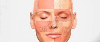
We, cosmetologists, work with superficial fat packets, which are also called compartments. These compartments on the face not only have a protective (buffer) function, protecting blood vessels and bone structures from external damage, but are also a source of nutrition.
Why do people with more pronounced subcutaneous fat age later than thin people? This is due to the fact that fat is a source of nutrients, for example, hormones - estrogens, and they, in turn, preserve youth and beauty, and also adipose tissue can quickly “retrain” into another type of tissue - chondrogenic, osteogenic, neurogenic. That is, if your body, when damaged, needs to “patch” holes in cartilage, bones or nerves, then embryonic cells of adipose tissue will participate in the repair.
Table of contents
- Etiology and pathogenesis
- Clinical manifestations
- Principles of treatment, hardware treatment methods
A change in the contours of the oval face (change in facial configuration, plump face, puffy cheeks) is a gradual disruption of the smooth contour of the face, usually observed in men and women after 45–55 years.
In our company you can purchase the following equipment for correcting facial contours:
- Thermage (Solta Medical)
- Maximus (Pollogen by Lumenis)
- GeneO+ (Pollogen by Lumenis)
During involutional changes, all layers of facial tissue are affected: the quality of the skin deteriorates, the ligamentous apparatus of the muscular aponeurotic layer (SMAS) weakens, the volume decreases and redistribution of subcutaneous adipose tissue occurs in the middle and lower third of the face, and bone tissue is resorbed. These changes are accompanied by the action of gravitational forces, which ultimately leads to ptosis of soft tissues.
Classic SMAS lifting
SMAS lifting - incision and detachment zones
- An incision is made along the red line.
- The skin is separated from the SMAS up to the blue dotted line and lifted to expose the SMAS layer.
SMAS lift, SMAS cutting and tension
- An L-shaped incision is made on the “bare” SMAS layer.
- Through this incision, all supporting ligaments in the cheek area are dissected.
- The free edge of the SMAS flap moves and, due to the fact that all ligaments have been cut, pulls the platysma - the anterior neck muscle.
- SMAS is fixed in the new position.
SMAS lift, removal of excess skin, suturing
After the SMAS returns to the position it was in 20 years ago, excess skin is formed. The skin flap is straightened, excess is removed, and sutured along the incision line.
As you can see, the SMAS lift not only eliminated jowls, but also tightened the sagging skin of the neck. This is how the SMAS lift compares favorably with lifting the lower face area.
Etiology and pathogenesis
The main reason for changes in facial contours is aging . All facial tissues undergo age-related changes - the epidermis, dermis, subcutaneous fat, underlying muscles and bones. In women, aging is often more pronounced, which is associated with hormonal changes and the onset of menopause. It accelerates the rate of bone resorption and the development of osteoporosis, which, in turn, leads to thinning and deformation of the bones of the facial skull, negatively affecting the contours of the face.
Gravity also contributes to the visible change in the contours of the oval of the face: areas with a significant amount of subcutaneous fat tend downward under the influence of gravity. In conditions of impaired elasticity and elasticity of the connective tissue, this gradually leads to sustainable changes in the contours.
Aging affects different levels:
- changes in skin density and extensibility;
- changes in volume and redistribution of adipose tissue;
- stretch of the superficial musculoaponeurotic system (SMAS);
- displacement of tissues relative to each other;
- violation of the tone of facial muscles;
- weakening of skeletal support for the soft tissues of the face due to selective bone resorption.
Epidermis
In the aging epidermis, a number of structural and functional changes are observed that have a negative impact on the condition of aging skin. However, changes in the dermis, subcutaneous fat and SMAS affect the oval of the face to a greater extent.
Dermis
The three main extracellular components of the dermis are collagen, elastin and hyaluronic acid - all of which begin to degrade with age. Collagen loss is approximately 2% per year, which significantly affects the structural integrity of the dermis. Basically, the volume of collagen decreases due to an increase in the activity of matrix metalloproteinases, which destroy its fibers. As damaged collagen fibers accumulate, the number of fibroblasts gradually decreases, which contributes to a further increase in the activity of matrix metalloproteinases and, thus, triggers a “vicious circle” of skin aging.
In turn, elastin, which provides skin elasticity, begins to calcify with age, and its fibers begin to degrade. The amount of glycosaminoglycans decreases, the production of hyaluronic acid by fibroblasts decreases. Loss of the structural integrity of the dermis leads to increased rigidity and decreased elasticity of the skin, and these changes occur more actively in women. Clinically, this can be recorded by applying pressure to the skin - in young people the surface is leveled in a few minutes, while with age this process can take up to 24 hours. The described pathogenetic processes contribute to changes in the contours of the oval of the face.
Hypodermis
With age, there is a decrease in the volume of subcutaneous fat against the background of an increase in the percentage of fat in the body. Superficial fat packets of the face shift diagonally medially and downwards. Atrophic processes in deep fat compartments and redistribution of the volumes of superficial fat packets in the middle and lower thirds of the face, together with weakening of the ligamentous apparatus, accentuate the existing grooves on the face and change its contour.
A redistribution phenomenon is observed when the fat layer on the face, arms and legs becomes thinner, and in the abdomen, waist and hips - thicker. At the same time, the content of visceral fat increases, which is associated with increased thermoregulatory function by isolating internal organs.
An important element of subcutaneous fat are connective tissue fibrous cords formed by bundles of collagen fibers with an admixture of elastic fibers that connect the dermis to the muscular aponeurotic layer (SMAS). Over time, collagen and elastin become less abundant, which gradually disrupts the structure of connective tissue cords. This weakens the “attachment” of the skin to the SMAS and thereby changes the contours of the face.
The time of appearance and rate of development of these changes is influenced by heredity, lifestyle, bad habits, ultraviolet radiation, stress, concomitant diseases and other factors.
Musculoaponeurotic layer (SMAS)
The musculoaponeurotic layer forms a three-dimensional network with connective tissue fibers that communicate with the dermis. As discussed above, changes in the viscoelastic properties of the SMAS and its three-dimensional mesh structure ultimately lead to tissue ptosis and changes in facial contours.
Clinical picture of wrinkles in the upper third of the face and basic facial patterns
Forehead wrinkles (wrinkles of surprise, attention; wrinkles of anxiety) - horizontal folds of the forehead;
their number can vary from one to several depending on the height of the forehead and the individual facial pattern. They are formed as a result of the work of m. frontalis, which is part of the occipitofrontalis muscle (m. occipitofrontalis) is the only levator in the upper third of the face: its function is to raise the eyebrows. M. frontalis is a wide, flat, vertically directed fan-shaped muscle layer extending from the scalp to the eyebrows. If this muscle is presented in the form of a continuous wide cord, then horizontal wrinkles are formed; if it is divided by a more or less pronounced aponeurosis into two abdomens, several arcuate wrinkles are formed.
The main facial patterns (types of persistent muscle response) in the forehead area (according to S.Yu. Shelekhov):
- straight, uniformly horizontal (photo 5) - characterized by uniform activity of m. frontalis with a highly located aponeurosis, forming straight horizontal facial wrinkles; more common in men;
- pattern of central activity (photo 6) - characterized by pronounced activity of the central part of m. frontalis and low-lying aponeurosis; wrinkles have a wavy shape;
- pattern of lateral activity (photo 7) - is distinguished by the most pronounced contractions of the lateral portions of m. frontalis. The main wrinkles are located in the lateral areas of the forehead and have an arched shape;
- pattern of medial activity (photo
 - characterized by pronounced activity of the medial parts of the m. frontalis in collaboration with the m. corrugator supercilii. As a result, wrinkles are formed in the form of a wave in the central part of the forehead.
Wrinkles in the area between the eyebrows (glabellae), or the so-called wrinkles of frowning and anger, most affect the perception of a person’s face.
Pronounced vertical wrinkles between the eyebrows indicate negative emotions and anger. These wrinkles make you perceive even a friendly person as dissatisfied and stern. The area between the eyebrows is perhaps one of the areas most in need of aesthetic correction. Glabella wrinkles are formed mainly as a result of the interaction of m. corrugator supercilii, m. procerus and m. depressor supercilii. Their number may vary depending on the anatomical features of these muscles. As a rule, there are from 1 to 2-4 vertical wrinkles and one or more horizontal wrinkles of the glabella.
Anatomically m. The corrugator supercilii is the deepest muscle of the glabella; it is a small (about 3 cm), but strong and dense muscle that starts from the frontal bone along the brow ridge, 0.5 cm from the midline of the forehead. Further, its fibers go obliquely upward and laterally, between portions of the orbicularis oculi muscle and are woven into the skin in the area of the medial (and sometimes middle) part of the eyebrow. This is a depressor muscle; it moves the inner part of the eyebrow inward and downward, forming vertical wrinkles between the eyebrows.
Photo 5 (left). Straight, uniformly horizontal facial expression pattern of the forehead. Photo 6 (right). Pattern of central activity of the frontalis muscle
Photo 7 (left). Pattern of lateral activity of the frontalis muscle Figure 8 (right). Pattern of medial frontalis muscle activity
M. procerus begins in the area of the aponeurosis of the transverse nasal muscle and the periosteum of the nasal bone and is woven into the skin of the glabellar region. This small, symmetrical, vertically directed muscle partially covers the m. corrugator supercilii and in its upper part is intertwined with the frontal muscle. In collaboration with m. corrugator supercilii it pulls the skin of the medial part of the eyebrow down to the bridge of the nose and contributes to the formation of transverse wrinkles in the area of the glabella.
A small thin muscle, m., also plays a role in the formation of wrinkles between the eyebrows. depressor supercilii. It is attached to the frontal bone 1 cm above the ligament of the inner corner of the eye and is woven into the skin of the head of the eyebrows. Its function is to lower the head of the eyebrow. During active work m. depressor supercilii, it is clear that vertical wrinkles continue quite low into the area of the upper eyelid. This is important to consider when correcting wrinkles between the eyebrows with preparations of botulinum neurotoxin type A (botulinum toxin, BTA): you need to use additional injection points in the m. depressor supercilii.
As a patient ages, forehead wrinkles may not be as important to the patient as maintaining eyebrow position without signs of drooping. Therefore, it is preferable to administer minimal doses of BTA at high injection points.
Thus, the anatomy of the muscles of the glabellar complex shows that they are all depressor muscles, which, being interconnected, contribute to the formation of wrinkles between the eyebrows and the lowering of the medial part of the eyebrow.
The main facial expression patterns of the eyebrow area (according to S.Yu. Shelekhov):
- straight horizontal (photo 9) - the movement vectors of the muscles that wrinkle the eyebrow are directed towards each other, while the activity of m. procerus; 1–2 vertical wrinkles are formed, the length of which depends on the width of the corrugator muscle;
- straight vertical (photo 10) - characterized by the fact that m. procerus is maximally active, contraction vectors m. corrugator supercili are weakly expressed; predominantly one or several horizontal wrinkles form above the bridge of the nose;
- uniformly active (photo 11) - m. corrugator supercili and m. procerus are equally active, which leads to the formation of both vertical and horizontal wrinkles between the eyebrows;
- friendly (melancholic) (photo 12) - motion vectors m. corrugator supercili are directed towards each other with a slight convergence down and towards
- to the center together with a friendly contraction of the central part of m. frontalis. This results in two vertical wrinkles between the eyebrows and a central horseshoe-shaped forehead wrinkle.
In some cases, superficial or deep wrinkles may appear directly above the eyebrows as a result of compensatory contraction of the lower parts of the m. frontalis after injection of BTA into the forehead area. This feature of muscle response should be taken into account when carrying out botulinum therapy for a gentler relaxation of the forehead muscles and possible additional correction using the BTA microinjection technique, as well as when strengthening this area with fillers.
Clinical manifestations
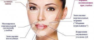
A change in the contours of the oval of the face is manifested by a violation of the clear line of its oval, loss of volume in the cheek-zygomatic region, the formation of deep nasolabial folds and “marionette lines”, drooping of the corners of the lips, deepening of the chin fold ( Fig. 1 ).
The face loses its even contour, begins to look loose and, as patients themselves sometimes say, “gradually flows down.”
Rice. 1. Brigitte Bardot aged 28 years (A) and about 80 years old (B). Changes in the contours of the oval of the face and other signs of aging are noticeable (Wikipedia.org, Pinterest.com)
| A | IN |
Principles of treatment, hardware treatment methods
Therapy is aimed at various tissue levels and solving a number of problems:
- improvement of quality and functional parameters of the skin;
- correction of fatty tissue volumes;
- reposition of soft tissues;
- myomodeling;
- replenishment of skeletal support for soft tissues.
To correct altered facial contours, the following cosmetic ingredients : cocoa butter (Cacao Butter), Nannochloropsis Oculata Extract, Kigelia Africana Extract, Goat Milk, Annuus (Sunflower) oil Seed Oil), etc.
You can replenish lost volumes, correct the shape of your face and improve the condition of your skin with injections of hyaluronic acid . They increase the volume of intercellular substance, help maintain the three-dimensional structure of the intercellular matrix, trigger biological stimulation of natural metabolism, moisturize the skin, participate in the transport and distribution of water in tissues, and enhance the protective function of the skin. A number of hyaluronic acid preparations allow you to mechanically even out the contour of the face, but this method only lasts for a few months.
You can make facial contours clearer with the help of lifting or reinforcing mesothreads . of botulinum toxin helps solve this problem (again, temporarily).
A powerful, but quite traumatic method of correcting changes in the oval of the face is plastic surgery (for example, a circular facelift). However, this solution is not indicated for all patients and has the potential for serious complications.
Hardware cosmetology today offers 2 main methods for improving the contours of the oval face: high-intensity focused ultrasound and RF lifting . Unlike laser techniques, they are able to act at a greater depth, triggering remodeling processes not only at the level of the dermis, but also in the subcutaneous fat and even SMAS. RF techniques based on deep heating, such as Thermage and TriPollar RF (Maximus and geneO+), can reduce the volume of local fat deposits by activating lipolysis.
RF and focused ultrasound cannot be considered as an alternative to plastic surgery - however, they can postpone plastic surgery for a while.
Questions from our users:
- facial contour correction
- facial contour correction cosmetology
- facial contour correction with fillers
- facial contour correction massage