The importance of facial anatomy for cosmetologists
Innovative cosmetic procedures should not be implemented without basic understanding of human anatomy: its structure, characteristics of the epidermis.
This information helps the cosmetologist build a correct program of work on age-related modifications of the epidermis and improve its condition. They are also necessary when choosing cosmetological effects and massage techniques.
Studying structural features, facial expressions, analysis using methodological tables, diagrams - basic information that a cosmetologist should follow. Without them, it is impossible to carry out a single aesthetic procedure: no massage, no Botox, no injection therapy.
Correct movements for classic facial massage
Classic massage uses the following types of massage movements:
- stroking
- trituration
- kneading
- vibration
The key here is stroking and rubbing, but to achieve the best effect you should alternate all types.
It is important not to exert excessive effort - in the technique of classic facial massage, the master works literally with his fingertips, with high smoothness and soft gliding - because the facial muscles are located close under the surface of the skin, plus there is a huge number of nerve endings on the face compared to other parts of the body.
The face responds very sensitively to massage - so even a light impact is enough for the desired effect, if applied correctly. But that is why excessively active influence easily crosses the line of reasonableness.
Classic massage is carried out without taking your hands off the client’s skin.
About facial muscles
They are elongated bundle-shaped branches with a thin muscle layer. They are located in the skin-connective head structures.
Unlike muscles located in other areas of the human body, facial muscles do not have double attachment to the bony skeleton. Instead, they are woven with one tip into the internal ligaments.
The only exception to this rule is a small muscle group located on the side of the face. Responsible for grinding food, it regulates the chewing process. All facial muscles (except for the orbicularis oris, nasalis and supracranial muscles) have a pair and differ in the depth of placement. So, they distinguish between superficial, deep, and medium.
Skin on the face
https://www.youtube.com/watch?v=0z8cU4gOfyw
The skin of the face performs the protective function of the body from the external environment. In order for this protection to occur in the best possible way, cosmetologists do their best to maintain the normal condition of the facial skin, because sagging, wrinkles, rashes or dryness are not only aesthetically unsightly, but also signs of deterioration in the motility of cellular metabolism, or a malfunction of the skin tissues.
Facial anatomy for cosmetologists describes in detail the structure of the facial skin, which consists of many cells, and their healthy condition affects a person’s appearance.
The vital activity of cells is very similar to the life of all creatures - they absorb oxygen, feed, and have the ability to reproduce. Although cells are the smallest living units, they contain a large number of organelles and elements that ensure the normal life cycle of each cell, and, accordingly, its owner:
- Ribosomes provide protein synthesis in the cell.
- The centrosome takes part in the regeneration of nutrients.
- Lysosomes are responsible for metabolism and absorption of nutrients.
- Cytoplasm - retains the activity of all useful substances in the cell, except for the nucleus.
- Microvilli are responsible for transporting substances from the cell through the membrane.
- The nucleus stores information about hereditary characteristics.
The epidermis is the first top layer of facial skin, serves as the main barrier of protection, and is responsible for tanning when exposed to sunlight. Almost all cosmetic procedures are aimed specifically at maintaining the elasticity and tone of this particular layer of skin. The epidermis in its structure has several layers of cells - lower, spinous, granular, flattering and horny.
The last layer of skin, the horny layer, is the topmost, and consists of dozens of corneocytes - cells that are the most mature on the face, and therefore any metabolic processes stop in them. These cells are already old, and therefore contain a small amount of water, keratin and do not have nuclei.
Their main function is to create a protective barrier for the skin from external factors. Usually, over the course of 28 days, the old cells are sloughed off, and new ones grow in their place - there is a constant process of the appearance of new cells and the exfoliation of old ones. Most mechanical and chemical peels operate at this level. The second layer of facial skin is the dermis.
It consists of two levels:
- The reticular layer is the level at which the networks of lymphatic and blood vessels, hair follicles, sebaceous glands and all fibers are located - they are responsible for the smoothness of the skin.
- The papillary layer concentrates nerve endings, processes and capillaries.
You can carry out any procedures on this layer of skin using deep-penetrating products with active ingredients. Most cosmetics are surface-acting products, so only special education will help you choose the composition of products that will penetrate through the epidermis to the dermis.
The dermis is responsible for the production of elastin and collagen in skin cells. Therefore, when deep wrinkles appear, there is an immediate need to act on this layer of skin, ensure its elasticity, and strengthen it.
The third, deepest layer, subcutaneous fat, is responsible for storing nutrients that directly affect the condition of the skin. This layer of skin consists of many nerve and blood vessels, as well as fat deposits. The need to act on this layer of skin arises with vitamin deficiency, when the face loses its healthy color.
About abbreviation
The work of the deep facial muscles is regulated by the central nervous system. The brain receives signals about current processes and communicates them to the facial muscle groups. That is, they translate the received information into abbreviations that display certain facial movements on the human face.
Any muscle movement is always a detailed reflection of transmitted nerve impulses.
Muscle contractions can be impaired due to traumatic and infectious diseases. Disorders are congenital or acquired (in this case permanent or temporary) in nature. One of the most significant pathologies is partial paralysis. It provokes an inability to correct muscle contraction, which is why a person experiences difficulty closing his eyes and jaw.
Contraindications for classical massage - when it is better to postpone the procedure
- A viral or bacterial disease in the active phase, a period of exacerbation of a chronic disease, elevated temperature. You should not undergo a facial massage at such a time - it is better for your body to concentrate its energy on solving more pressing problems. If you undergo a massage during an illness, it will either not be effective enough on its own, or may lead to a worsening of its course.
- The same applies especially to oncology and blood diseases. In such cases, first of all, you need to deal with this main problem, because local activation of blood flow in any area, which occurs under the influence of massage, can have undesirable consequences.
- With great caution and only with the direct, explicit and unequivocal permission of your attending physician, you can undergo facial massage if you have persistent hypertension with a pressure above 140/90 at rest.
- You should also refrain from any type of massage if there are unhealed lesions and inflammation in the massaged area.
How muscles are connected to massage lines
The facial muscles are an improvised frame that keeps the skin toned, maintaining its firmness and elasticity. Massage is a cosmetic manipulation that has a positive effect on them. Having a tightening effect, it allows you to transform your facial skin, making it fresher and more rested, get rid of redness and rashes, and create a clearly defined contour.
The procedure is performed strictly in accordance with the massage lines. These are the directions of movements that a cosmetologist must adhere to.
Massage lines are aligned in accordance with the flow of lymph, and under certain influences they can speed it up. This has a beneficial effect on the general condition of the skin, rids the body of waste and toxins, and relieves swelling.
Read on facesave.ru about the most popular and effective facial massage techniques in this section.
How to slow down facial aging?
Facial aging is a multifactorial process manifested in bone resorption, changes and sagging of the soft tissues of the face. Procedures for external influence on facial skin are essentially care products that do not bring the proper rejuvenating effect.
- Superficial treatments: creams, ointments, gels and other procedures moisturize and maintain skin condition.
- Injection techniques: fillers and botulinum toxin. Fillers based on hyaluronic acid help fill the volume of tissue that has undergone atrophy, and botulinum toxin relaxes the muscles that form wrinkles. The absolute advantage of these techniques is their reversibility of action. Typically, fillers and botulinum toxin preparations dissolve within 6 months.
- Surgical facial rejuvenation: the most effective method. There are different types of face lifting. It is necessary to approach the choice of face lifting method from the standpoint of existing facial changes, their severity, the patient’s wishes and the surgeon’s capabilities.
Fundamentally, the following types are distinguished: skin lifting; SMAS lifting; MACS-lifting; spacelifting.
- Skin lifting
In the mid-20th century, it was believed that it was skin changes that led to facial aging. However, the era of skin tightening showed its ineffectiveness due to the short-term results - the result lasted no more than 1 year. Modern cosmetology is again resorting to skin tightening using threads. Thread lifting gives short-term results and makes sense for initial facial changes. Pronounced age-related changes are not properly corrected with the help of threads, so their use must be strictly dosed.
- SMAS lifting
SMAS is a complex acronym that stands for the entire muscular aponeurotic complex of the face. After separating the skin layer from the muscle layer, an incision is made and the facial muscles are tightened and fixed in a new position. At the same time, the neck is lifted and jowls are eliminated. After the muscles are moved to the new position, excess skin is created, which is removed and the skin is sutured along the incision around the auricle. Therefore, this type of lifting is suitable for pronounced changes in the middle zone of the face, the presence of jowls and a flabby neck.
- MACS-lifting
It is considered a low-traumatic technique. Through a small incision near the auricle, the skin is peeled off and the muscles are tightened using purse-string sutures. The difference from SMAS is that the muscles do not move and are fixed, but are stretched like a drum, which will soon lead to drooping of the face. However, the commercial attractiveness of this method is very high. It is worth remembering that this method can only be good for initial changes - just like thread lifting.
- Spacelifting
The essence of the operation developed by Dr. Mendelson is to eliminate the facial spaces that stretch under the influence of gravity. Since these spaces exist only in certain areas of the face, spacelifting does not provide a comprehensive lift and does not allow for significant rejuvenation of the neck area. Therefore, spacelifting gives a good rejuvenating effect in the middle zone of the face with unexpressed changes in the neck.
Classification
In the human body there are over 100 facial muscles located on the head and neck. You can study each of them in more detail, based on the photographs, pictures, and descriptions posted below.
From an anatomical point of view, all facial muscles can be divided into 6 subgroups:
- Mimic;
- Cervical musculature;
- Chewable;
- Language;
- Oral cavity;
- Oculomotor.
Some may be classified into several subgroups at the same time.
Chewing group
This includes those responsible for regulating chewing movements. Namely:
- Temporalis muscle;
- Chewing muscle;
- Lateral pterygoid muscle;
- Medial pterygoid muscle.
The muscles listed provide minimal impact on the overall appearance of the face.
The chewing muscle group is responsible for the movement of the lower jaw during speech activity and grinding food. She is always in hypertonicity and does not need to perform specific exercises. Prone to spasms due to excessive clenching of teeth, it can negatively affect blood flow, activating photoaging processes in the designated area.
This can be said about the pterygoid muscles, whose main purpose is to grind solid food. Their care is selected according to the indications of a particular person. This is the only way to achieve positive dynamics: tighten the oval of the face, prevent the development of deep wrinkles, etc.
Mimic group
The functions of facial muscles are limited to displaying emotions. Due to the extensibility of the epidermis, the construction of folds that appear in the vertical direction, emotional manifestations are formed in relation to muscular contraction. Hence the rule - the more emotional a person is, the greater the likelihood of the formation of facial wrinkles in the face or neck area.
Facial muscles are divided into three categories:
- Upper facial;
- Median;
- Lower facial and cervical.
The former, when stretched, provoke the formation of vertical forehead wrinkles, diagonal folds in the bridge of the nose, as well as “crow’s feet” under the lower eyelids. The latter provide a feeling of “sunken cheeks”, outline nasolabial folds, and make wrinkles under the eyes and in the corners of the lips more noticeable. The facial muscles of the third group contribute to the protrusion of the lower lip forward and the lowering of the corners of the mouth.
All identified defects and problems can be easily removed by high-quality training of the entire muscle zone.
Neck band
By analogy with the facial muscles of the face and neck, the cervical muscles are also divided into three categories:
- Superficial cervical muscle group;
- Hyoid muscle group;
- Deep cervical muscles (this includes the posterior scalene, anterior and median).
If we talk about the differences, the cervical muscles, in comparison with the facial and chewing muscles, have five fascia (connective membranes covering them from the outside):
- Superficial;
- Own cervical;
- Scapuloclavicular;
- Inner cervical;
- Prevertebral.
Cervical spasms provoke the formation of skin laxity in a designated area of the body, lead to the development of wrinkles and folds, and trigger photoaging processes. In some cases, a double chin occurs, as well as transverse creases.
Platysma: what is it, where is it located?
Platysma is the thinnest superficial layer of the cervical muscles, responsible for its aesthetics and overall appearance. Located in the deep layers of the skin, it firmly fuses with it.
Although platysmal tissue is not involved in motor activities carried out by the neck and head, it is involved during strenuous exercise. This determines its role in facial expression.
Platysma has a number of significant differences from other facial muscle groups. It is more susceptible to modifications and quickly loses its plasticity. Therefore, it requires carefully selected care that would meet modern cosmetological requirements.
Correction of age-related changes
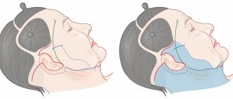
With age, multifactorial processes occur that determine the aging process of the face. These processes affect the facial skeleton and integumentary tissues. After 30 years, the beginning changes in the face are already visually noticeable. Changes in all tissue layers lead to their displacement and downward movement. More pronounced processes occur in the middle third of the face. The decisive factor in the drooping of the integumentary tissues of the face is a change in the bone surface.
- Forehead
Flattening of the frontal bone, combined with constant tension of the frontal muscle, leads to drooping of the integumentary tissues of the upper area of the face and the formation of transverse grooves in the forehead. The sliding mass of soft tissues of the forehead forms excess tissue in the area of the upper eyelids, the tails of the eyebrows droop and a “heavy” look is formed.
- Middle face area
Atrophy of bone tissue and fat packets in the midface area, combined with weakening of the muscles, leads to contusion of the ligaments and grooves. Pronounced bags under the eyes and eye bags form.
- Neck
The framework that holds the tissues of the lower zone of the face is the platysma - the subcutaneous muscle of the neck. Atrophy of the jaw bone, prolapse of the submandibular salivary gland, together with weakening and divergence of the platysma fibers, form an obtuse cervical-mental angle and a “double chin.” Thus, with age we observe: a reduced lower jaw; a stretched space at the bottom of which fat has accumulated; atrophied neck muscles that can no longer hold soft tissue and sag under gravity; the resulting jowls.
Fig.1 Aging according to Mendelssohn
Rice. 2 Bone atrophy according to Mendelssohn
Rice. 3 Facial aging: pronounced folds, jowls, “fat packets”
Map of facial muscles and their functions
Facial anatomy is a well-researched branch of medicine. Therefore, muscle functions do not need additional study - they have already been thoroughly analyzed and strictly defined for a long time. In some cases, muscle groups have a “telling” name, and therefore it is not difficult to guess about their functions.
The cranial vault muscle (also known as the “helmet tendon”) is responsible for raising the brow arches and gathering the skin on the forehead into transverse folds.
The occipital frontalis pyramidal muscle raises the eyebrows, leading to the formation of transverse folds and creases. It is paired - one above both eyebrows, which is why they move independently of each other. This activity affects the opening of the eyes, which gives the face different expressions.
The temporalis muscles coordinate jaw movements.
The fibers of the pride muscles are located in the area between the eyebrows and extend all the way to the forehead. Shifting the eyebrows, wrinkling the nose are the actions for which they are responsible. The structural features of the facial muscles cause the appearance of wrinkles between the eyebrows when they are tense.
The muscles that wrinkle the eyebrows set them in motion. They draw the inner brow line toward the midline in two directions: inward and upward, bringing the edges closer together. Hypertonicity provokes the formation of vertical creases between the eyebrows.
The orbicularis oculi muscles are responsible for closing the gaps between the eyes.
The nasal muscle , contracting, provokes motor activity of the wings of the nose. Its contraction causes expansion and contraction of the nasal passages.
The lacrimal muscles raise the upper lip and move the wings of the nose.
The infraorbital muscle is also responsible for raising the upper lip.
The large zygomatic is located in the lower area of the face, moving the corners of the mouth in different directions. It is she who is responsible for the smile and can provoke the formation of nasolabial folds.
The orbicularis oris muscle tightens and extends the lips, allowing them to be compressed.
Modioulos (in Latin) is the knot of the corner of the mouth. It is he who gives the aesthetics to the lower facial third.
The laughter muscle is needed to stretch the corners of the mouth. A number of people develop dimples on their cheeks during its contraction. In addition to facial functions, it is of particular importance in the overall aesthetics of the face and its modeling. Proper development of the laughter muscle allows you to make adjustments to the oval of the face and slightly raise the corners.
The cheek muscles are located immediately above the laugh muscles. They model the cheeks, spreading the mouth opening to the sides. When the mouth expands, the muscle becomes hypertonic. An interesting fact about the muscles: there is a layer of fat between the cheek muscle and the epidermis; in men it is narrower than in women. It is observed in the greatest extent in children.
Triangular muscle (descending oris) - it is responsible for lowering the corners of the mouth. Direction helps in expressing feelings of sadness. In a state of hypertonicity, the face takes on a negative expression.
The descending muscles pull the lips down, giving the face an expression of disgust.
The subcutaneous muscle is represented by two parts, which are located under the quadratus muscle of the lower lip. During contraction, a dimple may form on the chin.
The neck muscles are especially important when turning and tilting the head. Muscular thinning provokes the formation of a double chin, as well as a decrease in the plasticity and elasticity of the skin, and the acquisition of a gray complexion.
Lymphatic system
Facial anatomy for cosmetologists focuses on the important role of normal functioning of the lymphatic system on the condition of the skin.
This system is a very dense capillary network that is present in all organs and tissues of the body. Disruption of the lymphatic system often affects the condition of the skin of the body - it loses its beautiful color, elasticity and velvety. The loss of these qualities due to problems with lymph flow is doubly noticeable in the condition of the facial skin.
The lymphatic system refers to the vascular system of the body. Under its influence, lymph, a clear liquid, moves in the body, which, like blood, circulates throughout the human body.
But the lymphatic system does not have a pump, the function of which in the circulatory system is performed by the heart, and therefore the movement of lymph occurs very slowly - towards the large veins, at a speed of 0.3 mm / s . Therefore, it is always worth activating its work with mechanical influence - massages, baths and cosmetic procedures - such manipulations will speed up the work of the glands.
This system cleanses the body.
Important functions of the lymphatic system are:
- Distribution of fluid in the body.
- Transport of nutrients from tissues.
- Protecting the body from bacteria, supporting the immune system.
It consists of:
- Vessels
- Knots
- Duct.
- Tonsils, thymus.
In the human skull, the lymphatic system has 7 groups of nodes:
- Occipital.
- Cervical.
- BTE.
- Buccal.
- Submandibular, located in the triangle of the chin.
- Parotid.
- Chin.
Therefore, if the lymphatic vessels are clogged and the functioning of the system is disrupted, many skin diseases arise, which can manifest themselves in the form of acne, pimples, and other rashes.
If you regularly carry out lymphatic drainage procedures, then these manipulations will have a good effect on metabolic processes in the tissues of the body. For example, you can reduce facial swelling, improve its contours and elasticity, and normalize the tone of facial muscles with regular massage. It is very important for a cosmetologist to know the directions of lymph flow on the face.
Since this is a complex network of capillaries, lymph flows in several directions:
A) Lymph that flows through the facial tissues enters here with the help of superficial vessels. The lymph flow corresponds to the blood veins.
Superficial lymphatic vessels are grouped into anterior and posterior:
- The posterior vessels supply lymph to the back of the head. There they pass into another group of vessels - the occipital.
- The anterior vessels are located simultaneously from the forehead, eyelid, crown and temples. These vessels are connected to nodes near the ears, through which the lymph continues to move through the vessels down the neck.
B) The lymphatic network begins from the eyelids, from the nose, cheeks and lips, its movement is partially directed to the submandibular triangle, where the submandibular nodes are located. Another part of these vessels interrupts their circulation in the buccal nodes.
C) The mental lymph nodes, which are located under the hyoid bone, are supplied with lymph from the vessels near the lips and chin.
D) Deep vessels from the hard and soft palate direct their lymph flow to the deep nodes of the parotid gland.
Alternative classification
In addition to the described muscle groups, those that belong to the internal organs (uvula, palate, middle ear, etc.) are also distinguished. How do emotional manifestations and muscles relate?
All muscles included in the facial group are responsible for displaying a certain emotion:
- Frontal - raises the eyebrow arches, forms horizontal frontal wrinkles, thus expressing delight and amazement on the face.
- The orbicularis muscle allows you to close your eyes when very frightened, as well as roll them up or down. With its help, a person shows embarrassment and misunderstanding.
- The zygomatic major and minor muscles help produce a smile by lifting the corners of the mouth.
- The muscles responsible for lowering the corners of the mouth are active during negative emotions.
- The laughter muscles allow the corners of the lips to stretch horizontally, forming “pits” at the moment of smiling.
- The greatest activity of the orbicularis muscle is observed when blowing kisses.
- Disappointment, anger, confusion are the emotions that are displayed with the help of the mentalis muscle (it slightly raises the lower jaw, pushing it forward).
- Fear, disgust and other negative feelings are impossible without the superficial neck muscle, which works by straining.
More than a thousand different combinations of muscle contractions have been studied and recorded, reflecting one or another feeling on the human face.
Structure of the skull
The human skull is the main protection for the facial muscles and nerves that are responsible for facial mobility. In total, the skull contains 23 bones - that is, 8 paired and 7 unpaired. All of them are divided into 2 groups: facial and brain bones.
Facial bones are smaller paired bones:
- Nasal.
- Palatal.
- Zygomatic.
- Tearful.
- Upper jaw.
- Inferior turbinate.
Unpaired facial bones:
- Lattice.
- Sublingual.
- Opener.
- Lower jaw.
This group affects the normal functioning of the respiratory and digestive organs. The marrow bones in total consist of paired and unpaired bones.
They are located above the facial region and form some parts of the face, namely:
- Frontal tubercles.
- Eye sockets.
- Frontal zone.
- Whiskey.
- Nasal cavities.
The paired bones are the parietal and temporal small bones, and the unpaired bones are the frontal, occipital and sphenoid. All parts of the skull are connected to each other by special “sutures”.
Don't miss the most popular article in the section: Fashionable haircut for short hair. Photo, front and back views.
Photoaging: age-related muscle modifications
Over time, the muscle structure loses its elasticity and plasticity and changes in size and lengthens. The characteristics of the modifications depend on the nature of muscle manifestations in different situations. For example, under stress, during rest or work, in dialogues with people, etc. Factors such as lifestyle, proper care, and heredity become important.
In those areas where the muscles are not firmly attached to the skin surface, fatty hernias form over the years. They arise due to the pulling of ligaments under the influence of excessive loads. Where the ligaments remain able to hold the facial muscles, wrinkles, creases and folds appear.
Most people are convinced that age-related changes in tissues occur due to their hypertonicity or excessive relaxation, but the point is different - incessant muscle activity that provokes spasms. With frequent contraction, the muscles fall into folds, changing the texture of the skin and its structure.
For example, spasms in the area of the frontal muscle tissue provoke the development of horizontal and vertical folds in the specified facial area, increasing the tone of the orbicularis muscles. Gradually, all this leads to the appearance of “crow’s feet” and nasolabial furrows.
Epidermis
Rice.
2. Epidermis The epidermis is a constantly exfoliating epithelial layer of the skin, which consists mainly of 2 types of cells - keratinocytes and dendritic cells. Melanocytes, Langerhans cells, Merkel cells, and intraepidermal T lymphocytes are present in small quantities in the epidermis. Structurally, the epidermis is divided into 5 layers: basal, spinous, granular, shiny and horny, differing in the position and degree of differentiation of keratinocytes, the main cell population of the epidermis (Fig. 2).
Keratinization. As keratinocytes differentiate and move from the basal layer to the stratum corneum, their keratinization (keratinization) occurs - a process that begins with the phase of keratin synthesis by keratinocytes and ends with their cellular degradation. Keratin serves as a building block for intermediate filaments. Bundles of these filaments, reaching the cytoplasmic membrane, form desmosomes necessary for the formation of strong contacts between neighboring cells. Further, as epithelial differentiation progresses, epidermal cells enter the degradation phase. The nuclei and cytoplasmic organelles are destroyed and disappear, metabolism stops, and cell apoptosis occurs when it is completely keratinized (turns into horny scales).
The basal layer of the epidermis consists of a single row of mitotically active keratinocytes, which divide on average every 24 hours and give rise to new cells in the overlying epidermal layers. They are activated only in special cases, such as when a wound occurs. Next, a new cell, a keratinocyte, is pushed into the spinous layer, where it spends up to 2 weeks, gradually approaching the granular layer. The movement of the cell to the stratum corneum takes another 14 days. Thus, the lifespan of a keratinocyte is about 28 days.
It should be noted that not all cells of the basal layer divide at the same rate as keratinocytes. Epidermal stem cells under normal conditions form a long-lived population with a slow proliferation cycle.
The spinous layer of the epidermis consists of 5-10 layers of keratinocytes, differing in shape, structure and intracellular content, which is determined by the position of the cell. Thus, closer to the basal layer, the cells have a polyhedral shape and a round nucleus, but as the cells approach the granular layer, they become larger, acquire a flatter shape, and lamellar granules appear in them, containing various hydrolytic enzymes in abundance. Cells intensively synthesize keratin filaments, which, gathering into intermediate filaments, remain unconnected on the nuclear side, but participate in the formation of multiple desmosomes on the membrane side, forming connections with neighboring cells. The presence of a large number of desmosomes gives this layer a spiny appearance, for which it received the name “spinous.”
The granular layer of the epidermis is composed of still living keratinocytes, distinguished by their flattened shape and a large number of keratohyalin granules. The latter are responsible for the synthesis and modification of proteins involved in keratinization. The granular layer is the most keratogenic layer of the epidermis. In addition to keratohyalin granules, keratinocytes of this layer contain large quantities of lysosomal granules. Their enzymes break down cellular organelles during the transition of the keratinocyte to the phase of terminal differentiation and subsequent apoptosis. The thickness of the granular layer can vary; its value, proportional to the thickness of the overlying stratum corneum, is maximum in the skin of the palms and soles of the feet.
The shiny layer of the epidermis (so named for its special shine when viewing skin preparations on a light microscope) is thin, consists of flat keratinocytes, in which the nuclei and organelles are completely destroyed. The cells are filled with eleidin, an intermediate form of keratin. It is well developed only in some parts of the body - on the palms and soles.
The stratum corneum of the epidermis is composed of corneocytes (dead, terminally differentiated keratinocytes) with a high protein content. The cells are surrounded by a waterproof lipid matrix, the components of which contain compounds necessary for exfoliation of the stratum corneum (Fig. 3). The physical and biochemical properties of cells in the stratum corneum vary depending on the position of the cell within the layer, directing the exfoliation process outward. For example, cells in the middle layers of the stratum corneum have stronger water-binding properties due to the high concentration of free amino acids in their cytoplasm.
Rice. 3. Schematic representation of the stratum corneum with the underlying granular layer of the epidermis.
Regulation of proliferation and differentiation of epidermal keratinocytes. Being a continuously renewing tissue, the epidermis contains a relatively constant number of cells and regulates all interactions and contacts between them: adhesion between keratinocytes, interaction between keratinocytes and migrating cells, adhesion with the basement membrane and underlying dermis, the process of terminal differentiation into corneocytes. The main mechanism for regulating homeostasis in the epidermis is supported by a number of signaling molecules - hormones, growth factors and cytokines. In addition, epidermal morphogenesis and differentiation are partially regulated by the underlying dermis, which plays a critical role in maintaining postnatal skin structure and function.
Areas and lines of classic facial massage
Movements in the classic facial massage technique mainly correspond to the adjusted lines of the least tension of the skin and are directed from the center to the periphery of the face.
- From the center of the chin to the lower edges of the ears - and then down again, along the sides of the neck.
- From the corners of the mouth to the ears.
- From the wings of the nose to the temporal zone.
- From the center of the forehead to the top edges of the ears.
- Circular movements around the eyes - first from the temples to the nose along the lower eyelid, then from the nose to the temples along the upper.
- The forehead should also be further worked in the center and along the hairline.