How to recognize lentigo?
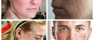
Treatment of this problem should begin with a correct diagnosis. The main symptom is a thickening of the stratum corneum of the skin, so that lentigo spots in most cases rise slightly above the surface. These spots are located both on the face and on the body.
Other characteristic symptoms:
• bright color of all shades of brown; • round or oval shape, clear boundaries; • visual sensation of swelling; • size from 1 to 3 mm, in rare cases the spot grows up to 2 cm; • severe itching with lentiginosis.
What is the clinical picture
When pigmentation is detected on the skin, most people do not associate it with any disease. Moreover, rarely does anyone know about lentigo and what it is.
This is due to the fact that the spots do not cause discomfort. The only thing the patient notes is a change in their color from light to darker (as in the photo).
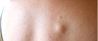
Sometimes the pigmented areas may thicken slightly or become bulging.
Such changes signal the degeneration of this neoplasm from benign to malignant. Then you need to urgently consult a doctor for diagnosis.
If your dermatologist has looked at the spots and determined that they are benign (not cancerous) and cannot turn into skin cancer, then he can begin to treat them. Sometimes we simply freeze the lesions with liquid nitrogen on the day of presentation. Laser treatment can make dark skin even darker if the wrong type of laser was used.
Dermatological surgeon Amy Paul
Causes
Lentigo is nothing more than an accumulation of excess melanin - a coloring pigment. These spots can either be inherited or appear unexpectedly in adulthood. Unfortunately, doctors have not yet fully studied this disease.
The following reasons for its appearance have been precisely defined: 1. Old age. Lentigo on the face is very common in people over 70 years of age, sometimes these spots begin to appear after 40. 2. Young age. The so-called juvenile lentigo in children and adolescents is known. Its causes are unclear; with the onset of adulthood, the spots disappear on their own. 3. Skin injuries. Occurs at the site of superficial wounds. 4. Excessive insolation. Excess ultraviolet radiation always has a negative effect on the skin, solar lentigo is proof of this. 5. Lentiginosis, or congenital lentigo, is inherited and manifests itself not only on the face, but also on the body together with other unpleasant pathologies: heart disease, congenital deafness, etc. 6. Malfunctions of the liver and intestines. There are cases where lentigo spots disappeared after surgical operations on the gastrointestinal tract.
The many faces of hyperpigmentation
Today, hyperpigmentation is considered one of the main signs of aging. Dark spots in the décolleté area, on the face and hands are perceived as an inevitable symptom of hormonal changes or liver disease. It is not surprising that women are as concerned about the appearance of characteristic spots as they are about new wrinkles.
The production of melanin (skin pigment) is a natural reaction of the skin that tries to prevent UV rays from penetrating into the nuclei of cells where DNA is located.
Hyperpigmentation occurs when, due to dysfunction of melanocytes (cells that produce melanin), melanin is synthesized many times more than necessary. This can happen for several reasons. Let's look at them in order.
Traumatic hyperpigmentation
This hyperpigmentation occurs due to the release of melanin in response to skin damage - as a kind of attempt by the body to protect itself.
This type of hyperpigmentation occurs more often in people predisposed to it, so if you have freckles on your face, be careful with peels, laser therapy, and squeezing pimples.
Help: The good news is that this type can only be treated with external remedies, i.e. ointments, creams and serums. Look for so-called “tyrosinase inhibitors” in the formulations - a protein that controls melanin synthesis: hydroquinone, arbutin, kojic acid, licorice root extract, nicotinamide, emblica extract, vitamin C, alpha-lipoic acid. As well as new products on the cosmetic market - synthetic peptides oligopeptide-34, oligopeptide-51.
Despite the fact that the mutagenic effect of hydroquinone has been actively discussed recently, cosmetologists have not yet developed a more effective whitening agent. Hydroquinone is particularly blamed for causing malignant tumors in rats. However, in the human body this substance is “neutralized” in the liver with the formation of non-toxic derivatives. Hydroquinone is approved for over-the-counter cosmetics at a concentration of 2%, but in some salons cosmetologists can make their own products containing a higher percentage. The main reason for limiting this drug for sale is various types of dermatitis due to the use of products with substance concentrations of 5% or higher.
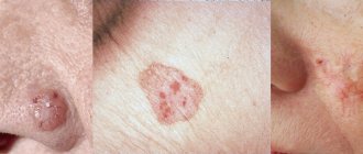
Pigmentation in the elderly (lentigo)
Occurs in older people as a result of repeated exposure to UV radiation
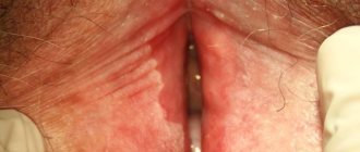
The so-called “solar lentigo” are small spots 1 cm in diameter with a smooth edge, very similar to freckles. They appear on the hands and face, but unlike freckles, they do not disappear in winter and do not become paler. Lentigines occur in areas exposed to UV radiation throughout life. This is why it is important to use sunscreen on your neck, décolleté and arms at all times, especially if you spend a lot of time in the sun or like to visit a tanning salon.
Help: There are many ways to reduce the appearance of lentigo, it all depends on how quickly you want to see results. Long-term treatment includes the use of retinoids and sunscreens, as well as serums with tyrosinase inhibitor agents. The best results are obtained by combining these substances with glycolic acid, which helps them penetrate deeper into the skin. For example, a combination of glycolic acid with nicotinamide (vitamin B3) and retinol or resorcinol is good. If you want quick results, peelings with trichloroacetic acid and laser correction (Fraxel, IPL laser) are included in the treatment.
Chloasma, or “mask of pregnancy”
Chloasma (melasma) is a fairly typical skin condition during pregnancy or when using contraceptives.
This is due to an increase in estrogen levels. There are 2 types of melasma: epidermal and dermal, although the mixed type is more common.
Epidermal melasma
appears in the form of spots from light to dark brown, in some cases even black, with jagged edges. This type is more amenable to correction, since melanin is located in the upper layers of the skin, which means that the whitening components from creams and serums can achieve their goal.
Dermal melasma
appears in the form of gray-blue spots, the pigment lies in the middle layers of the dermis, where the lightening components no longer penetrate.
To make a correct diagnosis, it is best to use a Wood’s lamp (“blue lamp”) - the brighter the spots in the light of the lamp (epidermal melasma), the easier the therapy will be. However, most women do not conduct such tests, but simply randomly buy “whitening serums” in the hope that the spots will go away on their own. Alas, such a strategy does not always lead to success. Chloasma is one of the most stubborn types of hyperpigmentation. Lasts for a very long time even after the end of pregnancy or discontinuation of COCs.
Help: Hydroquinone, azelaic acid, a combination of retinoids and corticosteroids, such as fluocinolone (Fluocinoloni acetonidum), will help you in treatment. Oddly enough, but in this case, azelaic acid at a 20% concentration may be even more effective than hydroquinone at 2-4%. To speed up the effect, the so-called Jessner peel (an alcohol solution of resorcinol, salicylic and lactic acid) or peeling with salicylic and glycolic acids is performed once every two weeks. If you add medications with kojic acid to this treatment, the effectiveness increases further. In some cases, IPL laser treatment or microdermabrasion may make sense.
If you want to correct the manifestations of chloasma during pregnancy, be sure to consult your doctor!
Whatever regimen you and your dermatologist choose, the main thing is to strictly follow the regimen. To make this routine easier for consumers, manufacturing companies such as Obagi and La Roche-Posay produce products in special “numbered” packaging.
Today, cosmetic options are such that hyperpigmentation is not necessarily “forever.” Reducing unpleasant stains is possible even in the most severe cases. Of course, for skin 30+, spots will not disappear with the wave of a magic wand, and here you should no longer rely on “green” cosmetics. But the right therapy will definitely help you!
Tatiana Morrison
Photo thinkstockphotos.com
Products by topic: (retinol), [product](fluocinolone), [product](salicylic acid), [product](achromine), [product](acids), [product](peptides)
Treatment of lentigo on the face
The connection between lentigo and certain diseases of internal organs and the risk of developing skin cancer is alarming. Therefore, if freckles and moles rarely pose a danger to our health, then if lentigo occurs on the face, treatment should begin immediately.
First of all, you need to reconsider your lifestyle: avoid direct sunlight, apply creams with a high level of SPF protection to your skin, and do not skimp on skin care in terms of nutrition and hydration. If necessary, a dermatologist will prescribe the use of certain medications.
In addition, it is worth following a diet that excludes possible allergens. You should also check your gastrointestinal tract and put it in order; if you are overweight, you will have to mercilessly say goodbye to it.
Lentigo therapy at home
You can treat lentigo yourself at home if it is clearly established that the pathology is benign.
All folk recipes are based on the use of natural lightening agents:
- lemon juice;
- hydrogen peroxide;
- ammonia;
- rowan;
- calendula;
- black currant;
- parsley;
- almonds;
- honey
Here are some productive recipes for homemade masks:
- The stems, leaves and roots of parsley are finely chopped to a paste. Then add honey. The resulting composition is applied to problem areas and, after leaving for half an hour, washed off with warm water.
- Place the lemon peel in boiling water and keep it warm for about 30 minutes. Then let the water sit and wipe the stains up to 3 times a day.
- Grind almonds in a coffee grinder and mix them in equal proportions with oatmeal. Add 5-7 g of milk powder and flavor with a small amount of water to form a mass similar to sour cream. Use this thickener to scrub the affected areas for about 5 minutes and wash off any remaining residue. Do this no more than 2 times a week.
- Dilute dry white clay (1 tbsp) with warm water until smooth and add a little lemon juice. Apply the composition to age spots and wash off after 20-25 minutes. This mask is used every day.
The suggested masks should be done in the evening, when you are not planning to go outside. This is due to the fact that after the procedure you cannot expose the skin to UV exposure.
As a treatment for lentigo, it is recommended to use pharmaceutical ointments with a whitening effect:
- zinc;
- sulfuric;
- Clotrimozole;
- salicylic.
Among the creams, the following brands are distinguished: Skinoren-gel, Melanativ, Clearvin. They should be applied to the skin twice a day.
I recommend using a combination of whitening cream (hydroquinone 4%) and tretinoin (vitamin A). But it will take months of treatment to achieve results. The laser is much faster.
Plastic surgeon Sam Naficy
Lentigo – treatment with the M22 device
Modern methods of getting rid of lentigo include photothermolysis - treatment with IPL light or laser radiation. The essence of this method is the pulsed effect of light beams on the skin in the affected area, during which excess melanin is destroyed without affecting healthy areas of the skin.
It is generally accepted that laser copes with this problem better. However, modern light devices, for example, the M22 multi-module platform, demonstrate a high rate of removal of various age spots. For each type of hyperpigmentation, including lentigo, a special program is provided, which significantly increases the effectiveness of M22 compared to other photosystems. M22 rays penetrate the skin, destroy excess melanocytes and keratinized cells, cause the synthesis of collagen and elastin fibers and other active substances. These processes promote rejuvenation and rapid regeneration of damaged areas, lightening of spots and smoothing of the relief. After just a few sessions, the spots cease to be noticeable and the skin acquires a healthy color.
There are solar lentigo and age-related (senile).
This type of pigmentation is very persistent, does not depend on the time of year and does not disappear on its own. Solar lentigo occurs in those who spend hours under the scorching sun on the beach and, of course, in tanning bed lovers.
Senile lentigo appears in old age and is often called “senile ripples” or senile keratomas. It is associated with a slowdown in the metabolic process during age-related hormonal changes in the body.
Birthmark (nevus ). This is perhaps the most common type of hyperpigmentation. Perhaps not one person is deprived of this type of pigment formation disorder. They are a collection of melanocytes. Nevi can appear at any age, but are especially active in adulthood.
Melasma (chloasma). Large pigment spots that do not have a clear shape or specific size. Mostly appear on the face of young women. Their favorite location is the face, and the main cause is hormonal changes, mainly such as pregnancy, lactation and oral contraceptives. In some cases, they disappear on their own when hormonal levels normalize.
Skin melanoma treatment stages 1, 2, 3. Symptoms, signs, metastases, prognosis.
Content:
- General information about melanoma
- What are the different forms of melanoma? Superficial spreading melanoma
- Nodular melanoma
- Malignant lentigo melanoma
- Acral melanoma
- Atypical (dysplastic) nevi
- Primary metastatic melanoma
- Surgical treatment of skin melanoma
What are the different forms of skin melanoma?
The main clinical forms of melanoma are:
- superficial spreading (39-75%);
- nodal;
- malignant lentigo melanoma;
- acral melanoma.
Superficial spreading melanoma (flat, radially growing melanoma)
The tumor develops equally often both on unchanged skin and from a pigmented nevus. It can be localized on open and closed areas of the skin, mainly on the lower extremities in women and the upper half of the back in men. It is a plaque of irregular configuration with a scalloped outline, areas of regression and discoloration, mosaic coloring, and keratosis on the surface. On average, after a few years, a node appears on the plaque, indicating the transition of horizontal growth to vertical.
Nodular melanoma
Nodular melanoma, accounting for 10-30% of all skin melanomas, is the most aggressive type of tumor. The neoplasm usually appears on unchanged skin. Clinically it is a node, less often a polyp-like formation on the skin. Patients note a rapid, within several months, doubling of the node's volume, its early ulceration and bleeding. The most common localization is the skin of the back, neck, head, and limbs. Histologically, invasion of atypical melanocytes to different depths of the dermis and subcutaneous fat is revealed.
Malignant lentigo melanoma
Melanoma of the lentigo maligna type accounts for about 10-13% of all melanomas and is characterized by a long horizontal growth phase. In typical cases, it occurs in older people on open areas of the skin of the face and neck in the form of black-brown spots or plaques. This type of melanoma is less aggressive than other flat melanomas.
Acral melanoma
Acral melanoma occurs in the nail bed and accounts for about 8% of all skin melanomas. It usually appears as a dark spot under the nail, which makes its timely diagnosis extremely difficult.
How is skin melanoma diagnosed?
The most effective method for early detection of skin melanoma is periodic self-examination of the skin. There is a kind of “melanoma alphabet” that describes a number of signs of degeneration of a mole, indicated by the first four letters of the Latin alphabet:
- A (asymmetry) - asymmetry: the shape of “good” moles is often symmetrical;
- B (border irregularity) - the edges of the mole are usually smooth and clear. An uneven, scalloped outline is more characteristic of melanoma;
- C (color) - benign nevi are colored more or less evenly. The unequal color of different parts of the neoplasm is more characteristic of a degenerated mole;
- D (diameter) - the diameter of a mole is more than 6 mm: the larger the mole, the greater the likelihood of its degeneration. Malignant degeneration is indicated by various kinds of changes in a pre-existing mole. It was found that pigmented formations on the skin that regularly changed shape and color turned out to be melanoma 4 times more often than those whose appearance remained unchanged. Therefore, to the first four letters of the “alphabet of melanoma,” a fifth was added;
- E (evolving) - the appearance of any external changes in the mole, which most often are: change in color (decrease or sharp increase in pigmentation); violation or complete absence of skin pattern in the area of the nevus, “varnish” surface or peeling; the appearance of an inflammatory areola around the mole (redness in the form of a corolla); change in configuration along the periphery, blurring the contour of the nevus; an increase in the size of the nevus (especially over the age of 30) and its compaction; itching, burning, tingling in the mole area; the appearance of cracks, ulcerations in the mole area, bleeding; loss of existing hairs on the mole; sudden disappearance of a mole (especially after tanning in the sun or in a solarium).
Very valuable additional clarifying information can be obtained by performing a dermoscopic examination of a pigmented skin tumor, which allows for a visual assessment of the tumor at 10-40x magnification.
Risk groups and factors predisposing to the development of skin melanoma.
Individuals at increased risk of developing melanoma include:
- with white skin, red hair, blue, gray or green eyes;
- constantly sunburned;
- those who have suffered sunburn and have been exposed to the sun for a long time, especially under the age of 20;
- having close relatives with melanoma (other skin cancer);
- having more than 100 moles on the body or more than 50 before the age of 20;
- having Dubreuil's melanosis (precancerous skin disease).
There are well-known cases of melanoma being diagnosed after injury to a pigmented nevus of the skin (mole). Melanoma often occurs after accidental or intentional (cutting, burning) injury to a mole. Sometimes 1-2 injuries are enough for melanoma to occur.
Trauma to the nevus can be chronic and occur unnoticed. For example, a well-starched shirt collar can injure moles on the neck, and bras can injure moles on the torso. Moles localized on the soles of the feet, palms, and perineum are chronically injured.
The influence of certain hormones on the development and clinical course of melanoma is assumed. Puberty, pregnancy and menopausal changes are regarded as risk factors for the development of melanoma from pigmented nevi.
Genetic predisposition plays a role (familial cases of melanoma).
Stages of skin melanoma
Establishment of the stage of melanoma is carried out on the basis of data from a morphological study of the remote primary tumor focus, regional lymph nodes (if they are metastatic) and data from instrumental examination.
- stage 0 melanoma in situ (I level of invasion according to Clark) (atypical melanocytic hyperplasia, severe melanocytic dysplasia, non-invasive malignant lesion);
- Stage I melanoma less than 1 mm thick and non-ulcerated melanoma up to 2 mm thick;
- Stage II melanoma with a thickness of more than 2 mm and ulcerated melanoma with a thickness of up to 2 mm.
Primary metastatic melanoma
- Stage III - all melanomas with the presence of metastases in regional lymph nodes;
- Stage IV - all melanomas with the presence of metastases in distant organs and tissues.
Observation and examination after treatment
After radical surgery, local tumor recurrences are extremely rare. However, given that melanoma metastases can develop many years after removal of the primary tumor, lifelong monitoring by an oncologist is necessary.
Therefore, patients who have completed treatment must undergo an examination, which includes:
- examination of all skin;
- palpation of regional lymph nodes;
- X-ray examination of the chest organs;
- Ultrasound of the abdominal organs;
- general and biochemical blood test;
- determination of the level of lactate dehydrogenase (LDH) in blood serum (for melanoma with metastases).
In some cases, it is necessary to use other examination methods (MRI of the brain, CT of the abdominal and thoracic cavities, osteoscintigraphy, etc.).
During the first two years, control examinations are carried out every 3-6 months. During the third year every 4-12 months, then annually.
Clinical manifestations of skin melanoma
Melanoma is one of the most dangerous types of malignant tumors. The insidiousness of melanoma is that, once it occurs, it can develop unnoticed in the superficial layers of the skin for several years, and then quickly spread through the lymphatic and blood vessels to other organs (lymph nodes, lungs, brain, liver), where they arise. new foci of its growth (metastases).
Melanoma usually appears as a painless, flat skin growth or nodule. Melanoma can vary in color: black-blue, brown or pink. Sometimes the tumor can have several shades at once (uneven distribution of pigment).
Thin melanomas without ulceration have a more favorable course. With nodular forms and the presence of ulceration on the surface of the tumor, the risk of developing metastases is very high.
At an early stage (thin and flat tumors), surgical excision can get rid of the tumor for a period of 5–10 years in more than 90% of patients. On the contrary, for tumors more than 4 mm thick and especially ulcerated, the five-year survival rate without recurrence of the disease is no more than 50%.
The chances of cure sharply decrease if you try to remove the tumor yourself (cutting, tying the “leg” of the tumor, burning with various chemicals, etc.).
What determines the success of treatment
The result of hyperpigmentation treatment depends on how deep the pigment lies.
Based on the depth of the pigment, all types of hyperpigmentation can be divided into two large groups. It is this classification that allows us to understand whether our treatment will be successful or not.
Epidemal hyperpigmentation is pigmentation with clear boundaries, sometimes protruding above the surface of the skin, usually dark brown in color. She is the one who responds well to treatment
Dermal hyperpigmentation is a dull light brown pigmentation, sometimes with a grayish tint, that does not have clear boundaries. This type of pigmentation is practically untreatable.
Mixed hyperpigmentation - pigmentation has areas of dark and light shades at the same time. Treatment in this case has a partial positive effect.
Some epidemiological aspects (statistics) of skin melanoma
Melanoma occurs approximately 10 times less frequently than skin cancer and accounts for 1–4% of the total structure of human malignant neoplasms. This tumor is one of the most malignant and is characterized by rapid growth and early rapid lymphogenous and hematogenous metastasis.
About half of melanoma cases occur in people aged 30–50 years. It is extremely rare that a tumor can develop in children. Melanoma can develop on the skin of any area of the body, but its favorite localization in women is the lower extremities (lower leg), and in men - the torso (back). In older people, the tumor is somewhat more often localized on the skin of the face.
Over the past ten years, the annual number of cases of skin melanoma in Belarus has increased 1.5 times: from 461 cases in 2001 to 676 in 2010. In general, every 6-8 years the number of patients with melanoma doubles in the world.
In approximately half of the cases, melanoma develops on apparently healthy skin, in other cases - on the site of congenital or acquired pigmented nevi and Dubreuil's melanosis.