Norm
Jaundice occurs due to disruption of bilirubin metabolism, its excretion and accumulation.
Bilirubin tends to form due to cytochromes, red blood cells and myoglobin, which have broken down.
Bilirubin comes in two forms:
- Incoherent. It is toxic, interacts with the protein albumin, and is transported to the liver area through the bloodstream;
- Connected. Cells that are in the liver area combine bilirubin and glucuronic acid, changing it into direct or conjugated bilirubin. Next, the liver produces bile, where bilirubin will be located, and it enters the intestines with it. Next, bilirubin is removed from the feces.
Addison's disease
The pathology is characterized by bilateral damage to the adrenal cortex. As a result, hormone secretion worsens or stops completely. Often, adrenal insufficiency is associated with autoimmune processes, in which the body essentially fights itself.
Addison's disease manifests itself in the form of the following symptoms:
- darkening of the skin and mucous membranes;
- weakness, fatigue;
- loss of appetite, weight loss;
- dizziness, arterial hypotension;
- nausea, vomiting, diarrhea;
- craving for salty foods;
- depressive states.
Patients undergo adrenal hormone replacement therapy. The diet is also adjusted. The menu should contain a sufficient amount of vitamins, proteins, fats, and carbohydrates. The prognosis of the disease is generally favorable.
Types of jaundice
The following types of jaundice are distinguished according to their nature:
- Physiological. It occurs most often in children if their liver tissue is immature. Usually occurs in a large percentage of newborns on the third or fourth days of life. Usually occurs in premature babies. This is due to the fact that the body has not yet had time to adapt to the habitable environment. Jaundice itself can disappear in seven to twenty-one days, and does not harm the child’s body.
- Hemolytic. It occurs when red blood cells are destroyed. Hemoglobin, when released, turns into bilirubin. This effect may occur due to anemia and changes in the composition of hemoglobin protein. It happens that red blood cells are destroyed due to the influence of poisons and various drugs on them. During pregnancy, this type of jaundice occurs due to conflict between the child and the expectant mother.
- Parenchymal (or hepatic). This type of jaundice occurs due to inflammatory processes in the liver tissue due to hypoxia, autoimmune diseases, toxins and hepatitis viruses, which are divided into A, B, C, D, E, F, G.
- Mechanical. This jaundice occurs due to disturbances in the bile flow from the liver to the duodenum. This jaundice can appear due to: stones in the ducts, inflammatory processes, narrowing of the ducts (the disease is dangerous due to complications), neoplasia of the pancreas and liver, roundworms and helminths in the gastrointestinal tract, due to which the bile ducts can be blocked and anemia develops.
Another classification of jaundice is based on its duration:
- Acute jaundice. Rapid simultaneous onset of symptoms;
- Chronic jaundice. Symptoms appear gradually, the speed of their manifestation is associated with the course of the disease that caused jaundice.
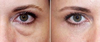
WHAT DO BAGS UNDER THE EYES TELL US?
You can be well-slept and rested, but if a person has bags under his eyes, he will still look tired and sick.
It would seem that the problem is not serious. However, bags under the eyes can signal significant health problems. Most often, swelling under the eyes is considered a sign of kidney disease, but this is not always true. There are many reasons for swelling under the eyes.
Kidney diseases
Kidney diseases are the first to be suspected, and this is no coincidence. The fact is that with serious pathologies (glomerulonephritis, severe stage of pyelonephritis, kidney tumors) protein is lost.
“With urine, the body loses a huge amount of large molecular protein compounds. That is, the kidney filter, which normally does not allow large compounds to exit with urine and retains them in the bloodstream, collapses, explains nephrologist at the Aflo-Center medical clinic network, Irina Anatolyevna Kotryakhova. “Low levels of protein in the blood cause some of the fluid to leak into the tissues.”
Renal edema usually does not occur without a large amount of protein in the urine.
Even doctors at district clinics sometimes get lost in determining whether there is “a lot or a little protein in the urine.” It happens that after medical examination the patient is urgently referred to a urologist or nephrologist to treat renal pathology. In fact, protein can be lost in small quantities for completely natural reasons. For example, if on the eve of the tests a person worked out hard in the gym. “If the daily loss of protein exceeds 3 grams per liter (this is very much) and swelling occurs, which is most pronounced in the morning, this is the kidneys. Such patients are sent to the hands of nephrologists,” says Irina Kotryakhova about the intricacies of diagnosis.
Urological patients can be seen immediately. Their face is swollen like a ball. Most often, a morning puffy look has nothing to do with kidney disease. Especially when it comes to women over 30 years old.
“As practice shows, 8 out of 10 young and middle-aged patients who complain of “bags” under their eyes are patients of plastic surgeons,” concludes Yuri Vladimirovich Goloviznin, a urologist at the Aflo-Center medical clinic network.
Heart problems
If, in addition to “bags” under the eyes, the patient is bothered by chest pain, rapid heartbeat, shortness of breath and swelling of the legs, the cause may be cardiovascular pathology. An ECG will show it first.
“In heart failure, swelling first appears in the feet and legs. If proper treatment is not carried out, they will rise higher and we will see “bags” under the eyes. But these “bags” are of a completely different kind, and not the swelling of the eyelids that we are used to seeing after sleep, explains Anton Ryabov, a cardiologist-arrhythmologist at the Aflo-Center medical clinic network. — Edema itself is not a symptom of cardiac disease. Therefore, self-diagnosis based only on “bags” under the eyes is strange, to say the least. Better yet, get some sleep. Perhaps the swelling will go away.”
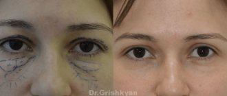
Aging
A little theory. The eyeball is separated from the orbit by adipose tissue - periorbital tissue, which is held in place by a connecting membrane - the orbital septum. With age, the volume of periorbital fiber increases (for some, more, for others, less). The septum cannot withstand the pressure, and the fatty tissue extends beyond the eye sockets. Genetic predisposition plays an important role in this case.
As Igor Popov, an ophthalmologist at the Aflo-Center medical clinic network, explains, the majority of those who observe unflattering “bags” are candidates for blepharoplasty. All kinds of creams that promise instant results will not help reduce the volume of periorbital tissue.
Liver diseases
Unlike “bags,” bruises are associated precisely with a decrease in subcutaneous fat. Due to lack of sleep or sudden weight loss, the eyeballs literally fall into their sockets. This is a common and non-threatening condition. According to Zhanna Krupina, a gastroenterologist at the Aflo-Center medical clinic network, “unnatural” brownish-yellow shades should alert you. Coupled with swelling of the hands (especially fingers), heartburn, nausea and pain in the right hypochondrium, a shadow cast under the eyes may indicate liver pathology.
Edema
There are more obvious reasons for the appearance of bags under the eyes, and, as a rule, they do not go unnoticed. As Nadezhda Shubina, an allergist-immunologist at the Aflo-Center medical clinic network, explained, allergic diseases of the conjunctiva, inflammatory diseases of the paranasal sinuses, ARVI - all conditions in most cases are manifested by edema. Not to mention chronic fatigue and a severe hangover - everything is clear here.
It is important to remember: physiological “bags” under the eyes as a result of swelling are “conspicuous” after sleep and gradually fade away by the end of the day.
Pathological growth of fatty tissue does not depend on the time of day. Such “bags” do not go away after a long rest and practically cannot be corrected at home or in cosmetologist’s offices. Much less often, swelling of the eyelids is a signal of serious pathologies. But in beauty salons it is not customary to talk about this.
Drug-induced edema
Some medications also cause fluid retention in the body; most often, patients complain of side effects from oral contraceptives.
Combined hormonal contraceptives are based on two female hormones - estrogen and progesterone. The amount of estrogen is almost always the same, the only differences are in the amount of progesterone. Each patient reacts to this proportion differently.
“Differences in progesterone levels are reflected in subjective sensations. Then we select a drug with a different composition for the patient, which reduces the accumulation of fluid in the body,” says Valentina Smirnova, a gynecologist at the Aflo-Center medical clinic network.
Allergic reactions
Allergic swelling comes with the onset of spring and is usually accompanied by itching and redness. Those who have encountered this problem will not confuse such “bags” with anything else.
Thyroid diseases
Another cause of edema, according to Tatyana Sokolova, an endocrinologist at the Aflo-Center medical clinic network, is hypothyroidism. In this case, swelling appears not only under the eyes, but also on the legs, arms, and there may be swelling of the internal organs. Problems with the thyroid gland are accompanied by a number of other symptoms, and “bags” only confirm the diagnosis.
And in conclusion
In any case, you should take care of your eye health and consult a doctor at the first alarming symptoms.
In the Aflo-Center network of medical clinics, you can undergo a comprehensive examination, carry out the necessary diagnostics and treatment of existing diseases.
Make an appointment with a doctor by phone: (8332) 497-003,
Electronic registration: online.afflow.ru
What you need to know
Yellow skin color also occurs due to carotene oversaturation. This happens if a person often eats vegetables and fruits that contain it in large quantities.
How to distinguish such yellowing from jaundice? With it, only the skin is stained, but not the mucous membranes.
It happens that the skin turns yellow due to taking medications containing acryquine or picric acid.
Further, the skin turns yellow. If you consume excessive amounts of spices, spicy foods, fast for a long time, or frequently use alcohol or drugs.
Table of contents:
Manifestation of xanthelasma
Causes of xanthelasma of the eyelids Diagnosis of the disease Methods for removing xanthelasma of the eyelids Stages of the fight and prevention of xanthelasma
Skin formations are not only a cosmetic defect, but also a signal of an existing disease. Therefore, you should not ignore their appearance. Yellowish plaques, single or multiple, appear on the upper eyelid and are called xanthelasma. Xanthomas protrude on the lower eyelid or other parts of the body. These are benign formations, soft to the touch, with a characteristic color. As a rule, degeneration into malignant does not occur. They are usually diagnosed by a visual examination by a doctor. If necessary, he prescribes additional tests and examinations so as not to confuse them with tumors that pose a threat to health.
Diagnostics
To identify jaundice and determine what kind of disease a person has, you need to carry out the following diagnostics:
- CBC, where hemoglobin is determined;
- Blood biochemistry, which shows total bilirubin and its secretions;
- Lipid profile study;
- Test for thyroid hormones;
- Testing for tumor markers;
- Test for roundworms;
- TAM, which determines the level of bilirubin and its secretions;
- Immunological analysis to detect antibodies to viral hepatitis;
- PCR research;
- Antiglobulin test (for newborns);
- Fibrogastroduodenoscopy, which shows inflammation, neoplasia and bile duct stones.
Dyspnea
Diagnostic procedures for each patient are selected individually by the doctor.
Symptoms
In the early stages, pigmentation appears in the form of small spots of a light shade. The mechanism of occurrence of the pathological area is associated with the destruction of melanocytes. The area under the eyes is susceptible to vitiligo, as it is the most sensitive area.
At the initial stage of development, the disease is observed by the appearance of small white zones. As the disease progresses, the spots unite together, which externally forms large-scale affected areas. Patients are often diagnosed with a chronic form of the disease, but exacerbations of the pathology are possible in the spring and autumn. It is advisable to protect the affected areas from exposure to ultraviolet radiation.
Symptomatically, vitiligo is accompanied by:
- changeable pigmentation under the visual apparatus;
- redness in this area;
- intensive production of sebaceous secretions;
- peeling;
- areas with thickenings;
- discoloration of hairs on the skin;
- sweating disorders.
The patient's general condition remains normal. A patient with vitiligo experiences psycho-emotional discomfort in society.
How to treat yellow spots?
Treatment of yellow spots on the eyes is individual and depends on the nature of the formation. That is why timely diagnosis is especially important. After the examination, the doctor will determine the cause of the pathology and prescribe treatment.
Pterygium and pinguecula in the initial stages are treated with drops and gels that moisturize the conjunctiva. If the disease is accompanied by eye irritation and redness, anti-inflammatory therapy is additionally prescribed. Surgery is indicated only in cases where long-term therapy has not led to the expected result. When diagnosing a nevus, the patient needs dynamic observation by an ophthalmologist.
Advanced cases of conjunctival cysts can only be treated surgically. There are two options for the operation: cauterization and laser exposure. Treatment of Horner-Trantas spots is carried out through drug therapy aimed at eliminating the underlying disease.
In our modern “Vision Restoration Center” you can undergo diagnostics using the latest equipment. Based on the results of the examination, you will receive a consultation with an ophthalmologist with more than 10 years of experience. We employ doctors with scientific degrees and personal awards.