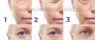
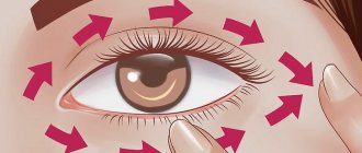
Eliminating a lower eyelid hernia will make you 5-7 years younger. Think for yourself how much more beautiful and younger you need to look!
A hernia of the lower eyelid is usually called a protruding swelling or protrusion. This cosmetic defect is very widespread. Most often it occurs in both eyes, but it can also occur in only one.
Fortunately, modern cosmetology and plastic surgery have several techniques that can effectively combat this problem.
Hernia of the eyelids - what is it?
A hernia
is a fairly common defect of the eyelids. Hernias make the look heavier, create bags under the eyes, age the face and, most importantly, can contribute to deterioration of vision.
An eyelid hernia itself is the filling of the space under the upper or lower eyelid with fatty tissue. As a result, the skin bulges, blood circulation and lymph flow are disrupted, and swelling forms around the eye. Depending on the location of formation, there are two types of hernias - lower and upper eyelid hernias.
Fat-sparing blepharoplasty with redistribution of fat bags
Fat-saving (fat-preserving) blepharoplasty is very popular among patients who want to completely maintain their usual appearance, only slightly adjusting their appearance. The innovative technique is not aimed at removing excess fat deposits, but at partially redistributing them.
Blepharoplasty with redistribution of fat bags prevents the possibility of the “sunken eyes” effect. High-quality separation of the eyelids and cheeks, elimination of tear grooves harmoniously transforms the appearance, making the appearance young, bright, and contrasting.
The essence of the fat-sparing blepharoplasty technique
The classic approach recommends that the plastic surgeon completely remove the maximum possible amount of excess fat and excess skin that spoils the appearance. Today, such operations are performed using the transconjunctival method, which does not involve external incisions on the lower eyelids.
Transconjunctival lower blepharoplasty does not violate the integrity of the anatomy (the orbital septum and orbicularis oculi muscle are not intersected), as with external percutaneous blepharoplasty.
- With transconjunctival blepharoplasty, the frequency of fat movement and rounding of the outer corner of the eye is significantly less than with external blepharoplasty.
- Understanding the anatomy of the lower eyelid and proper incision placement (4 to 5 mm below the eyelid cartilage) is critical to achieving successful results.
- Fat can be removed or retained (transferred) depending on the patient's wishes.
- Fat repositioning (moving) is an advanced technique performed with a detailed knowledge of the anatomy of the eyelids.
- To obtain excellent results, it is necessary to know and practice various surgical nuances.
- Adding various procedures (canthoplasty, canthopexy, fat transfer, skin removal, etc.) as needed can improve the final results.
- The patient receives excellent results when the surgical technique is mastered and refined.
Lower eyelid rejuvenation with fat redistribution is a complex aesthetic procedure. One of the key aspects of lower fat-sparing blepharoplasty is the surgical approach through which orbital fat hernias are exposed. There are two standard approaches to the fat pads of the lower eyelid: percutaneous (through the skin and muscles) and transconjunctivally (through the inside of the eyelid).
Percutaneous lower blepharoplasty is a more traditional method in which the skin, orbicularis oculi muscle and orbital septum are divided. This approach carries a higher complication rate, including lower eyelid retraction, ectropion, and lower eyelid rounding. This is primarily due to lack of eyelid support, excessive skin removal, and disruption of the orbital septum (with subsequent scarring) and orbicularis oculi muscle (with subsequent muscle weakness) when accessing fatty hernias.
With transconjunctival access to the orbital fat, an incision is made on the inner (conjunctival) surface of the eyelid, thereby maintaining the integrity of the orbital septum and the orbicularis oculi muscle. This is a very important step in preventing malposition of the lower eyelids, since there is no distortion of the anatomy of the eyelid and postoperative gross scarring.
For this reason, transconjunctival lower blepharoplasty with fat redistribution has become widespread in aesthetic surgery.
Transconjunctival blepharoplasty does not remove excess skin. This requires additional procedures. However, if necessary, excess skin can be removed using pinch blepharoplasty.
Adding these additional procedures to transconjunctival blepharoplasty does not result in the same rate of eyelid malposition complications seen with percutaneous lower blepharoplasty.
The advent of volumetric fat preservation through fat redistribution or grafting has become an important adjunct to lower eyelid blepharoplasty and can be used to improve cosmetic results.
Anatomy of the lower eyelid
The incision for transconjunctival lower blepharoplasty is made from inside the eyelid. This can make anatomical orientation and surgical maneuvering difficult because the operative field is small and narrow compared to an open percutaneous approach. For this reason, performing this operation requires the surgeon to have a thorough knowledge of the anatomy of the lower eyelid.
The lower eyelid is divided into three layers or plates (lamellae).
- AL - anterior lamella (skin/muscle)
- PL - posterior lamella (cartilage/conjunctiva and lower eyelid retractors)
- ML - middle lamella (orbital septum) and fatty hernias.
- FP point of fusion of the lower eyelid retractors and the orbital septum,
- FP is critical for incision placement during surgery
Cartilage forms the skeleton of the eyelid, giving it both support and flexibility. Its dimensions are 4-5 mm in height, 30 mm in length and 1 mm in thickness. It is connected to the conjunctiva on its inner surface and to the orbicularis oculi muscle and skin on the outer surface.
Below the cartilage, the conjunctiva continues to the lower fornix. The lower eyelid retractors, also called capsulopalpebral fascia, are located just anterior to and closely connected to the conjunctiva. This sympathetically controlled muscle (similar to the Müller muscle of the upper eyelid) originates from the tendon of the inferior rectus muscle and continues to fuse with the inferior border of the cartilage. There is fatty tissue just in front of the retractors.
In front of the adipose tissue is the orbital septum, which limits them anteriorly. This is a layer of connective tissue originating from the periosteum on the orbital rim (arcus marginalis) and inserting into or connecting with the capsulopalpebral fascia 2-3 mm below the lower edge of the eyelid cartilage.
A conjunctival incision just below the cartilage will separate the conjunctiva and lower eyelid retractors from the cartilage.
This plane is best suited for surgical intervention needed in the fascial plane between the orbicularis oculi muscle and the orbital septum, such as accessing the orbital floor to repair an orbital fracture. Because the dissection in this plane occurs anterior to the septum, fat does not protrude into the surgical field.
A transconjunctival incision below the junction of the orbital septum and lower eyelid retractors (4 to 5 mm below the cartilage) is the preferred approach for transconjunctival blepharoplasty as it is a direct route to fatty hernias.
There are three fatty hernias on the lower eyelid: nasal, central and lateral. Nasal and central fat hernias are separated by the inferior oblique muscle
Care must be taken not to damage this muscle during surgery.
The central fatty hernia is separated from the lateral inferior arcuate ligament.
The separation of the central and temporal (lateral) fatty hernias by the arcuate ligament can often be seen clinically by asking the patient to look upward. But at the same time, the separation between the nasal and central fat pads is not pronounced, since hernias tend to overlap the inferior oblique muscle and dull this separation (this is visible during surgery).
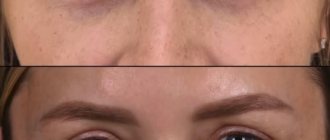
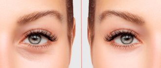
Fat-sparing blepharoplasty of the lower eyelids is based on the rational correction of fatty hernias. They are not removed, but evenly distributed. This allows you to maintain the required volume, getting rid of unsightly “bags under gases”.
Comparative analysis of traditional and fat-saving blepharoplasty
Long gone are the methods in which plastic surgeons used simple skin tension to eliminate aging changes. This technology made the face lifeless, depriving it of facial expressions. Today, the face is perceived as a 3D relief object that requires more subtle surface treatment. It is necessary not only to get rid of the furrows and wrinkles that appear with aging, but also to preserve the usual relief characteristic of a young person. Redistribution blepharoplasty corrects volume, making the eyes look youthful.
With the old traditional approach, the eyelids were completely devoid of fat deposits. The consequence was a tight fit of the tissue to the orbit. Outwardly, the eye looked like it had sunk inward, which revealed the work of a surgeon. It is enough to look at photos of famous artists, athletes or politicians from past years to notice which of them used traditional blepharoplasty. Today, only specialists can determine the work of a plastic surgeon.
The main disadvantage of the traditional method and significant advantages of the new technique
The work of a plastic surgeon may be completely satisfactory for the patient, but after the operation the eyes will look the same as those of other people. Individuality is completely lost, zest is lost. Today, such work is considered unsatisfactory, sometimes requiring rework.
Volume adds charm to the look, making the look unique, beautiful, and bright. This is possible due to the high quality of work of a plastic specialist who uses new methods.
Fat-preserving blepharoplasty makes the eyelid voluminous, preserving the natural beauty of the eyes, its individual attractiveness, completely eliminating age-related skin changes. The periorbital area gains a second youth, without looking like it has been operated on, which is important for public people. The maximum, long-term effect is achieved to maintain beauty, youth, and health.
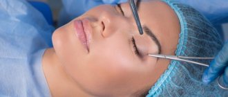
Surgical technique of fat-sparing blepharoplasty
The eyelid is infiltrated transconjunctivally with 2 ml of 1% xylocaine with adrenaline 1: 100: 000. In order to perform fat transfer, the tear trough (the area into which the fat will be transferred) is marked before the operation.
1-2 ml of the same local anesthetic is injected into the area of the tear trough. Usually the nasal and central hernias move.
After the time required for hemostasis and anesthesia, the lower eyelid is moved with the index finger of the non-dominant hand, while the second finger of the same hand moves the eyeball slightly back. This maneuver elevates and exposes (inflates forward) the incision site on the transconjunctival surface of the eyelid.
The free dominant hand is used to make an incision 5 mm below the cartilage to directly enter the fat compartment of the lower eyelid.
An electric knife (Valleylab) with a Colorado tip or surgical scissors can be used for this incision.
The orbital septum is attached to the capsulopalpebral fascia 2–3 mm below the cartilage. An incision below this insertion point enters the retroseptal space, provides direct access to fatty hernias and preserves the integrity of the orbital septum. This is a critically important point, since it allows you to leave the orbital septum intact and avoid severe scarring.
The conjunctiva is then sutured with a 4-0 suture, which is pulled upward and protects the eyeball during surgery.
Next, all three packages of fatty hernias are highlighted
To eliminate the tear trough, the nasal and median hernia are moved to its projection, after placing separate sutures on them, which are brought out to the skin and a knot is tied on the roller.
Postoperative care and rehabilitation
After surgery, patients are given the following recommendations. Necessary
- Apply ice compresses ten minutes per hour while awake for 2 days.
- Instill eye drops containing an antibiotic and a steroid (Tobradex) three times daily for the first week after surgery
- Sleep with your head elevated (on two pillows) for the first week.
- Avoid strenuous activities for the first 10 days after surgery.
In the postoperative period, bruising and swelling usually resolve quickly. Watery eyes usually disappear within the first week. The stitches are removed on the fourth day.
By giving priority to the new technique, the patient receives an excellent result that meets international standards.
- After the operation, the patient returns to his usual lifestyle in 3-6 days.
- You can fully evaluate the result of a plastic surgeon’s work in 1-2 months.
- Strict adherence to the specialist’s recommendations during the rehabilitation period will speed up the recovery process.
- The shape and appearance of the periorbital area will remain for 5-7 years.
Complications
Most surgical complications associated with fat removal or transfer are similar. These typically include excessive bruising and swelling, chemosis, and subconjunctival hemorrhage.
- Edema. To reduce swelling, in the absence of medical contraindications, a higher dose of prednisone may be prescribed. In cases of prolonged or refractory edema, when the patient violates the diet and consumes excessive salt, a change in diet is required.
- Chemosis is subconjunctival swelling that can occur when tissue is excessively cauterized during surgery. There are different treatments for chemosis depending on the severity.
- Subconjunctival hemorrhage - bleeding under the conjunctiva, can be very distressing for the patient, which usually reaches its maximum extent a few days after surgery and resolves within 1-2 weeks.
- Excessive fat removal. Dangerous and may cause sunken eyes.
- Incorrect position of the eyelids. Not typical for transconjunctival blepharoplasty.
- Entropion. May occur with a transconjunctival incision. Usually it is spastic, cicatricial. Correction depends on the etiology. Postoperative ectropion occurs rarely in cases of undiagnosed preoperative decrease in eyelid tone.
- Finally, in rare cases, postoperative trichasis may develop. Most likely, it is associated with impaired blood supply to the eyelids and/or post-operative scarring.
When fat transfer is performed, complications unique to this procedure may occur. These include diplopia (in the postoperative period), fatty granulomas, prolonged swelling, unevenness in the projection of the tear trough, pigmentation in the area of the suture of the roller.
- Diplopia is usually temporary and is associated with anesthetic injection, swelling, or injury to the inferior oblique muscle. Treated with higher doses of oral steroids, which should be tapered over 10 days.
- Prolonged swelling is treated similarly to diplopia with higher doses of steroids.
- Irregularities in the projection of the tear trough are usually caused by granulomas and are resolved with steroid injections (Kenalog).
- Temporary hyperpigmentation may occur at the site where the skin prolene suture exits.
New plastic surgery techniques make it possible to quickly and radically change one’s appearance, eliminating congenital or acquired defects. The operation takes little time, but the results will last for years. To learn more about the work and make an appointment, call 89673108595 or fill out an application on the website.
Hernia of the eyelids: causes
The skin of the eyelids is practically devoid of subcutaneous tissue and is therefore prone to stretching. The orbicularis oculi muscle and the skin of the eyelids weaken early and lose their elasticity, mainly due to repeated contractions during blinking.
The main reasons for the formation of an eyelid hernia are age-related changes, heredity and constitutional features. The formation of hernias is also accelerated by stress, lack of sleep, hormonal changes, constant eye fatigue, ultraviolet radiation, smoking and alcohol consumption.
Eyelid hernia: symptoms
A characteristic feature of a hernia of the upper eyelid is the overhang of the swollen area over the eye, and the lower eyelid has “bags” under the eyes.
At first, it is difficult to notice the formation of a defect: the functions of the eye are not impaired, and cosmetic changes at the stage of hernia growth are not so noticeable. However, over time, the formations become more distinct and, first of all, disturb the proportions, age the face and change the expression of the gaze. Sometimes a hernia can cause excessive tearing and cause blurred vision.
A hernia of the lower eyelid makes itself felt by “bags” and swelling under the eyes. But these external changes can also be a physiological “bagginess” of the cheekbones, which requires different treatment.
A hernia of the lower eyelid can be detected if you close your eyes and lightly press your fingers on the eyeball: with a hernia, the formation under the eyelid will increase.
It is quite difficult to distinguish fatty hernias from swelling around the eyes. Edema may be associated with diseases of the kidneys, endocrine or cardiovascular system. Therefore, to determine an eyelid hernia and exclude possible diseases, it is better to consult a professional ophthalmologist.
Contraindications
The following factors may be contraindications to the plastic procedure:
- acute diseases, chronic diseases in the acute stage;
- oncological diseases;
- diabetes;
- problems with blood clotting, anemia;
- arterial hypertension, increased intracranial pressure;
- myopathy;
- recovery after any eye surgery;
- blepharitis, keratitis;
- infectious lesions on the cornea;
- for women - menstrual period, pregnancy.
Treatment methods for eyelid hernia
An ophthalmologist must diagnose a hernia of the eyelid. When this disease is detected, we most often recommend surgery to the patient.
Although hernias themselves do not pose a health hazard and can only in exceptional cases contribute to vision impairment, they are an unpleasant cosmetic problem. Surgery to eliminate an eyelid hernia eliminates this pathology and helps correct age-related skin changes.
Surgical treatment of eyelid hernia is carried out in two main ways:
- Percutaneous approach
- when the surgeon makes an incision along the outer skin surface of the eyelid, excising excess skin and fatty tissue. Small wounds after this operation heal quickly and remain invisible - Transconjunctival access
- excess fat is removed through the conjunctiva, the inner surface of the eyelid. With this method, no external traces are left.
Removal of an eyelid hernia is painless for the patient on an outpatient basis. If you follow all the doctor’s recommendations before and after surgery, the success of the treatment is guaranteed, and the problem will be eliminated forever.
At Dr. Belikova's Eye Clinic, you can undergo a comprehensive vision examination and receive detailed recommendations from our specialists.