There are a huge number of types of moles. According to the World Health Organization (WHO) classification there are several hundred of them. How can an ordinary person who has nothing to do with medicine figure out how dangerous his mole is? At least roughly? There are several signs that we will talk about today.
I would like to point out right away that this article is not intended to help you diagnose yourself. Its main purpose is to reduce any tension you may have that has arisen after independently studying information about melanoma on the Internet.
What to do with moles?
If there are no clear signs of a malignant process, then you can go in 2 ways:
The first way is observation. In this case, we take a picture, a photo of the mole, warn the person about the signs in case the size, shape, symmetry changes, and schedule a return visit in 3-6 months, depending on how much we fear for this formation.
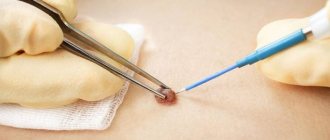
The second way is removal of the formation with histological examination. We even resort to this path more often. There is a common belief that a mole should not be touched. In fact, there is no danger in removal. Moreover, this is even a preferable option, since in this case a suspicious formation can be sent for histological examination. So, we excise the formation, sometimes if it is a suspicious mole, then not with a laser, but with a scalpel, with minimal grip (1-2 mm is enough) and send the material for histological examination.
If we receive the result that it is a benign formation, then we have removed it as benign and no further action is required.
Good signs
A pedunculated mole, the thickness of which is no more than 2-3 mm
A distinctive feature of malignant tumors is the unlimited division of their cells. This property does not allow such neoplasms to have a “leg”. Unfortunately, this sign cannot guarantee that the formation is benign. In the literature, about once every 10 years there are indications of pedunculated melanoma.
Long-term existence of a mole without changes
If a mole exists from birth or 5, 10, 20, 30 or more years and at the same time! nothing! has not changed - with a high probability, everything is fine with her. This sign is not unconditional. It must be remembered that a long-existing mole is not immune from turning into melanoma, but its chances are, in any case, less.
Presence of hair on the surface of the mole
In fact, hair does not grow from a mole, but grows through it. Hair roots (hair follicles) are usually located deeper. If hair grows from a mole, this means that the cells of the mole still retain their functions. They can form “channels” for hair growth and “pass” it through the mole. This sign also does not 100% “insure” that the mole is actually melanoma. Fortunately, in my practice there was only one exception to this rule.
The skin pattern on the surface of the mole is preserved
This sign of benignity has the same roots as the previous one. It means that the mole is covered by normal skin cells, not tumor cells. Unfortunately, like all previous signs, it is NOT always reliable.
Flesh-colored mole
Most melanomas are brown or black. Therefore, if the color of a mole is flesh-colored, it is highly likely to be benign. Answering a possible question about the non-pigmented form of melanoma, I will say that it is! Disappearing! a small part of the total number of this type of tumor.
Soft consistency
If a mole is softer than the surrounding skin, it is most likely benign. This is due to properties common to all malignant tumors. If cells divide uncontrollably, in most cases they form a compaction that stands out against the background of the surrounding tissues.
Symmetry along at least one axis, smooth edge.
More than enough has already been said about these signs; I will not repeat them.
Melanoma and dysplasia
If, according to histology, we receive a result - severe dysplasia or early melanoma, then this can also be regarded as a positive result, since the disease was detected in the initial stage. In this case, we refer the patient to the oncology clinic, where he undergoes further treatment.
Early (thin) melanoma is very treatable, with a good survival prognosis. In contrast to melanomas, which are left untouched, which grow and patients treat them at stages 3-4, when effective treatment is no longer possible.
Briefly about the main thing:
You should not try to make a diagnosis yourself; this should still be done by an oncologist. On the other hand, it’s also not worth making a mountain out of nothing out of the blue.
If your mole does not have a single “bad” sign, but has several good ones, you should still see an oncologist. Calmly, without haste and fuss, as time permits.
The main point of this article is not for you to be able to understand for yourself whether a mole is malignant or not. My goal is to help you not go crazy because of the horror stories you have previously read on the Internet.
If doubts still overcome you, you can ask me any question in the format of an online consultation.
Self-examination
It is useful for people of any age to regularly examine their skin for new growths. Of course, it is not necessary to spend a lot of time and run to doctors with every mole that you find. But there are some symptoms that will help you pay attention to changes occurring in the tissues when examining moles. A mole that is not yet going to turn into melanoma:
- symmetrical;
- has clear outlines;
- evenly colored;
- retains its size, does not increase or decrease;
- It does not itch and does not cause any discomfort.
Accordingly, if the nevus begins to change somehow (be it a blurring of the border or a change in color), itches, hurts or causes any other unpleasant sensations, then it’s time to consult a doctor - this is a mole that needs to be observed professionally. Remember that a healthy organ does not remind you of itself.
Where does this myth come from?
The Internet is full of stories like “he removed a mole and died.” It is enough to type this query in Yandex or Google, and you will find a sad story almost immediately. There are hundreds, if not thousands of them. In one of my articles, I cited possible misjudgments of people in such situations, as well as examples of reliable and dubious sources of information. Among the incorrect speculations, generalizations and hyperboles, there are only 2 real scenarios when removing a mole can actually lead to death.
“I removed a mole and died”: type 1 – removal of moles without histology
The experience of online consultations that I have suggests that in our country 50% (or more) of skin lesions are removed without histology. Often it is oncologists (!) who say phrases like these:
“Everything will be fine, it’s definitely benign...”;
“We can’t do histology here – it’s too small...”;
“I’ll just take a scraping before removing it – that’s enough”;
“I have 100,500 years of experience - do you think I won’t see melanoma?!”;
“I’ll do a dermatoscopy - histology is not needed here”;
“You don’t need to overpay for histology - everything is fine here.”
Meanwhile, according to research, the accuracy of diagnosing melanoma with a simple visual examination is about 60% (!!!) [1]. With dermatoscopy and cytological examination of scrapings - no more than 95%. [2] And only histology approaches 100%. [2]
And then everything is simple. If melanoma is removed without adequately capturing healthy tissue, a recurrence of this tumor develops. If non-radically removed melanoma recurs, the patient’s chances of surviving for 5 years are halved [3].
The moral here is simple - remove moles only with histology. Discuss this with your doctor in advance and do not give in to any persuasion such as those indicated above.
Only histology, only hardcore 
“I removed a mole and died”: type 2 – mistakes made by a pathologist
If you turn again to the domestic Internet and type in the search the phrase “melanoma histology errors”, you get the impression that no one here makes this diagnosis correctly. Mistakes, criminal negligence, killer doctors, ignoramuses, etc.
I can’t agree with this, because to draw conclusions about the situation in the country based on 10–20 mistakes made by morphologists, in my opinion, means judging the whole based on a very small part (approximately 0.0025% of the total number of diagnoses per year - 8,000).
Morality. How to remove a mole and not die as a result of a pathologist’s mistake? The answer is simple - histology should be carried out in a large laboratory, which has: firstly, morphologists specializing in melanoma, and secondly, the possibility of consultations with colleagues in doubtful cases. If you have the slightest doubt about the correctness of the diagnosis, take the samples of the mole and have it reviewed in one, or better yet, two other laboratories.
So we figured out that the myth “there are moles that cannot be removed”, the legs grow from the myth “he removed a mole and died.” And we were convinced that melanoma could NOT develop after removal of a nevus, the benignity of which was confirmed histologically. Now, taking this data into account, let’s try to finally answer the question posed in the title of the article.
Is it time to sound the alarm?
Moles, or, in scientific terms, nevi, are present on the body of every person. It is believed that the bulk of them, up to 90%, appear by the age of 25. But they can also arise later - under the influence of various events. For example, a typical phenomenon is when they literally break out during pregnancy. Sometimes moles, on the contrary, disappear over time. They can be yellow, brown, black. This is all within normal limits and there is nothing to worry about.
But it happens that a mole begins to grow unevenly or changes color, its surface becomes “polished” or it begins to bleed - that is, changes occur in it. Another option is the appearance of a new mole of an unusual appearance. It is precisely such incomprehensible neoplasms that require close attention.
Diagnostics
Dermatologists and oncologists are involved in determining the type of nevus.
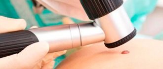
Using a dermatoscope, the doctor examines the formation and determines its nature (benign or malignant). Sometimes a histological examination (scraping method) is required.
Biopsy (tissue sampling) for nevi is not used due to their trauma during this procedure. And as you know, it’s better not to touch moles again!
Unusual moles – when to see a doctor?
If birthmarks on the skin cause concern, you should definitely consult a dermatologist or oncologist.
Urgent consultation is necessary if there are symptoms such as:
- visible swelling or redness around the birthmark;
- itching of the skin around the mole;
- bleeding in the area of the mole;
- noticeable molar asymmetry and/or irregular coloration.
Melanoma
Pigmented epithelioid cell melanoma without ulceration
During your visit, your doctor will carefully examine the birthmark and evaluate whether further testing is necessary. One element of this process is dermatoscopy - a study using a dermatoscope (surface microscope), which allows you to accurately examine the border of the birthmark and the upper layers of the dermis.
A dermatologist, having noticed alarming symptoms, will prescribe surgical removal of moles - this is a simple procedure that is performed under local anesthesia. The whole process takes no more than 30 minutes. The suspicious section is then sent for histopathological examination. If there is a reasonable suspicion that a skin cancer has developed, the patient is immediately sent to the oncology center for further examination.
Mole removal is carried out using different methods - laser, radio wave or using a scalpel.
- The laser is used to eliminate cosmetic imperfections when there is no doubt about the benign quality of the birthmark. It “evaporates” the stained cells, leaving virtually no traces.
- A radioknife and surgical scalpel are the option of choice in cases where it is necessary to preserve cut tissue for analysis.
Rehabilitation period
After removal of papilloma, lipoma, atheroma, hemangioma and other neoplasm, the recovery period begins. At this stage, you must adhere to the following recommendations:
- avoid contact of the wound with tap water (the area around the removed nevus is cleaned in the usual way);
- protect the problem area from direct sunlight - with the doctor’s permission, you can use products with an SPF filter;
- treat the wound with medications prescribed by the surgeon - medications with antibacterial, regenerating and softening properties are used;
- Avoid injuring the area with the removed mole.
Plastic surgeon Pavel Ivanov, a specialist in cellular technologies, will excise the nevus effectively and painlessly. Sign up for a consultation now.
Classification and photo
1. A flat nevus or birthmark is a pigmented island of skin with clearly defined boundaries. It can take the form of lentigo - multiple brown or brownish formations in the upper layers of the epidermis.
2. Convex nevus or mole. It has a diameter of up to a centimeter, a smooth or lumpy surface and rises above the level of the skin. Its color varies from beige to black, and a hair is usually located in the center of such a formation.
3. Blue nevus or blue mole. It looks like a smooth hemisphere, slightly raised above the skin, sometimes reaching a size of 2 cm. The color of this benign formation ranges from blue to dark blue.
4. Giant nevus. It is a large spot on the body, gray, bright beige (sometimes brick), black or brown.
Dangerous nevi that must be removed
Moles, as a rule, do not pose a danger; in most cases they are harmless. In rare cases, they degenerate into a malignant formation, which is a pathology and must be removed and treated.
Normally, moles are brown in color, have smooth edges, are small in size, do not grow, and do not change over time. Some nevi are suspicious and require constant monitoring and observation. These include:
• Moles with a diameter of more than 1 cm.
• Nevi acquired in adulthood.
• Moles that change color, shape, size over time.
If the formation is suspicious, you cannot do without consulting a specialist. It is imperative to seek help from professionals if you notice some changes in the mole:
• Formation of nodules, blisters or ulcers on the surface of the nevus.
• Change in the structure of the mole. For example, it becomes hard, dense or soft.
• Inflammatory processes that occur in the mole or the tissues around it.
• The appearance of unpleasant sensations at the site of formation - pain, itching, burning.
• Changes in the size and shape of the mole.
• The appearance of bleeding at the site of formation, dry crusts or gloss on its surface.
• Changes in pigmentation, nevus color, surface pattern.
There are cases when moles need to be removed for cosmetic reasons. Most often, this procedure affects nevi on the face or areas of the skin that are not hidden by clothing. In addition, it is recommended to remove moles if they are constantly injured due to their location (for example, by a chain, bodice harnesses).
What methods of mole removal are there?
There are several ways to remove moles. Here are some of them:
Surgical method.
It is the most common. It is preferable when removing deep and large skin diseases, and is also used in case of oncology. The disadvantage of this method is the scars that remain at the surgical site. If the mole is located on the face, this method is undesirable.
Cryodestruction method (using liquid nitrogen).
The mole is removed using cold. The advantage of this method is that the tissue destroyed by the cold is not removed, but remains in place, forming a dry crust. This protects the wound from infections and does not require treatment. Healthy tissue forms under the crust. The disadvantage of this method is that due to the inability to determine the exact area of action of nitrogen, healthy cells may suffer. There is also a possibility of incomplete elimination of the tumor. In this case, a repeat procedure is required. This method of nevus removal is used extremely rarely.
Electrocoagulation method.
This method involves exposing the desired area to a high-frequency current, resulting in thermal damage to the neoplasm. The advantage of this method is the ability to send the tumor for examination in order to determine its composition and the absence or presence of pathological cells (biopsy). However, after this exposure, small scars remain on the skin.
Laser.
This method is characterized by a small diameter of the treated surface and a certain depth of exposure of the laser beam to the desired location, as a result of which the surrounding tissue is almost not damaged. When small moles are removed, no traces are left, but when large moles are removed, a colorless spot may appear in their place.
Radiosurgery.
The most effective method for removing skin lesions at the moment. The procedure is carried out using a radiocoagulator, which allows almost no damage to healthy tissue and can be used to remove both benign and malignant formations. Due to the fact that this device simultaneously cuts, stops bleeding and disinfects the site of exposure, there are practically no traces left after the procedure. After surgery, a crust remains at the site of exposure, which prevents infection from entering the wound and the formation of scar tissue. Also, the removed mole can then be sent for a biopsy.
Remember that only a specialist can determine what type of treatment you need to choose and what type of mole you have. You can get the necessary advice on removing tumors, improving the condition of the skin, correcting wrinkles and other issues in the field of dermatology at the medical office.
| before the procedure | after a course of procedures |
Author: Lyudmila Shamkolovich
Where to run?
If you notice an unusual mole on your body, be sure to go to a dermato-oncologist. If this doctor is not available, you can consult a dermatologist, surgeon or oncologist. An experienced specialist can visually determine the nature of the nevus. A special device called a dermatoscope helps him with this. Essentially, this is a powerful magnifying glass; by examining a mole through it, the doctor can notice the smallest details that are simply impossible to see with the naked eye. If there is doubt about the diagnosis, the doctor will conduct a histological examination to determine the characteristic signs of benign, premalignant and malignant neoplasms.
By the way, if you want to remove a mole for aesthetic reasons, then the decision should also be made by a dermato-oncologist after dermatoscopy, which allows you to choose the optimal method, determine the boundaries and depth of removal.
Even if degeneration into melanoma has not occurred, for medical reasons those moles that are subject to constant friction, pressure, and injury are removed. And also those that are in the groin area and under the arms, under the chest, on the belt, and in men - on the face at the shaving site.
If a mole that has degenerated into melanoma is removed at an early stage, the probability of complete recovery reaches 95%; if time is lost, it is only 20%.
Why do they appear?
Nevi are essentially benign skin formations.
Some people get scared when they see many moles on their body. What does this mean for a specialist? Only that the patient’s body is prone to accumulation (accumulation) of melanin in the surface layers of the epidermis.
The reasons for the appearance of numerous or single nevi are varied.:
1. External:
- exposure to ultraviolet radiation, one of the most common factors in the formation of moles due to increased melanin levels during tanning;
- traumatic damage to the epidermis, systematic violations of the integrity of the skin contribute to the appearance of pathological changes in it;
- exposure to radiation, which rapidly changes normal skin cells;
- consumption of harmful products (GMOs, fast food, alcohol) and smoking, these habits negatively affect metabolic processes in the body.
2. Internal:
- endocrine disorders and diseases, any changes in hormonal levels can cause the appearance of skin pathologies, pigmentation, moles;
- hereditary predisposition, the presence of various nevi in the family.
The appearance of moles can be triggered by inflammatory and autoimmune skin diseases, toxic lesions, burns and frostbite, as well as the constant use of low-quality cosmetics and preparations for face and body care.
When the sun is the enemy
Probably everyone already knows that a chocolate tan has little to do with health. Ultraviolet radiation causes DNA damage and is the main cause of malignant skin tumors. The more time a person spends exposing his body to the rays, the higher the likelihood of unpleasant consequences. The activity of the sun is also important. It is no coincidence that dermatologists strongly advise not to be on the beach between 11 and 16 hours of the day, when it is most aggressive.
By the way, many scientists associate the risk of developing melanoma with the fact that in childhood and adolescence a person often received sunburn. They cause changes in melanocytes, the pigment cells of the skin, which over time lead to malignant conditions. This also explains the fact that in almost 50% of cases, melanoma develops on the legs - they are always open in children in the summer and are most exposed to solar radiation.
Lately, there has been more and more talk about the dangers of “artificial sun” – solariums. According to some reports, one session here (which is usually 5-10 minutes) is equal to a whole day on the beach! Some European countries (Germany, France and Austria) and the state of California (USA) have even banned the use of solariums for children under 18 years of age.
Of course, the sun is a source of vitamin D and a remedy for depression, but in large quantities it is the real enemy. Otherwise, the World Health Organization would not recognize ultraviolet radiation as a carcinogen on a par with arsenic, smoking and asbestos.
What does the appearance of red moles on the body mean?
If red moles appear on the body, this may be a simple cosmetic defect, or it may indicate pathological processes occurring in the body. In some cases, the occurrence of angiomas indicates poor nutrition, prolonged exposure to the sun, and a sludge buildup in the body.
Red dots like moles may indicate increased estrogen levels in the body, liver dysfunction and increased insulin in the blood. In addition, in some cases, angiomas appear as a response of the body to a lack of iodine, magnesium, chromium, vitamins C and K.