The nose (Latin: nasus) is the entrance to the respiratory tract. It has the shape of a triangular pyramid. The highest point closest to the head is the root of the nose, from which the nasal dorsum extends to the tip. This is the least studied organ. Scientists have discovered that when a person loses their sense of smell, they lose some taste buds, and in some cases, taste disappears completely. People who have lost their sense of smell are more susceptible to depression than others.
The organ is divided into the external nose and sinuses. Functionally and anatomically, the nose is connected to the system of paranasal cavities. The external nose is a triangular pyramid formed by a bone and cartilaginous skeleton. The skin of the nose is covered with sebaceous glands. In the bony part the skin is mobile, and in the cartilaginous part it is firmly connected to the cartilage. The functioning of the surrounding muscles is similar to the sphincters.
What is epicanthus?
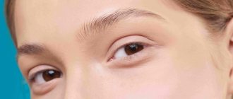
It is worth noting! The epicanthus is a vertical skin formation, which is also called the “Mongolian fold”.
This piece of skin covers the lacrimal tubercle and visually makes the palpebral fissure narrower.
The fold runs from the bridge of the nose to the upper eyelid and bends in the shape of a crescent. This element is typical for representatives of Asian peoples , residents of the Far North, Tatars, Kazakhs and Yakuts.
Epicanthus in anthropology
Anthropologists who study the origins of the human species give two versions of the appearance of epicanthus.
According to the first of them, this fold protects the lacrimal canal from hypothermia, dust , harmful organisms and from exposure to smoke and other external factors.
This theory is not supported by the fact that epicanthus may be present in peoples who live in conditions where there are no such natural external stimuli.
Thus, the theory about the physiological origin of this fold, which appears in people with a wide bridge of the nose, .
In such cases, tension occurs on the skin in this area, forming an epicanthus.
It should be noted! In any case, the fold disappears with age in most people. Usually, even among Mongoloids, this formation does not persist until old age.
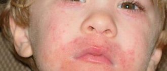
If epicanthus occurs in representatives of the Caucasian race, this can be considered normal up to 6-7 years .
The manifestation of such a fold at an older age almost always has a pathological etiology.
What is nasion? What it is, its meaning and influence on the length of the nose. This chapter is devoted to this issue.Nasion (nasion, medical term) is the point of intersection of the nasofrontal suture with the sagittal plane. Simply put, the nasion is the bridge of the nose. The woman shown in the photo above had a significantly protruding nose bridge before surgery. The shape of her nose was corrected, the nasolabial angle was increased and the bridge of her nose was moved down.
In the photograph of the skull, the nasion area is outlined with a red line. In the right photo, a red arrow points to this place.
The suture between the frontal and nasal bones is indicated by a blue line. The right nasal bone is colored green.
Often the bridge of the nose protrudes significantly forward. The profile line is highlighted in the upper right diagram. The bottom two diagrams show digital photo editing. On the left, the removal of the hump and the reduction of the tip of the nose are modeled. On the right, the shape of the bridge of the nose has been additionally corrected.
In the left image, the bridge of the nose is almost an extension of the line of the forehead. It is difficult to determine where the forehead ends and the nose begins.
On the right, where the shape of the bridge of the nose has been corrected, the nose looks quite natural.
The line of the forehead of the girl shown in the photograph goes forward to the most protruding part at the level of the eyebrows (indicated by a black arrow). The profile then rushes back (indicated by the red arrow). Below the level of the eyelashes, the profile line again rushes forward along the bridge of the nose.
The place indicated by the red arrow is the nasion. In the lower right photo, the red arrow also points to the bridge of the nose. The blue arrow points to the most prominent part of the eyebrows.
If you put the patient on his back and place a ball on the area between the eyebrows and nose, then after some time it will stabilize at the lowest point. This point is the nasion.
In the woman on the left, the nasion is almost not pronounced, and the forehead smoothly merges into the nose. But besides the fact that nasion visually increases the length of the nose, it is a reference point in the perception of nose length.
Here is a photo from the section on raising the tip of the nose. It defined the concept of a point identifying the tip of the nose. After all, it is this point that allows you to estimate the length of the nose.
To determine the length of the nose, two points must be determined. This is the tip of the nose and the deepest part of the nasion. In the diagram they are indicated by red and blue arrows, respectively.
When assessing the length of the nose, the gaze goes from the nasion down to the tip of the nose. The length of the path between these two points is indicated by a blue arrow. In the photo, the length of the girl’s nose is quite normal.
The length of the woman's nose in the top photo is estimated from the level of her eyebrows. This is due to the unclear bridge of the nose.
On the left diagram is a picture before the operation. On the right side, computer correction of the nasal profile was performed. On it, the nasion is deepened and moved to the level of the eyelashes.
As a result, the length of the nose was significantly reduced. There was practically a visual change in the length of the nose without an actual change in its size. Further, as a result of surgical enlargement of the nasolabial angle and elevation of the tip of the nose, the nose will look even shorter and more attractive.
In front of you is a piece of nasion cut from the nose. On the left is the outer surface of the bone, which you can feel with your finger by touching the bridge of your nose. On the right is the inner surface of the same bone.
These photographs show the same piece of bone in comparison with the skull. The suture connecting the nasal bones together is clearly visible. In the diagram it is red.
Here again is a photograph of the patient before and after surgery. This patient had the tip of her nose shortened and raised, the bridge of her nose slightly deepened, and the hump removed. The diagram below shows the length of the nose from the tip to the deepest point of the nasion.
Deepening the bridge of the nose or removing part of the nasion is the third method, after raising the base of the nose and increasing the nasolabial angle (raising the tip), to reduce the length of the nose.
In front of you are instruments called osteotomes. The osteotome, which is used to remove bone in a nasion, is highlighted in the photo with a red rectangle. The working part of the tool is bent at an angle of 20 degrees.
The lowest chisel is used to remove the nasal hump. The top two are for narrowing the nasal bones.
Here is an osteotome in action when cutting bone in a nasion. The projection inside the nose is shown in red. The blue line shows the location of the osteotome if it were not curved. Thanks to this, it becomes clear that the skin of the nose would not be able to stretch to this level, therefore, it is possible to work in this area only with a curved osteotome.
The photo above shows bone being cut with a chisel. Usually it looks like thin shavings. Just as when removing a hump, it is necessary to cut off the bone in parts and little by little.
In cases where the nasion is too large, it is customary to remove a fairly large amount of bone at once. The pictures show two huge parts of the patient's nasal root.
But at the same time, a fairly large part of the nasal root can be removed through multiple passes of the osteotome. The photo above shows the bone removed in sections.
These pieces of bone are also cut from the nasion of one nose.
The next chapter is devoted to the varieties of sizes and shapes of cartilage at the tips of the nose.
Features of Russians and Europeans
Epicanthus is not a characteristic feature of residents of Russia, Europe and America , but with a flat and wide bridge of the nose, this fold is not considered pathological in representatives of such nationalities.
But in most cases, in such people, a fold forms as a complication after injuries or abnormal development of the eyelids .
Sometimes such a disorder appears in people who live in environmentally unfavorable areas, where there may be an increased radiation or toxic background.
Causes
Note! Despite the protective function of the epicanthus, it can appear as a result of the following pathological disorders and factors:
- Injuries to the skin around the eyes , leading to the formation of large areas of scar tissue.
- Infectious diseases transmitted from mother to child at the stage of intrauterine development . These can be both infectious pathologies and endocrinological diseases.
- Chromosomal disorders.
- Abuse of alcohol, smoking and junk food by the pregnant mother. Against the background of such disorders, various fetal anomalies develop, including epicanthus.
- Dyne's syndrome.
- Multiple pregnancy.
- Premature birth.
In most cases, this is a congenital disease, which mainly appears in children under unfavorable gestation conditions and the presence of various physiological or genetic diseases in the mother.
Kinds
Keep in mind! Epicanthus as a disorder can be congenital or acquired.
And in the first case, the disease can be classified depending on the characteristics of the skin fold:
- straight (formed on the upper eyelid and runs from the eyebrow to the lower eyelid, closing the tear duct);
- reverse (the main part of the fold is localized on the lower eyelid);
- palpebral (folds are symmetrical and short, matching the width of the palpebral fissure);
- superciliary (folds also remain symmetrical and are the same in size as the circumference of the eye).
Acquired epicanthus can have different external configurations and is formed from pathological scar tissue.
Moreover, unlike the congenital form, the fold can occur due to injuries only on the damaged eyelid.
With endocrine disorders, sweet spots may appear in both eyes , but epicanthus may differ in shape and size.
Anatomical features of the nasal cavity
The nasal cavity is the area where the olfactory receptors are located. There is a distinction between the vestibule and the immediate cavity. Both of these parts differ in both mucous membrane and epithelium.
- The vestibule extends from the nostrils to the upper edge of the cartilage and is characterized by stratified “slab” epithelium. There is a hairy skin around its perimeter, the purpose of which is to prevent large dust particles from entering the nasal cavity.
- The immediate nasal cavity is divided by the septum into the right and left parts.
The nasal septum has several parts depending on the type of tissue. This is the bone and cartilaginous part adjacent to the anterior bone. The septum continues with the ligamentous part and ends in the visible, skin-covered part between the nostrils.
The nostrils or nasal openings are the entrance to the nasal cavity. The internal nostrils (choanae) are openings at its posterior end connected to the nasopharynx. The right and left choana are separated by a vomer (part of the septum), limiting them.
Part of the nasal cavities are the passages:
- upper - located above the middle concha (between it and the upper part of the nasal cavity);
- middle - located between the middle and lower shell;
- lower - located under the lower concha (between it and the lower part of the nasal cavity).
Behind the shells, near the choanae, there is the space of the nasopharynx, where all 3 passages converge. The superior passage is higher in the front than in the back, where the body of the sphenoid bone is located above the olfactory bone.
Complications
If the epicanthus is a pathological formation , in the absence of measures to eliminate it, its progression is possible , which, in turn, can lead to the following complications and consequences:
- visual fatigue and decreased visual acuity;
- drooping of the upper eyelid, leading to a narrowing of the palpebral fissure;
- the appearance of progressive strabismus;
- increased pressure of the eyelid skin on the eyeball due to skin tightening;
- incomplete closure of the eyelids;
- uncontrollable strong tearing.
Treatment
This problem requires only surgical treatment.
Moreover, if the presence of a fold does not lead to complications and does not cause an unpleasant reaction in the person himself from an aesthetic point of view, the formation can be left untouched.
Important! Also, the operation is impossible if the patient is under seven years of age: sometimes, upon reaching this age, the fold disappears by itself.
If this does not happen, an operation is performed during which the skin is excised along the ciliary lines, and then the adipose tissue that makes up the epicanthus is removed from under the skin.
Next, if necessary, a new fold of a smaller size is formed and the muscles of the eyelids are tightened.
To avoid any accidental reflex movements of the eyelids, surgery is performed under general anesthesia .
Can epicanthus go away without treatment?
approximately 80% of cases in newborns, the formation goes away on its own by the age of 6-7 , so until this age nothing is even done in terms of treatment.
Children whose problem has not disappeared after reaching 7 years of age should be examined by an ophthalmologist , who may prescribe surgical intervention.
This is usually necessary if the fold not only does not disappear, but also increases in size, which is additionally accompanied by pain, discomfort in the eyes (burning, dryness, itching) and the inability to close the eyelids tightly.
Possible side effects after surgery
Usually such operations do not have negative consequences.
But if the rules regarding rehabilitation and prevention , patients may develop the following side effects :
- divergence of sutures left after surgery;
- the formation of a subcutaneous hematoma, which may not resolve over time;
- eversion or drooping of the upper eyelid;
- the appearance of conjunctivitis;
- burning sensation in the eyes;
- severe pathological lacrimation.
Treatment and rehabilitation period
Keep in mind! It is impossible to eliminate such a formation with medication; surgical intervention is required, which follows the following algorithm:
- The fold in the eyelid area is excised towards the eyelash line.
- To reduce the size of the formation, part of its fat layer is removed.
- A new fold is formed, which allows the specialist to adjust its size and the shape of the eye.
- If necessary, the eyelid muscles are tightened, after which sutures are placed on the incisions.
Usually, after such an operation, the rehabilitation period for children is very short : from several days to a week, and during this day the incisions heal without leaving scars.
During the rehabilitation period, the child should not walk or strain his eyes by spending time at the computer or reading books.