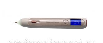
The equipment is made as convenient as possible to use: images are copied via a USB port in a couple of minutes, the doctor can select the most suitable option for comparison (“before and after” on one screen, slider or silhouette) in an intuitive interface, the results are easily exported as a high-quality image resolution or as video. The 3D camera comes in three versions: for the face (Mini), for the body (Body), for the face and body (Infinity). The equipment does not require special premises and is easily portable. Dual flash always ensures perfect illumination, and a stereo vision system with two aiming beams allows you to select the exact distance and position of the camera without additional fixation.
Parallel polarized light (PPL) photography
The technique was created for an objective assessment of light reflected from the surface of the skin. Photos taken with green light-emitting diodes (LEDs) can be useful for analyzing diseases such as atopic dermatitis, rosacea and xerotic dermatitis.
In 2022, Korean scientists successfully used the technique to quantitatively and objectively assess rosacea in a clinical setting. 3 The two main subtypes of rosacea, erythematotelangiectatic (ETR) and papulopustular (PPR), were visually and optically identified differently during the study, allowing accurately differentiate the diagnosis.
Estimation of skin moisture content
Water balance in the skin is one of the main indicators of its condition. It is most often assessed by corneometry. Using a special device - a corneometer (Fig. 1) the electrical resistance of the skin is measured. The sensor for corneometry is a capacitor under a glass cover. A current with a frequency of 0.9–1.2 MHz passes through it, which forms an electric field that penetrates the skin. Typically, the design of the sensor is selected in such a way that the depth of penetration of the electric field into the skin does not exceed 10–20 nm and affects only the stratum corneum, since the stratum corneum is most sensitive to changes in water balance. The capacitance of the capacitor will depend on the dielectric constant of the stratum corneum, which changes depending on the water content in the epidermis. The higher the hydration, the lower the resistance to electrical current. Modern corneometers are usually equipped with an LCD display and control buttons, and can work both autonomously and when connected to a computer. There are also combined devices with replaceable sensors that allow you to measure not only the moisture content in the skin, but also other indicators - the amount of sebum and pH. Recently, manufacturers of special equipment have begun producing portable corneometers that are suitable even for home use.
Rice. 1. Corneometer® CM 825, Courage&Khazaka
To monitor skin hydration, a test that determines transepidermal water loss (TEWL) is often used - TEWL test or tevametry. It allows us to judge the state of the lipid barrier of the stratum corneum and can be useful to confirm the effectiveness of moisturizing and protective cosmetics, as well as products aimed at strengthening the skin barrier. Assessment of transepidermal water loss can be carried out using the so-called. “open” (Fig. 2) or “closed” measurement system (Fig. 3). The operating principle of the first system is as follows. In the measuring cell of the device, which is applied to the skin, there are two sensors located one below the other. When the sensor is installed on the skin, a density gradient of water vapor evaporating from the surface is established in its cavity. This gradient is measured using two pairs of sensors located in the sensor cavity (one pair measures temperature and the other measures relative humidity). The data is analyzed using a microprocessor. As a result, the amount of water evaporated per unit time (TEL) is calculated.
Rice. 2. Tewameter® TM 300, Courage&Khazaka
The second method for estimating TEWL is based on measuring the partial pressure of water in a closed chamber with a single humidity sensor. When the camera is applied to the skin, there is a gradual increase in the partial pressure of water in it. The increase in partial pressure in the chamber over a certain period of time is measured. The TEVP assessment method is the most objective for assessing the lipid barrier in relation to fluid conservation. The disadvantage of this method is that it is susceptible to factors such as ambient temperature and humidity.
Rice. 3. VapoMeter, Delphin Technologies
Cohesiometry is a study of skin peeling, which also helps to indirectly assess the moisture content of the skin. Peeling of the skin depends primarily on the rate of proliferation of the epidermis, as well as on the water balance. Normally, desquamated corneocytes are approximately the same small size. A large number of large cells indicates damage and dehydration of the skin. Cohesiometry is based on the use of transparent adhesive tapes, to which corneocytes adhere when applied to the skin. After this, the tapes are painted and photographed in transmitted light. Then the image is processed and the peeling index is calculated. The equipment used for the study is the same as for optical profilometry.
The water content in the deeper layers of the skin can be assessed using ultrasound and OCT methods, which will be discussed below.
Multispectral Imaging Technology
It is used in dermatology mainly for the diagnosis of melanoma. However, there are reports of the use of the method for assessing blood oxygen saturation based on the spectral characteristics of oxy- and deoxyhemoglobin and diagnosing other skin diseases 1, 4.
The idea behind the technique is that inflammatory diseases have subtle color changes that are difficult to reproduce using conventional RGB imaging. Determined differential spectral reflectance wavelength can also be used to quantify and classify skin diseases. In some types of skin diseases, irregular spectral features are observed because the optical properties of the tissue are altered compared to normal skin. Spectral images contain not only information about the distribution of dyes, but also the histological structure of skin diseases.
3. Laboratory research
Laboratory tests are especially important if skin manifestations are suspected to be due to the presence of other, non-dermatological diseases. If this is confirmed, symptomatic treatment is prescribed, and the main measures focus on the treatment of somatic disease. The connection between skin pathology and other diseases can be identified by:
- general blood analysis;
- general urine analysis;
- blood chemistry;
- stool analysis;
- immunogram.
If necessary, the dermatologist refers the patient for consultation to other specialists who can prescribe a number of more specific studies.
About our clinic Chistye Prudy metro station Medintercom page!
Computer capillaroscopy of skin
Capillaroscopy is a non-invasive tool for studying microcirculation 5. The method first appeared at the beginning of the 20th century, has undergone many improvements and today is increasingly used in dermatology, angiology and rheumatology to assess the activity of the vascular bed.
Recently, the technique has been enhanced by videocapillaroscopy - the results of the study can be saved on a computer and the dynamics of changes can be monitored.
Capillaroscopy can also be used for the differential diagnosis of connective tissue diseases.
Assessment of microcirculation in the skin
Skin temperature is related to blood microcirculation, so skin temperature measurement data can be a tool for promoting cosmetic products that affect skin blood supply. Recently, thermal imaging methods using infrared video cameras have become widespread. In the thermograms obtained in this way, the skin shimmers with all the colors of the rainbow - from blue-violet to red (Fig. 7). Orange and red zones are high temperatures, blue zones are low. It is logical to assume that blood circulation is slower in the blue zones. However, heat transfer primarily depends on the temperature of the external environment, therefore, when conducting such studies, it is necessary to carefully control the room climate, and measurements should be carried out after a person has adapted to them.
Rice. 7. Thermograms obtained using infrared video cameras
Spectrophotometry
The spectrophotometer has a wide range of applications in dermatology.
It can evaluate the effectiveness of sunscreens in vitro, measures light transmittance at 1-nanometer intervals and is equipped with an ultraviolet light source, combining an integrating sphere and monochromatic light capable of transmitting energy flux from 290 to 400 nm.
In addition, the device allows you to objectively assess and determine the skin color of patients and measure color coordinates and indicators of melanin and erythema 6.
We draw up a procedure protocol based on ultrasound scanning of the skin by zone
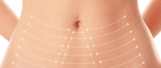
During the scan, the patient lies down with his head thrown back, since this is the position in which the procedure itself is performed in this area. Therefore, scanning must also be carried out in this position of the patient. It is necessary to determine the thickness of the subcutaneous fat, the presence of connective tissue in it and its quantity, determine the depth of the superficial fascia of the neck (fascia superficialis colli), deeper than which you should not work due to the risk of damage to the submandibular salivary gland (glandula submandibularis) (Fig. 1. ).
In this case, we see that to achieve optimal results in this area, a 4.5 mm handpiece will be used, since it hits the fascia, promoting its maximum contraction and strengthening.
Rice. 1. Scanning of the submental area. |
The main task of this scanning point is to determine at what depth the body of the lower jaw (corpus mandibulae) is located in the most protruding area, along the lower edge (Fig. 2.). This measurement is necessary to determine the lower limit of work for the safe implementation of the procedure. In this case, the periosteum begins at a depth of 3 mm, therefore, the lower limit for the lifting tips (3 mm M7 and 4.5 mm D4) should be located higher so as not to injure the periosteum. In this area you can work with a 1.5 mm S7 handpiece.
Rice. 2. Scanning in the area of the body of the lower jaw (the most protruding part of the bone). |
Next, a scan is performed to determine the depth of the SMAS layer, the total content of connective tissue in the subcutaneous fat in the buccal and parotid-masticatory areas (Fig. 3.). It is at this stage that it is determined which attachments will be used to lift the cheek area, reduce the severity of jowls, marionette lines, and nasolabial folds. This patient has a large amount of connective tissue in the subcutaneous fat and pronounced SMAS - good prognostic signs. In the procedure protocol, it is important to use both the M7 3 mm handpiece - to compact and reduce collagen in the sac, and the 4.5 mm D4 handpiece - to strengthen the SMAS connective tissue.
Rice. 3. Scanning the buccal area. |
In the zygomatic region, it is necessary to determine the depth of the bone arch (Arcus zygomaticus) so as not to injure the periosteum and neurovascular bundles emerging at this depth (Fig. 4.). If necessary, do not enter this area with a manipulator of appropriate depth. In this case, you can work with 3 mm M7 and 4.5 mm D4 maniples up to the upper edge of the zygomatic arch, since the periosteum is at a depth of 7.5 mm.
Rice. 4. Scanning the zygomatic arch. |
The forehead area is an area where incorrect preparation of the procedure protocol is often encountered due to overestimation of the depth of possible work (Fig. 5.). The purpose of ultrasound scanning is to determine at what depth the soft tissue ends and the frontal bone (os frontale) begins. For example, this scan shows that the frontal bone begins at a depth of 2.8 mm. Therefore, a 1.5 mm S7 handpiece will be used for efficient and safe operation.
Rice. 5. Scanning the forehead area. |
Spectrocolorimetry
Modern spectrocolorimeters are portable devices that fit easily into your pocket.
The spectrocolorimeter makes it possible to measure pigment changes in tissues and assess the degree of differentiation of normal skin. It is more accurate and reliable than the human eye, which perceives color individually. In addition, lighting conditions affect color accuracy, and color reference scales can become damaged over time by exposure to light or other physical factors.
A spectrocolorimeter eliminates these problems.
The essence of the method is to register color according to several values in a given three-dimensional space.
The first value, brightness (L), expresses the brightness of color changes across the spectrum from black to white.
The second value (a) is the color tone from red (+) to green (-). When a rash appears on the skin, the value becomes positive.
The third value (b) represents the color tone from blue (-) to yellow (+). If hyperpigmentation appears, the value becomes positive.
The research results showed that when comparing the assessment accuracy between a person and a machine, the reliability of the spectrocolorimeter was significantly higher 7, 8.
2. Examination of the patient
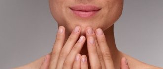
When answers to the basic questions are received, a skin examination is performed, which begins with the affected area and necessarily covers the entire body. The most objective results are obtained by examining the skin in diffuse daylight. A magnifying glass is often used. In many diseases, lesions on the skin have a certain, “recognizable” shape, structure and color.
The first two stages of skin diagnosis, as a rule, allow the dermatologist to make a preliminary diagnosis or suggest several possible ones. In addition, the nature of the disease becomes clear - acute or chronic condition, hereditary or acquired; The stage of development of the disease and the degree of damage to the skin are also determined.
Visit our Dermatology page
Ultrasound examination of the skin
Ultrasound uses high-frequency sound waves and their echoes to capture images of tissue structures. Modern ultrasound machines offer excellent image resolution in a portable device, which has allowed the method to come to dermatology.
Ultrasound imaging has been used in dermatology for almost 30 years. Based on differences in keratin, collagen and water content, ultrasonic waves are reflected to the transducer and converted into a black and white image.
The method has a wide range of applications: diagnosis of dermatological conditions, including melanoma and non-melanoma skin cancer, benign tumors, inflammatory diseases, lipoablation, etc. 9
Ultrasound is also integrated into non-invasive techniques such as continuous wave Dopplerography, ultrasound elastography, and ultrasonography.
In particular, ultrasonography is used by doctors to identify the structural layers of facial skin when performing botulinum toxin injections in order to correctly determine the depth of penetration of the needle 10.
4.Special diagnostic methods
Dermatological diagnostics proper includes a number of methods and techniques that complement visual examination of the skin:
- palpation (to assess the elasticity and structure of the skin);
- skin allergy tests (to determine allergens);
- sowing the affected area (growing microorganisms from the surface of the skin in a special medium);
- microscopy of scrapings (examination of parts of the skin under a microscope);
- scraping (to identify peeling);
- diascopy (assessment of skin reaction when pressed with a glass slide);
- histology (examination for the presence of cancer cells).
Most diagnostic techniques in dermatology are safe and painless. Early detection of the disease gives a better chance of recovery. Even if skin rashes do not bother you with pain or itching, but only attract attention as something new on the body, you should not put off visiting a doctor.
Sign up for a consultation
Photoacoustic methods
This is a new biomedical imaging technique that allows visualization of tissue using acoustic detectors (light signal - sound output).
This technique has enormous potential because it produces images with high resolution, sufficient imaging depth, various endogenous and exogenous contrasts, and is free from ionizing radiation.
The method makes it possible to diagnose psoriasis and other skin lesions, including melanoma 11.
Measuring skin lipid balance
Lipid balance of the skin is an important condition for maintaining water balance in the body. The functional state of the lipid barrier can be assessed using the TEWL test.
The method for determining skin oiliness is sebumetry. The readings of this method depend on the activity of the sebaceous glands and reflect the amount of sebum produced. The method is based on the photometric assessment of greasy stains that remain on the adhesive film after its contact with the skin. This special film is sensitive to fats and changes its optical density depending on the amount of fat. A measuring cassette with a film at the end is applied to the skin for approximately 30 seconds. The film cassette is then placed into a device that operates on the principle of spectrophotometry. The optical transmittance of the tape is measured and the fat content per cm2 is calculated. Sebumetry allows you to evaluate the effectiveness of anti-acne products, cleansing cosmetics for hair and skin (Fig. 5).
Rice. 5. Sebumeter® SM 815, Courage&Khazaka
Neural networks
Futuristic forecasts say that soon neural networks will be able to diagnose diseases with greater accuracy and eliminate the human factor.
Recently, a study was presented on the effectiveness of a neural network in recognizing acne, and the startup ModiFace, acquired by giant L'Oréal, announced the launch of digital skin aging diagnostics for users 12, 13.
This new technology is based on an artificial intelligence-based algorithm developed by ModiFace and based on L'Oréal's expertise in skin aging and a photo database. Using deep learning, the algorithm was trained on 6,000 images from the L'Oréal database, selected using skin aging atlases, and then created a new model from more than 4,500 selfies for three groups of women (Asian, Caucasian and African American) under different conditions lighting.
The results, supplemented by dermatologists, made it possible to achieve a highly accurate assessment of skin condition.
1. Anamnesis collection
Methods for diagnosing dermatological diseases
no less diverse and complex than skin diseases themselves. Sometimes a visual examination is enough, and sometimes a comprehensive examination of the entire body is necessary. Skin diseases sometimes affect the general condition of a person, and vice versa - diseases of other systems affect the condition of the skin. When a person consults a dermatologist with complaints, it is important to understand whether skin manifestations are the cause or consequence of the disease.
It would seem that the skin is the largest organ in the human body in area and the most accessible for diagnostics. But it is the skin that serves as the first protective barrier against all kinds of harmful environmental influences; In addition, skin diseases are so diverse that their diagnosis is sometimes a more complex problem than analyzing the condition of internal organs.
Accuracy in making a dermatological diagnosis is largely determined by the “human factor”: the attentiveness and experience of the doctor, his ability for logical analysis and synthesis. Diagnosis of any dermatological disease includes several stages.
The first stage of a dermatological examination is taking an anamnesis, which involves an external examination and a conversation with the patient, and sometimes with his relatives. It is necessary to find out the possible causes and presence of factors contributing to the occurrence of skin diseases, the presence of comorbid (concomitant or background) diseases, information about heredity, lifestyle, nutrition, psychological state, and medications taken. Sometimes, with the same observed symptoms, the presence or absence of itching, the persistence of skin rashes, the frequency and area of their appearance can be very important.
Sign up for a consultation
A must read! Help with treatment and hospitalization!
Facial asymmetry – SMAS asymmetry
In this patient, during the scan, the cause of facial asymmetry was identified. On the scanogram from the left half of the face, the connective tissue in the pancreas and on the SMAS is clearly visible; the pancreas itself is thin.
The image from the right half of the face shows a more pronounced layer of subcutaneous fat with a low content of connective tissue.
As can be seen in the photograph, these ultrastructural changes caused facial asymmetry, with more pronounced sagging on the right side. To compensate for asymmetry, it will be necessary to make a larger number of lines on the right, for a more pronounced formation and contraction of connective tissue and for the effect of adipocytolysis, which will reduce the severity of asymmetry and create a uniform lifting effect.
References
- Basal cell skin cancer. Clinical recommendations. Association of Oncologists of Russia, 2022. - 85 p.
- Squamous cell skin cancer. Clinical recommendations. Association of Oncologists of Russia, 2022. - 31 p.
- Kumar, V., Abbas, A., Fausto, N. et al. Robbins and Cotran Pathologic Basis of Disease, 2014. - Vol. 1(7), 3(25) — 1464 p.
- Ferrante di Ruffano, L., Dinnes, J., Chuchu, N. et al. Exfoliative cytology for diagnosing basal cell carcinoma and other skin cancers in adults. Cochrane Database of Systematic Reviews, 2018 - Vol 12.