Women have long been aware that they need to regularly conduct self-examination of the breast, and if changes in the skin, retraction of the nipples or lumps in the structure are detected, it is necessary to urgently consult with a breast oncologist. However, now doctors are talking about another feature of the breast structure that can cause cancer - the “dense” mammary gland.
Malignant tumors of the reproductive system in women account for about 40% of all tumors, and half of them, that is, every fifth tumor, is breast cancer. For early detection of breast tumors, doctors recommend regular screening mammography, which every woman after 40 years must undergo once every 2 years. However, there are factors that can complicate diagnosis. In particular, the “dense” mammary gland. About what it is and what diagnostic methods should be used for this condition, AiF. ru told Candidate of Medical Sciences, Associate Professor of the Department of Radiation Diagnostics and Radiation Therapy of North-Western State Medical University named after. I. I. Mechnikova Irina Solntseva .
Your breasts have increased
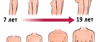
This fact may indicate that you have simply gained weight. After puberty ends, breasts grow only when all the dimensions of your body increase. Start watching your diet and try to move as much as possible.
Sometimes breasts become larger without gaining weight. This condition indicates that you are about to start your period or have started taking new birth control pills. Hormone levels directly affect this condition. If you do not observe asymmetry and your two breasts have increased in size equally, this should not bother you.
Why should every woman know whether she has dense breasts?
Since 2022, the world's leading oncology societies have included the “dense” gland in the list of risk factors for breast cancer. Here it is worth understanding that the denser the gland tissue, the higher the risk of developing cancer. With C-type density, notes Irina Solntseva, the risks of developing breast cancer increase by about 2 times, and with D-type - by 4-6 times. And this is a really serious problem, says the specialist, because dense breast structure occurs in 60% of young women and 40% of postmenopausal women. Moreover, the risks of cancer are equally high both in youth and in adulthood.
If, according to the results of a mammography, it turns out that a woman has a “dense” mammary gland, the oncologist-mammologist evaluates what other risk factors for cancer are present in the patient. Among them:
- early onset of menstruation;
- late onset of menopause;
- long-term use of hormone replacement therapy;
- benign proliferative diseases of the mammary gland;
- hereditary factor.
In a situation where the patient has “dense” breasts, and there are also other risk factors for cancer, the oncologist can recommend an individual monitoring regimen for her. For example, reduce the period between screening examinations by scheduling mammography not once every two years, but once a year. In addition, for women with “dense” breasts, mammography alone is not enough, so the oncologist will definitely prescribe a further examination in the form of an ultrasound of the mammary glands.
Angelina Jolie syndrome. Who should have their breasts checked by a geneticist Read more
One of your breasts is larger than the other
In most cases this is completely normal. No one has two absolutely identical and symmetrical breasts, so slight differences in size and shape should not cause panic.
In some cases, however, if the asymmetry is very noticeable and one breast is much larger, it may be a symptom of cancer. Be sure to check with a specialist.
Diagnostics
If a woman has characteristic complaints, a number of diagnostic studies are performed:
- X-ray of the chest. Mammography is a screening diagnostic method that allows you to confirm the presence of a pathological focus in the mammary gland. Ductography is an X-ray contrast study that allows you to visualize the duct system of the breast.
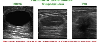
- Ultrasound examination of the mammary glands. Helps determine the location, shape and extent of pathological compaction in the breast tissue. The method is safe and can be performed on pregnant or breastfeeding women.
- Breast biopsy. Histological examination of the breast lump helps to reliably determine the nature of the disease and distinguish a benign tumor from a malignant one.
- Thermography of the chest area - registration of thermal radiation from tissues. The method is based on the fact that actively dividing malignant cells emit more energy and appear redder in the image than surrounding healthy tissue.
To diagnose lumps and pain in the breast, it is mandatory to consult an oncologist-mammologist. An endocrinologist, gynecologist and other specialists may be involved.
You have irritation on the skin under your breasts
Most often, this is a symptom of an allergy, which may have been caused by the fabric of your bra or soap. Sometimes irritation occurs due to wearing tight clothing, heat and excessive sweating. Try using anti-allergy cream. If the problem does not go away within several days, then consult a doctor.
It could also be diaper rash. This condition often appears in the summer, especially if you choose the wrong bra size. Special creams can help with this.
Wash your laundry thoroughly. Otherwise, bacteria can multiply on it, which causes such an unpleasant reaction.
Treatment
Treatment tactics depend on the cause of the lump and pain in the chest.
With lactostasis, the main task is to empty the mammary gland. To do this, a breast massage is performed; the woman is advised to avoid hypothermia and to put the baby to the breast in a timely manner. Adequate rest and good nutrition will not be superfluous. Night sleep is recommended in the side position. In some cases, oxytocin is prescribed, which stimulates the process of lactation and emptying of the mammary gland.
Antibiotic therapy plays a major role in the treatment of mastitis. The seal usually resolves with rational treatment. The development of purulent mastitis is an indication for surgical intervention.
Multiple or nodular mastopathy is subject to conservative therapy. Drugs are used that normalize hormonal levels. These include combined oral contraceptives, estrogen receptor inhibitors and other drugs.
A benign seal is usually treated surgically, since conservative methods are ineffective. Enucleation or sectoral resection of the mammary gland is used. In the first case, surgeons remove the tumor. There are usually no cosmetic breast defects left with this procedure. Sectoral resection is the excision of a tumor within healthy tissue. After this intervention, a slight deformation of the breast remains, which can be corrected with plastic surgery.
A compaction of a malignant nature is subject to surgical treatment. Mastectomy with removal of regional lymph nodes is recommended. In some cases, excision of the breast tumor within healthy tissue may be performed. The scope of the operation is determined by the oncologist, taking into account the extent of the tumor process. Alternative treatment methods include chemotherapy, radiation therapy, and targeted therapy. They are prescribed in various combinations with each other.
Your nipples are itchy
Most likely, the reason lies in the remnants of soap or shampoo that you did not wash off well. Wash your nipples with water and apply moisturizer. You will immediately feel better.
It could also indicate an allergic reaction caused by your clothing or cosmetics. Observe yourself and find out when exactly the itching occurs. Eliminate irritants.
Sometimes this problem also appears during menstruation. After a few days everything goes away on its own.
If, in addition to itching, you also have dry and slightly yellow skin, and perhaps even bruising, then consult a doctor immediately, as this may be a symptom of cancer.
Survey technologies
If a mammogram reveals a “dense” mammary gland in a patient (density types C and D), doctors recommend undergoing an additional ultrasound of the mammary glands or a new technique for automated three-dimensional ultrasound examination. This technique allows you to obtain a three-dimensional image of breast tissue, which eliminates one of the most important disadvantages of the ultrasound method - operator dependence.
“The three-dimensional ultrasound technique is automated, which guarantees a complete examination of the entire mammary gland, without missing any area, even when examining large breasts or in the presence of implants,” says Irina Solntseva.
The sensitivity of three-dimensional ultrasound in detecting cancer against the background of a dense breast is 98-99%, therefore, it is currently the only method of ultrasound screening for breast cancer approved in the world, which is carried out additionally after mammography in cases of detection of a “dense” breast structure (types of density C and D) and the absence of focal pathology. In addition, the study is recommended for young women (under 40 years of age) who do not have any symptoms, but have risk factors for breast cancer, such as a BRCA ½ gene mutation and a family history.
Carrying out this procedure in addition to mammography against a background of dense breast tissue can detect 2.6 times more malignant tumors and 55% more invasive breast tumors.
Question answer
Can injury cause breast cancer?
Your breasts are leaking fluid
Breasts are designed for feeding, so if they have been stimulated, even if you are not pregnant or have children, something like this may be released from them. If this causes you some inconvenience, consult your doctor for special tablets.
Sometimes this problem appears after taking antidepressants or any psychotropic drugs. This affects hormones, which leads to such discharge. This process is normal and should not bother you.
What is a “dense” mammary gland?
“Dense” mammary gland is an X-ray concept. “The structure of the mammary gland consists of different types of tissue: adipose and fibroglandular (connective and glandular). Fibroglandular tissue gives a shadow on mammographic images, which is why it is called “radiologically dense,” says Irina Solntseva.
When there is more fibroglandular tissue in the structure of the mammary gland than fatty tissue, this condition is called “dense” mammary gland. “Doctors distinguish four types of density - A, B, C and D. The first two are non-dense types of structure, and they do not cause any concern. Types C and D are those options in which dense tissue predominates in the structure of the mammary gland. With the C-type it is in slightly smaller quantities, but with the D-type it occupies almost the entire volume of the gland,” explains the specialist.
It's written in the family. How to avoid hereditary cancer Read more
You may feel a lump in one breast
It may be a benign cyst. This is especially indicated by the fact that it seems smooth and round to the touch. Sometimes it appears and goes away on its own until the next cycle. If you are concerned, ask your doctor to check you.
Sometimes it can still be cancer or something that can become one over time. In any case, you need to contact a specialist.
Pay attention to such symptoms, and if you notice warning signs or are unsure about something, it is better to play it safe and seek medical help. In some cases, time is the key factor on the path to a quick and painless recovery.
Found a violation? Report content
How to protect yourself from breast cancer?
Currently, medicine does not know how to prevent the development of breast cancer, and therefore the most important and pressing task is its early diagnosis, at the stage when it is almost completely curable.
If a woman is regularly examined, the risk of missing the onset of tumor development is significantly reduced. After 40 years, all women need to undergo mammography regularly, once every 2 years, and in the case of “dense” breasts, additionally undergo ultrasound of the mammary glands with a frequency determined by a mammologist-oncologist. If a woman has complaints or there have been cases of malignant tumors in the family, then regular examinations, including examination by a mammologist and ultrasound, should begin from the age of 25-30 years. Do not forget that early diagnosis of breast cancer is a joint task between the doctor and the patient, and timely examination can save lives.
There are contraindications, you should consult a doctor
What to do if asymmetry appears
If breast asymmetry is diagnosed after mammoplasty, surgical correction is indicated. Depending on the cause of the complication, its purpose:
- use of implants of different sizes;
- molding a new pocket for the implant;
- elimination of fibrosis compressing the implant.
If the patient’s general condition is satisfactory, the procedure is performed no earlier than six months after the initial operation. If there is necrosis, hemorrhage, seroma or other specific complications, the implant is removed immediately.
Prevention
To prevent breast diseases accompanied by thickening and pain, women should adhere to a healthy lifestyle: eat rationally, maintain a work and rest schedule, exercise regularly, avoid stressful situations, stop smoking and abusing alcoholic beverages. If you are overweight, you need to lose weight. You should also choose a comfortable bra that does not squeeze or injure your breasts.
Book a consultation 24 hours a day
+7+7+78