Choosing a method for breast surgery
Most patients are primarily concerned about what the scar will look like after surgery. And it is this criterion that they are guided by when choosing a type of bust enlargement surgery - including breast augmentation through the armpit. At the same time, plastic surgeons, when talking about choosing a method for mammoplasty, pay attention to other priorities:
- size of the selected implant;
- the necessary set of measures for breast lift or areola formation in a new place;
- the condition of the tissue in the place where the incision will be made.
The widespread concern among patients about the scar that may remain after surgery has no basis - in modern plastic surgery, the technique of microsurgical suture is practiced. Such a seam is practically invisible - even if the incision is sutured again.
Many women consider breast augmentation through the axillary approach to be the preferred method, since the scar left after such an operation will be located in an inconspicuous place.
In any case, the choice of the type of breast augmentation surgery must be agreed with the doctor, who examines the patient and analyzes her body parameters. Depending on the goals that the patient and the doctor determine during the consultation, the type of surgical intervention that is most suitable for the task is selected - including breast augmentation using an axillary approach.
Why is fibrocystic mastopathy dangerous?
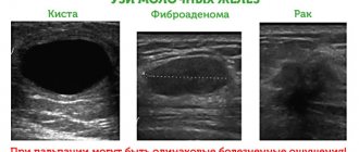
Mastopathy is always characterized by the formation of both cysts and fibrosis. When this process is balanced - equal parts of both, and it is distributed throughout the entire gland - then they speak of fibrocystic, diffuse or mixed mastopathy.
Fibrous mastopathy
Fibrous mastopathy is characterized by a predominance of the fibrous component over the cystic component in the development of fibrocystic mastopathy.
Increased formation of connective tissue at the site of former proliferation makes the breast gradually denser and more painful. It becomes difficult to evaluate her with mammography and even place her in a mammograph (due to pain). CESM and MRI are more suitable for diagnosis and screening .
Nodular mastopathy
Sometimes, with fibrocystic mastopathy, denser nodes appear in the breast tissue, often in the upper outer quadrants (where there is initially more glandular tissue). They often look suspicious for cancer upon examination and on mammograms and require MRI (or CESM ) and biopsy for definitive diagnosis.
Mastopathy compaction
Often such patients are even offered surgery—sectoral resection for localized fibroadenomotosis—as cancer is suspected. The operation can be combined with a breast lift, reduction or breast augmentation with implants.
Cystic mastopathy
Cystic mastopathy is characterized by the predominance of the process of cyst formation over fibrosis in the development of fibrocystic mastopathy.
These cysts are always multiple. At first they are small, but over time they can merge, reaching large sizes. puncture brings relief .
If the cysts recur, it is proposed to remove them, which can be combined with lifting, reducing or enlarging the mammary glands with implants.
Regarding mastopathy, monitoring for cysts and for preventive examinations of the mammary glands, we recommend that you consult a doctor:
Subbotina Olga Yurievna , AF-clinic, Spassky lane 11, M. Sadovaya, Sennaya, Spasskaya, tel. 310-00-33 and
Sokolova Valentina Ivanovna , Nova Vita Clinic, intersection of Engels and Thorez Ave., tel. 8(911)007-20-02
Dushina Irina Ilyinichna , SMT Clinic, Moskovsky Ave. 22, tel. 777-9-777
Raevskaya Natalya Aleksandrovna , Polyclinic No. 83, M. Sportivnaya, tel. 498-09-22 and 8(921)944-10-45.
Features of endoscopic implant installation
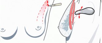
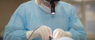
This type of mammoplasty is an enlargement of the mammary gland through access under the armpit. It is carried out in a low-traumatic way, which is based on the endoscopic method of surgical intervention. During the operation, the specialist makes a small incision (maximum 4 cm) in the armpit area, after which he creates a pocket for the implant.
Mammoplasty through an incision under the armpit is a plastic surgery that can only be performed by the most experienced, highly qualified surgeons. The complexity of this method is due to the fact that near the incision site there are large bundles of nerves, damage to which can lead to loss of sensation in certain parts of the body.
Creating a pocket for installing an implant is also not an easy task, given that the doctor forms it using endoscopic techniques. How securely the implant will stay in place depends on how well the surgeon has given the pocket for the implant the correct shape. If there is an error, the implant may become dislodged some time after surgery.
Possible causes of pathological processes
Enlarged lymph nodes in the armpits are a fairly common occurrence with mastopathy. However, there are other reasons that can cause this pathology:
- Failure to comply with hygiene standards or use of low-quality deodorant. In the second case, the product can clog the sweat glands, resulting in inflammation of the lymphoid tissue.
- Enlargement of the lymph system organs is observed in some pregnant women. In this case, this condition does not go beyond the norm and does not require treatment, since it goes away on its own after delivery.
- Lactation is another possible reason. Lactostasis or inflammatory processes in the area of the milk ducts can lead to enlarged lymph nodes. Drug therapy and massage help eliminate engorgement and tenderness of the mammary glands.
- Another possible reason is the presence of boils. Abscesses or suppuration in the shoulder area, arms or chest.
- Hyperplasia is sometimes diagnosed in children who have had measles or chickenpox.
- Lymphatic tissues can change size in diseases such as syphilis, tuberculosis, rheumatism, as well as malignant tumors.
- Axillary lymphadenitis, which is characterized by chills, weakness and pyrexia, can also lead to swollen nodes.
Possible complications with breast surgery
Mammoplasty through the armpit can in some cases cause a number of complications. For clarity, we divide them into two groups:
- Complications associated with the physiological characteristics of the patient. These include a tendency to form keloid scars and lymph nodes located close to the incision site.
- Complications caused by surgeon error. An insufficiently experienced specialist may incorrectly form a pocket for the implant, while it is this factor that has a decisive influence on how successful mammoplasty through the armpit will be (reviews about this type of operation can be read here). An error in the pocket creation process can lead to subsequent displacement of the implant, damage to nerve fibers or lymph nodes. If this operation is performed by a surgeon with insufficient experience, using outdated techniques, then a noticeable and unsightly scar may remain at the site of the incision.
Endoscopic mammoplasty in Moscow, performed at the Beauty Doctor clinic, is performed only by experienced, highly qualified surgeons. Before the operation, specialists conduct a thorough examination of the patient and perform the procedure only for direct indications.
Forecasts
Hormone-dependent carcinomas have a better prognosis if detected early; for example, with stage 1 lobular cancer, the vast majority of women are cured; with stage 2, eight out of ten who received radical treatment live long. The prognosis for stage 3 is sad for half.
When cancer is no longer a fear from your dreams, but a reality and your present with the future, it is important to search for a good clinic, where they know the problem to the smallest detail and have everything for optimal diagnosis and proper treatment. There are such clinics in Russia, and ours is the best representative of private oncology care.
| More information about breast cancer treatment at Euroonco: | |
| Surgeons-oncologists-mammologists | from 5,100 rub. |
| Removal of a breast tumor | from 101,200 rub. |
| Emergency oncology care | from 12,100 rub. |
| Chemotherapy appointment | RUB 6,900 |
Book a consultation 24 hours a day
+7+7+78
Bibliography:
- Semiglazov V.V./Carcinoma in situ of the mammary gland - morphological and clinical problems// Pract. oncology; 2002; No. 1; v.3.
- Semiglazova V.F., Semiglazov V.V., Manikhas A.G. Breast cancer. Chemotherapy and targeted therapy//-M.: Medpress-inform; 2012
- Hussain M., Cunnick GH / Management of lobular carcinoma in-situ and atypical lobular hyperplasia of the breast—a review // Eur. J. Surg. Oncol.; 2011 Apr; 37(4).
- Litière S., Werutsky G., Fentiman IS, et al./ Breast conserving therapy versus mastectomy for stage I-II breast cancer: 20 year follow-up of the EORTC 10801 phase 3 randomized trial // Lancet Oncol.; 2012 Apr; 13(4).
- Louis-Sylvestre C., Clough KB, Falcou MC., et al. /A randomized trial comparing axillary dissection and axillary radiotherapy for early breast cancer: 15 year results// Breast Cancer Research Treat. Special Issue: 24th Annual San Antonio Breast Cancer Symposium; 2001; 69.
Rehabilitation time after mammoplasty
The recovery time after this operation does not differ from the recovery time after breast augmentation performed using other surgical techniques.
The patient spends 2-3 days in the hospital - during this time she receives the necessary medications and is under the constant supervision of doctors. After discharge, the woman can return to everyday life, following a number of instructions - for a month it is necessary to wear compression garments and exclude physical activity.
Full recovery usually takes a month. This is also true in cases where repeated mammoplasty was performed through the axillary approach.
Despite the relatively quick rehabilitation, the patient who underwent endoscopic mammoplasty in Moscow must regularly visit a plastic surgeon for a preventive examination. The first consultation should be held no later than one month after the operation. In the future, it is recommended to visit a doctor every six months.
Diagnostic methods
Lobular cancer in the early stage is very difficult to detect: it is not visible either by touch, or by mammography, or by ultrasound; it is visualized only by MRI.
Invasive carcinoma can already be palpated, but mammography is also not easy - there is no clear separation from normal glandular tissue and calcium deposits in the vessels are very rare, helping to identify “evil”. The standard of examination is bilateral breast MRI.
Even when planning, at the first stage of the operation, a biopsy of the breast tumor is necessarily performed, the so-called “core biopsy” - a cylindrical column of pathological tissue is cut off with a thick needle. Cancer tissue is required for testing for cell type, hormone receptor content, HER2 and Ki67. All these tests are required to plan adequate treatment, since the choice of the medicinal component is determined not by the cellular structure, but exclusively by the molecular biological type of the tumor.
After the biopsy, they begin to search for distant metastases, performing CT scans of the chest and abdominal cavities, bone scintigraphy for complaints of bone pain, and MRI of the head for neurological symptoms.
Tumor markers in breast cancer are not of fundamental importance, since they increase in one and a half dozen completely benign diseases, and are used to monitor the effectiveness of antitumor chemotherapy.
Features of plastic surgery at the Beauty Doctor clinic
Axillary access to the mammary gland is performed by the most professional and trained surgeons, since endoscopic manipulations to create a pocket for an endoprosthesis require high qualifications.
In our clinic, located in Moscow, operations using endoscopic technology are performed by experienced and distinguished plastic surgeons. They work together with anesthesiologists who use the safest and most modern types of general anesthesia and local anesthesia.
Symptoms of mastopathy
Pain is the main symptom of mastopathy.
Connective tissue can swell and hold liquid (like white films on meat dipped in water). Completely similar - overformed connective tissue in the mammary glands during mastopathy - swells with excess fluid in the body. The swollen tissue mechanically affects the nerve endings in the mammary gland tissue, causing pain.
This happens physiologically in phase II of the cycle - when progestins (ovarian hormones) promote fluid retention in the body (normally). Therefore, pain in the mammary glands with mastopathy also occurs before menstruation.
In addition to pain with mastopathy, women find vague lumps in the mammary glands, usually painful and during the period of premenstrual tension. Ultrasound, mammography and MRI usually show nothing, or multiple cysts are found.