Temporal regions
Injections into the temporozygomatic branch of the trigeminal nerve are given in an "anatomical" rather than a "targeted" manner because the sensitivity reported by patients in this area is usually more diffuse in nature rather than specific to the anatomical location of the nerve's exit point from the temporalis fascia. Since the nerve anatomy in this area is very stable, an "anatomical" injection is sufficient. This point, described by Totonchi et al. [12], is usually located 1.7 mm lateral and 6 mm above the lateral canthus. This injection, originally described by Guyon et al. [16], infiltrates fan-shaped around the nerve into the temporal muscle. We also inject a small amount of Botox into the space just above the point where the nerve exits the temporalis fascia to ensure that the superficial portion of the nerve that lies outside the temporal region is adequately exposed to Botox (Figure 3). In our experience, the major drawback of the current neuropathology injection paradigm is that this nerve is not injected at the correct depth and location.
Figure 3
.
Injection sites in the temporal region. Image showing the connection of the auriculotemporal nerve (bright yellow), the temporozygomatic branch of the trigeminal nerve (dark yellow), and the anterior and posterior branches of the superficial temporal artery (red) to injection sites in the temporal region. The temporozygomatic branch of the trigeminal nerve is located in a different fascial plane compared to the auriculotemporal nerve, and the superficial temporal artery lies more lateral. 1. The area of injection into the temporomygomatic branch of the trigeminal nerve, which is usually located 1.5 cm behind the exit of this nerve from the deep temporal fascia; 2. Proximal to the auriculotemporal nerve – the point corresponding to the fascial compression bundles; 3. Distal to the auriculotemporal nerve - the point corresponding to the anterior temporal artery crossing the auriculotemporal nerve.
The auriculotemporal nerve is injected more “targeted” in multiple areas, based on sensitivity in all areas of nerve distribution, which may or may not roughly correspond to known anatomical areas of compression [7,9] (Video 2). (See Video, Supplemental Digital Content 2, which demonstrates the complete temporal ART injection technique. This video is available in the "Related Videos" section of the full text article on PRSGlobalOpen.com or available at https://www.com/PRSGO/A346 ). The main point of compression is the point where the anterior superficial temporal artery crosses the distal branch of the nerve (section 5A) [35]. The second most common sensitive area, in our experience, is located above the compressive fiber bundles on the auriculotemporal nerve just above the temporomandibular joint (site 5B). The tender point here should not be confused with pain in the joints of the lower jaw, which requires different treatment. As recently described [36,37], to confirm compression of the 5A artery, we do not routinely use Doppler to detect this point when injecting Botox. Although the use of Doppler can be very useful for intraoperative arterial localization and academic exercises, we believe that the additional injection time in migraine patients causing anxiety does not add value to the routine use of Doppler. Both distal and proximal injection sites are based on identifying the point of maximum tenderness during clinical examination rather than a strict anatomical location (Video 3). (See Video, Supplemental Digital Content 3, which demonstrates the author's injection of the proximal and distal regions of the auriculotemporal nerve. This video is available in the Related Videos section of the full-text article on PRSGlobalOpen.com or available at https://links.lww.com /PRSGO/A347)
Video graphics 2
.
Temporal injection technique. See Video, Supplemental Digital Content 2, which demonstrates the complete technique of ART injection in the temporal region. This video is available in the Related Videos section of the full text article on PRSGlobalOpen.com or available at https://links.lww.com/PRSGO/A346.
Video graphics 3
.
Auriculotemporal injection technique. Watch video, Supplemental Digital Content 3, which demonstrates the author's injection of proximal and distal AT regions. This video is available in the Related Videos section of the full text article on PRSGlobalOpen.com or available at https://links.lww.com/PRSGO/A347.
As we described in a previous publication [35], these areas may differ slightly and rely on point sensing for more efficient drug delivery.
Occipital region
The greater occipital nerve is injected in both an "anatomical" and "targeted" manner, since the point of exit of the nerve from the semispinalis muscle (circular diameter is 1.5 cm approximately 3 cm inferior and 1.5 cm lateral to the occipital protuberance) is well described [30] and may correlate with the point of maximum sensitivity. However, in our experience, the area of tenderness is usually located 0.5-1 cm lateral to this exit point, probably due to further irritation and compression by the nuchal line and larger nuchal vessels. When this nerve is injected, the needle must first be inserted deep enough to puncture the trapezius fascia, delivering the medication to the same space where the nerve is located. This injection is located much deeper than described in the PREEMPT occipital injection paradigm and has another important advantage of the ART technique. Instead of a 30G needle, a longer, stronger 27G needle is used to make piercing the trapezius fascia easier. LON is similarly injected in both an “anatomical” and “targeted” fashion using an injection modified according to the point of maximum sensitivity, usually within 0.5 cm of the described anatomical orientation behind the sternocleidomastoid muscle [11]. Additionally, there may be tender points in the distal "tail" of the greater or lesser occipital nerves as they pass over the mastoid and superior/medial areas of the ear, where they may intertwine with the terminal branches of the occipital artery (we have had no problems injecting near vessels of the occipital region; in case of minor bleeding, direct pressure is used for a few minutes, in contrast to the corrugator region, where aggressive direct pressure should be avoided to prevent ptosis). This injection is done in a more “targeted” way, affecting the sensory tail of the greater or lesser occipital nerve. The third occipital nerve is injected much less frequently, also in an “anatomical” [8] and “targeted” way, depending on the severity of the headache localized in this area (Fig. 4). Recent studies indicate that the third occipital nerve is much less likely to be involved in the pathogenesis of migraines [38]. Unlike the PREEMPT paradigm, injections are not given into the trapezius muscle lower in the neck because there are no actual nerve sites there (Video 4 (See Video Supplemental Digital Content 4, which demonstrates the complete ART injection technique in the occipital region. This video is available at (Related Videos section of the full text article on PRSGlobalOpen.com or available at https://www.com/PRSGO/A349).
Figure 4
.
Occipital injections. Image showing injection areas in the occipital region. Occipital protuberance (triangle), greater occipital nerve (black oval), lesser occipital nerve tail (black square), lesser occipital nerve (gray oval), lesser occipital nerve tail (gray square), third occipital nerve (white circle). The third occipital nerve is rarely injected because the patient usually has no sensation in this area.
Video graphics 4
.
Occipital injection. Watch video, Supplemental Digital Content 4, which demonstrates the complete ART injection technique in the occipital region. This video is available in the Related Videos section of the full text article on PRSGlobalOpen.com and is also available at https://links.lww.com/PRSGO/A349.
Most of our patients receive the standard PREEMPT dose of 155 units of Botox, but in a more effective way. In selected patients who only have one trigger point or one “region,” we will use less than the standard dose and target only what is needed. However, we still adhere to the site-specific injection modalities above rather than simply “following the pain” if it does not fit the ART injection paradigm described above.
The effect of botulinum toxin injections
Successful
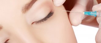
Which Botox will be considered successful:
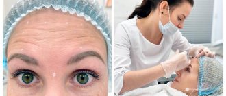
Expression wrinkles disappear.- Skin folds and creases are smoothed out and restored.
- The effectiveness of the procedure increases within 10-14 days.
- At the injection site, the skin becomes more elastic and resistant.
- Damage from injections is almost invisible and heals quickly.
- Minor swelling and redness that disappear 10-15 hours after the procedure.
- The effect of the procedure should last about 3-4 months, repeated administration is required only after 6-7 months. However, some people clear the drug more quickly. The persistence of botulinum taxin in the body is influenced by a person’s metabolism and lifestyle.
Unsuccessful
The Botox injection procedure is considered unsuccessful if:
- Severe allergic reactions.
- Prolonged migraines.
- Painful sensations at the injection sites.
- Drooping of the upper eyelids and eyebrows.
- Complete lack of results from the injection.
In rare cases, complications may occur after the procedure:
- Various respiratory infections.
- Frequent attacks of vomiting and nausea.
- The face turns into a completely immobilized mask.
Such complications arise in case of excessive use of botulinum toxin injections or contacting an illiterate cosmetologist.
You will find tips on removing Botox from the body after an unsuccessful procedure in our material.
conclusions
The ART technique of Botox injections is, in our opinion, the first technique that describes a comprehensive strategy for the treatment of migraine and targets Botox injections around the nerve and different areas of the nerve rather than on loosely distributed muscle groups [6,31]. The ART injection technique is less targeted than that described in the PREEMPT trial [6], but is more comprehensive than the targeted approach originally described by Guyon for screening purposes. The ART injection paradigm is not a screening tool, and both the surgeon and the patient should understand that the purpose of this method is the comprehensive treatment of migraine with Botox.
By injecting closer to the nerves to target known anatomical sites of nerve irritation and focus on regional areas of pain, Botox injection can provide migraine patients with significant pain relief, reduced complications from over-injections, potentially less tolerance to Botox, overall increased patient satisfaction with treatment, and reduced exhaustion due to complications and potentially poorer response from diffuse injections. This injection method may be effective even for episodic migraine, which is currently considered to be less responsive to the techniques described in the PREEMPT paradigm. Future prospective studies comparing this method with the PREEMPT method will definitively determine whether the senior authors' paradigm is validated for long-term use. Our current retrospective data, which are currently being considered for publication, can serve as experimental guidance for any future large prospective studies.
Recommendations on how to reduce risks
Take seriously the choice of a qualified and competent specialist. Do not give injections at home or from a self-taught cosmetologist.- After the injection, you must refrain from sleep for 5 hours.
- Try not to touch your face with dirty hands for 24 hours.
- Do not tilt your head down for 48 hours.
- Do not visit the bathhouse, sauna or solarium. Do not stay in the open sun for a long time. This condition must be observed for 7 days. We talked about why a bath after Botox is dangerous and the possible consequences here.
- Avoid heavy physical activity for 48 hours (read about sports after Botox here).
- In the evening, reduce fluid intake. Because it can increase swelling.
- Avoid alcoholic beverages for 3 days before and after Botox injection.
- You cannot combine Botox injections with medications that affect blood clotting, antibiotics, analgesics and psychotropic substances.
- Before you start injections, you should familiarize yourself with the contraindications. Botulinum toxin preparations are contraindicated for pregnant and nursing mothers, people suffering from protein allergies, eye diseases, etc.
We invite you to watch a video about recommendations for care after the Botox procedure:
