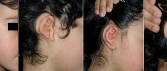
With microtia, the auricle is underdeveloped or completely absent. Atresia of the external auditory canal is a pathology of the development of the external auditory canal, its complete or partial absence. These anomalies are often combined with hearing impairment in the form of conductive or mixed hearing loss of degrees I-III. Since the development of the ear occurs sequentially, atresia is usually combined with anomalies of the auricle (in approximately 75% of cases). Microtia and atresia can be unilateral or bilateral.
These diagnoses imply a step-by-step approach to eliminating defects and are divided into hearing restoration and aesthetics - the formation of the auricle.
The initial and most important aspect should be HEARING. From a very early age, a child must be fitted with bone conduction hearing aids on a bandage, which transmits sounds through the bone directly to the cochlea of the inner ear, regardless of the condition of the auditory canal, outer and middle ear.
This can be done using the Ponto bone conduction hearing aid, which is a leader in the hearing aid market not only in Russia, but also in the world. Today, Ponto hearing aids have the most modern and advanced technologies compared to other analogues of bone conduction hearing systems. They offer more power, a wider frequency range, and ease and comfort in everyday use.
At the age of 4-5 years, the growth stage of the temporal bone is completed, and here parents are faced with the choice of implanting a bone conduction hearing aid or opening the ear canal.
Opening the ear canal is a complex, hours-long and painstaking operation that carries a large number of risks of complications.
At the same time, you need to understand that it will not be able to completely restore hearing, but can only improve it, and you will still have to wear a hearing aid for the rest of your life. If we look at statistics and comments on the Internet, we will understand that this method is not a panacea, and a large number of people lose their hearing again after the operated passage closes again. In addition, do not forget that the created channel is not capable of cleaning itself and periodic cleaning is required. Implantation of a bone conduction hearing aid is a minimally invasive operation that lasts no more than 20 minutes. During this time, an implant no larger than 4 mm in size is implanted into the temporal bone; after complete healing (2-3 months), the patient can wear Ponto on the implant support.
What is auditory canal atresia?
Atresia is a congenital absence or acquired closure of natural openings and canals in the body (Wikipedia). Atresia of the auditory canal is, accordingly, the absence of the auditory canal. In simple terms - the absence of a “hole” in the ear (solid bone instead).
Atresia of the auditory canal can be unilateral or bilateral .
According to ICD-10, it belongs to section Q16.1 “Congenital absence, atresia and stricture of the auditory canal (external).”
Acquired atresia of the external auditory canal (EA) is a rare pathology, the prevalence of which is 0.6 cases per 100,000 population [1]. Surgical treatment of acquired NSP atresia is difficult and is accompanied by frequent recurrence. The disease is also known as post-inflammatory fibrosis of the medial portion of the auditory canal [2-4], post-inflammatory acquired atresia [5], chronic stenotic otitis externa, obliterating otitis externa, acquired medial fibrosis of the auditory canal [6], idiopathic inflammatory fibrosing otitis of the medial auditory canal [7, 8].
Although the term stenosis is used to describe cases of acquired atresia [9, 10], these are two different concepts. According to the Great Medical Encyclopedia, atresia is the complete absence of a lumen or natural opening in an organ that has a tube structure [11], and stenosis (synonymous with “stricture”) is an organic narrowing of a hollow organ, vessel, duct or canal, accompanied by partial or complete disruption of it patency [12]. Acquired atresia of the ESP is represented by a conglomerate of soft tissue in its bony part, fused to the outer surface of the tympanic membrane (ET). With atresia, the auditory canal ends blindly, and a characteristic clinical sign of a “false bottom” or “blind sac” is formed [13].
Stenosis is a narrowing of the external auditory canal over some extent, which is caused either by congenital malformations of the EAS [14], or is acquired, secondary, with exostosis, persistent external otitis [15], trauma (surgical or non-surgical), with malignant neoplasms [16 , 17] or irradiation [18]. Cases of the development of LES stenosis after long-term use of boric alcohol drops have been described [19]. In some of these conditions, stenosis is formed by thickening of the subcutaneous tissue.
The purpose of this review is to describe the methods and results of treatment of both acquired atresia and acquired stenosis of the left joint.
The most elegant classification of acquired NSP atresia is presented by M. Tos [1, 20] (see table).
Classification of acquired atresias of the external auditory canal according to M. Tos
The etiological factor in the development of atresia can also be trauma [3, 6], surgery [21], tumor [22] or inflammatory processes [1, 4, 23-25]. The nature of the surgical interventions undergone can be different: tympanoplasty, removal of exostoses of the ESP, unsuccessful elimination of congenital atresia of the ESP [26]. In most cases, this is the final stage of granulating otitis externa. Some authors attach an important role in the development of acquired atresia to concomitant skin diseases [27]. Thus, in a study by I. Dhooge et al. [28] of 17 examined and operated patients with acquired atresia, 9 (52.9%) were diagnosed with various dermatological diseases.
Pathogenesis of acquired atresia of the NSP
The exact nature of inflammation and the processes that contribute to the development of atresia remains controversial because no precise experimental animal model exists [29]. Most authors agree that external or otitis media (in the presence of perforation of the BP), accompanied by the formation of granulations, is one of the reasons in the pathogenesis of atresia of the NSP. Loss of squamous epithelium from the lateral surface of the PD results in exposure of the fibrous layer. Under conditions of constant inflammation, healing occurs through the production of immature granulation tissue covering the de-epidermalized BP. Granulation myringitis was first described by J. Toynbee back in 1860 [30], since then some new data have appeared regarding the nature of this process. Granulations can form both on the whole BP and in the presence of perforation. Granulations can diffusely cover the entire PD or be localized in one quadrant while others are intact. The source of inflammation can be in the middle or outer ear. P. Stoney et al. [31] found no evidence that the development of granulation myringitis is associated with a specific type of bacterial or fungal infection. P. Bonding and M. Tos [5] observed one patient for 5 years in whom gradually progressive granulation myringitis led to fibrous thickening of the BP. Granulation inflammation can worsen, which is characterized by periodic otorrhea and ear congestion (wet stage). During bacteriological examination of discharge, a number of different organisms are cultured, most often Pseudomonas
spp.
and Proteus
spp., both of which are nonspecific and usually occur in chronic otitis externa and media [29]. Granulations are formed in deep-epidermalized areas of the PD and medial sections of the ESP as a result of trauma and inflammation. In the narrow anterior meatotympanic angle, granulations with the BP and from the anterior wall of the NSP can contact, and subsequently they epithelialize, which leads to blunting of the meatotympanic angle [6]. With further progression of this process, the PD gradually thickens, and the medial section of the ESP is filled with dense fibrous tissue [19]. Finally, if the process is not resolved, atresia of the ESP and hearing loss with a pronounced conductive component are formed (dry stage). P. Bonding and M. Tos [5] propose this process as a model for the development of post-inflammatory atresia of the external auditory canal. The pathogenesis diagram is shown in the figure.
Pathogenetic chain of development of acquired atresia of the external auditory canal.
The pathological process usually continues until the transition point between the bone and cartilaginous parts. The result of replacement of the entire squamous epithelium in the deep sections of the ESP and on the lateral surface of the PD with granulation tissue can be simple or, according to the classification of P. Bonding and M. Tos [5], solid atresia. The literature also describes a case of acquired bone atresia of the auditory canal, which was formed due to heterotopic ossification against the background of fibrosing otitis externa [32].
If, with intact PD, skin damage and granulation inflammation occur along the perimeter in a certain area of the NSP, epithelialization occurs from all walls of the NSP, and a membrane is formed. Such atresia is called membranous [5]. In membranous atresia, keratin accumulates behind the atretic membrane and cholesteatotic inclusions may be present in the proximal portions of the ear canal.
Histological examination of atresia reveals fibrous tissue with numerous blood vessels and nonspecific chronic inflammatory cell infiltration.
Clinical manifestations of acquired atresia of the NSP
As noted above, there are two distinct phases in the development of this condition. In the first, or wet, stage, the ear is in a state of episodic inflammation, when granulations either form or heal with the progressive development of fibrous atresia. The second, or dry, stage is characterized by persistent atresia of the NSP with severe conductive hearing loss. In most cases, patients have a long history of recurrent otorrhea, often accompanied by a feeling of fullness in the ear. The examination reveals the blind end of the external auditory canal in the form of a bag without signs of ongoing active inflammation. An audiometric examination typically shows an air-bone interval (ABI) of about 30-40 dB and a type B tympanogram. Conductive hearing loss gradually progresses as fibrous tissue develops from the medial to the lateral sections of the EAS. P. Bonding and M. Tos [5] described an increase in CVI from 15 to 40 dB over 8 years. If there is a suspicion of concomitant disease of the middle ear, then a computed tomography (CT) scan of the temporal bones is indicated. On a typical CT scan, soft tissues filling the ESP are visualized, while the tympanic cavity is usually intact and airy [14]. On K.T. temporal bones in the presence of atresia of the medial parts of the ESP, one should look for manifestations of chronic purulent inflammation in the middle ear, such as bone erosion or soft tissue formations in the middle ear or mastoid cells [14]. In addition, according to CT data, the anatomical structure of the external auditory canal is assessed: the width of the bony part of the external auditory canal, the degree of severity of the sutures (tympanosquamous and tympanomastoid), the degree of overhang of the anterior wall of the external auditory canal and the severity of the bony isthmus, the size of the short process of the malleus (parameters affecting distance from the BP to the anterior wall of the external auditory canal), which is necessary for adequate planning of surgical intervention [33, 34]. The lesion can be bilateral, its incidence varies from 10% [9] to 67% [29]. The disease occurs on average in the 5th decade of life, although J. Keohane [29] reports two cases of acquired atresia that occurred in children.
Clinical manifestations of acquired LES stenosis
In contrast to acquired atresia, the detection of stenosis of the LES is based on the signs accompanying this disease. Chronic otorrhea often occurs, which is difficult to treat with medication. In some cases, this is hearing loss, although it is not a characteristic feature [18, 19]. However, in all cases of stenosis, in contrast to atresia, a lumen of larger or smaller diameter remains in the area of narrowing of the ESP, through which the PD can often be visualized.
When differentially diagnosing both acquired atresia and acquired stenosis of the ulnar joint, it is necessary to exclude histiocytosis, tuberculosis, sarcoidosis, tertiary syphilis, lupus and malignancy.
Treatment of acquired atresia and acquired stenosis of the left joint
P. Stoney et al. [31] suggested that regular ear cleansing, the use of topical antibiotics and steroids, and gentle applications of alkali (caustic soda, caustic soda) to the granulation tissue during the “wet” stage help to achieve success in resolving most cases. This optimistic point of view is not shared by other authors, who believe that in some cases the development of atresia is irreversible, and medical treatment seems ineffective [24, 29]. Among drug treatments, the topical use of immunosuppressive drugs, such as tacrolimus, which has been successfully used in the treatment of treatment-resistant chronic otitis externa, also deserves attention [35].
By the time fibrous tissue fills the NSP, surgery remains the only effective treatment. The goals of surgical treatment of acquired atresia are to obtain a consistently wide lumen of the ear canal, prevent blunting of the anterior meatotympanic angle, and improve the patient's hearing. The surgical technique was first described by M. Paparella and J. Kurkjian in 1966 [36, 37]. Since then, the basic technique has changed little, but several modifications have appeared [25, 38]. Although M. Paparella and J. Kurkjian initially described the use of a postauricular approach, other authors suggested performing endaural operations [5, 6, 29]. Subsequently, some authors abandoned the endaural approach in favor of the postauricular one due to a large number of unsatisfactory results [6]. The choice of approach and the use of skin graft may vary depending on the experience of the surgeon [13, 39–42].
Whatever the approach, the next step is to remove the fibrous (atretic) tissue forming the atresia. In most cases, it is possible to remove atretic tissue while preserving the deep layers of the BP. If B.P. If it cannot be saved, myringoplasty is performed using autologous tissue. Most authors emphasize the need for complete removal of all atretic tissue, especially in the anterior meatotympanic corner, since relapses are associated with incomplete resection [4, 5, 6, 20].
Regardless of the method used, during the operation there is a deficiency of the skin of the external auditory canal in the medial sections. Various methods have been proposed to cover the exposed areas of the ear canal with skin, using full-thickness or split-thickness skin grafts or vascularized skin flaps. I. Dhooge et al. [28] completely exteriorized the fibrous tissue with the skin of the ESP, then carefully separated the skin from the fibrous tissue in such a way as to later cover part of the ear canal with preserved skin. C. Birman and P. Fagan [43] followed a similar strategy, preserving the skin from the lateral surface of the atresia. D. Katzke and D. Pohl [2] also suggest carefully separating the skin from the atretic tissue in such a way as to bring the flap down to cover the deep sections of the ESP; then they placed a split skin graft over the BP. T. McDonald et al. [9] followed the opposite tactics: they excised all the skin of the external auditory canal and completely replaced it with split skin grafts sutured to the skin of the auricle cup. D. Bell [10] describes the technique of a double displaced vascularized flap, in which a relegated flap is placed on the anterior wall, cut from the anterior surface of the tragus using an extended intraauricular incision, and from below - a flap cut from the postauricular region, which is passed through the tunnel under the auricle into the external auditory canal. According to the author, the inevitable surgical fistula does not cause any problems. But I. Dhooge et al. [39], who also used a similar method in the early stages of their practice, abandoned it due to the psychological discomfort that inevitably arises in patients due to the fistula and preauricular scar. In further practice, to cover the remaining exposed medial areas of the ESP, they used a full-thickness skin graft of an elliptical shape from the postauricular area [28]. According to the authors, a full-thickness graft, unlike a split-thickness graft, does not shrink and heals better. Maximum preservation of NSP skin is preferred because none of the grafts have the natural migratory ability of normal NSP skin. In addition, healing on bare bone is always associated with known difficulties (necrosis), so partial loss of skin graft areas can lead to healing by secondary intention.
Expansion of the bony part of the external auditory canal, or bone canaloplasty, in the surgical treatment of acquired atresia is recommended by many authors, although some of them suggest doing this only in cases where there are indications [4, 5, 9, 23, 24, 29-31, 37, 43-45]. In our opinion, it is always necessary to create a wide ESP and perform canaloplasty until the entire tympanic ring is completely visualized.
In contrast to acquired atresia, the goal of surgical treatment of stenosis is to obtain a dry wide ESP by expanding the bone and cartilaginous sections of the ESP and excision of compacted subcutaneous tissues [19]. To expand the lateral sections of the ESP in case of stenosis, some authors suggest performing meatoplasty according to Koerner, with resection of part of the cartilage of the auricle and the cartilaginous section of the auditory canal [46]. The use of skin flaps or grafts for stenosis of the left joint is not required, unlike acquired atresia. Thus, studies have shown that to treat lumbar stenosis, it is sufficient to perform only meatoplasty. J. Lavy and P. Fagan [13] performed canaloplasty/meatoplasty in 84 patients with LES stenosis, and had a relapse in 4 cases. Analysis showed that these cases had acquired atresia, and these patients subsequently underwent surgery using a skin graft. Similarly, U. Fisch et al. [47] reported on 49 patients with LES stenosis who underwent meatoplasty. In all cases, a stable cosmetic result was obtained, without re-stenosis.
Another approach to the correction of acquired stenoses of the left joint is proposed by G. Tirelli et al. [48]. It consists of using steel tubes of gradually increasing diameter to stretch the ear canal, which has been narrowed due to radiation therapy or surgical interventions (using the type of step-by-step bougienage).
ESP tamponade is used to ensure adequate and stable placement of vascularized skin flaps and/or grafts and to create proper contact with the underlying bone to promote good healing. Silastic films, gelatin sponge and antibiotic-impregnated gauze tape are commonly used. Tampons are usually removed after 2 weeks to perform otoscopy and monitor the condition of the formed ear canal. T. Soliman [44] uses a fragment of a rubber tube as a tamponade, which is installed for 6 weeks. R. Herdman and J. Wright [23] emphasize the importance of early cauterization of granulations during the healing phase to prevent further development of atresia.
On average, good anatomical results after surgery to eliminate acquired atresia of the NSP are approximately 60%, while the relapse rate is approximately 20% [3, 8, 18, 19, 24, 35, 41, 49-51]. Regardless of the surgical method used, relapse, according to different authors, can be observed after 6 months [19], 3 years [22, 23] and 9 years [23], but mostly re-formation of atresia occurs during the first year after surgery [2 , 29, 52]. Patients with concomitant NSP skin pathology and those whose atresia was caused by chronic otitis externa are more prone to relapse [35]. The main factor determining the outcome is the surgical technique. Simple removal of the conglomerate of atretic tissue leads to recurrence in almost all cases [18, 53].
If we talk about the surgical treatment of acquired stenosis of the left joint, it does not cause much difficulty for otosurgeons, and the results, as described above, are almost always satisfactory.
Restenosis is the only complication that is mentioned by many authors, although M. Tos and V. Balle [20] and C. Birman and P. Fagan [43] also report late perforation of the B.P. These same authors also refer to the possibility of damage to the facial nerve due to excessive canaloplasty, although they do not note a single such case in their series.
Conclusion
Acquired atresia of the external auditory canal is easily diagnosed, but treatment of this disease is challenging. Success depends not only on the choice of adequate surgical technique, but also on careful and long-term postoperative monitoring of the patient. In addition, given the high prevalence of dermatological diseases associated with atresia, a dermatological evaluation of each patient with ESP atresia secondary to chronic otitis externa is recommended.
The authors declare no conflict of interest.
Pathologies associated with atresia
Atresia is almost always accompanied by microtia (a smaller, deformed or absent ear). I have come across information that the only case of atresia without concomitant manifestations of microtia is the syndrome of chromosome 18q-.
And also in some cases, combined developmental defects may be detected, such as anomalies of the heart, lungs, kidneys, spine (additional vertebrae), limbs (polydactyly, ectrodactyly) and the central nervous system; some syndromes with underdevelopment of maxillofacial structures: hemifacial microsomia (1st and 2nd branchial arches, including Goldenhar), Treacher Collins-Francheschetti, etc.
Unilateral atresia and microtia in the absence of other associated pathologies is called Konigsmarck syndrome in the literature.
Treatment process
If we are talking about atresia of the external auditory canal and middle ear, then in this case surgery is used to restore hearing - thus ensuring much clearer audibility of sounds than when using hearing aids or the BAHA system.
In 2022, the JSC Medicine clinic plans to begin performing operations with the participation of Ashesh Bhumkar, an ENT surgeon with many years of experience.
Dr. Ashesh Bhumkar immediately restores the damaged ear canal and concha (if it is deformed due to microtia), and also reconstructs the bones of the middle ear (if they are missing or do not have the ability to correctly perform the necessary function).
In case of ear microtia, the patient’s own material is used to restore it in 95% of cases. For these purposes, a small incision (about 5 cm) is made in the area of the sternum, from which the required amount of intercostal cartilage is taken to treat microtia. It is used to create a new ear for the child, which is then covered with the patient's skin. This version of the implant has the highest degree of survival, and the ear after rehabilitation retains sensitivity so much that the child even feels the movement of cold air through the shell of the ear.
After treatment for auricular microtia and with age, the ear, unlike silicone prostheses, also undergoes age-related changes. It can grow in size and age along with its owner. No synthetic material can achieve this effect. In addition, ear implants are usually produced only in average standard shapes, which can disrupt the appearance of the patient, unlike those ear shells that are modeled individually for the patient.
However, to restore hearing by performing just such an operation to treat atresia and microtia of the auricle, the child must be at least 4 years old, and his chest circumference must be at least 61 cm.
Unilateral atresia
Atresia affects one side in 90% of cases of this defect, moreover, it appears on the right twice as often as on the left.
Difficulties:
- Overall hearing loss.
- The localization of the sound source is impaired - binaurality (Wikipedia), which, according to some data, in the absence of bilateral auditory load with age, can be irretrievably lost.
- Poor speech recognition in noisy environments.
I would like to point out separately that children with unilateral atresia develop normally with a healthy second hearing ear. If there is a slight delay in speech development, then it is largely an individual characteristic of the child. Until 4-5 years there is practically no inconvenience. Before this age, the problem may be this: the child does not hear where his name is coming from, which direction the car is coming from, etc. Also, in the case of otitis, a temporary deterioration in hearing may occur in the main hearing ear - the situation is not pleasant. When the age of active independent interaction with people in different settings comes, difficulties begin with not hearing and catching the thread of the conversation, and then psychological problems arise. Paul Stanley, a member of the band KISS, who had difficulty communicating and went through a long journey of self-acceptance due to unilateral microtia and atresia, wrote about this quite openly. I recommend reading Paul Stanley's book, or fragments of it related to this topic. It is sad that domestic audiologists assure that people with one hearing ear do not have any problems, thereby devaluing it.
When is the best time to undergo surgical treatment?
The best option for performing surgery to treat ear atresia is the preschool period. First of all, because in the lower grades, classmates of a child who has suffered from microtia and ear atresia are not ready to put themselves in the position of a child who looks “different from everyone else.” Often they can unknowingly inflict deep psycho-emotional trauma on him, from which, undoubtedly, we would like to protect an already injured child. In order to protect children from such situations, it is better if the process of surgery and rehabilitation is carried out before starting school. However, the older the child, the better it is to harvest the cartilage and model the ear; this should also be taken into account when planning treatment for atresia. Surgeries are possible not only for children: the maximum age of a patient operated on by Dr. Bhumkar to restore hearing is 50 years.
Hearing with atresia
The hearing organ consists of three main sections:
- External (ear, ear canal, eardrum).
- Middle (tympanic cavity, auditory ossicles: stapes, malleus and incus).
- Internal (labyrinth: cochlea, vestibule, semicircular canals).
Structure of the ear and parts of the hearing organ
Hearing impairment is a complete (deafness) or partial (hard of hearing) decrease in the ability to detect and understand sounds. (Wikipedia). Hearing loss can be conductive, sensorineural or mixed.
Sensorineural hearing loss (sensorineural hearing loss) is a hearing loss caused by damage to the sound-receiving apparatus: the structures of the inner ear and the central parts of the auditory analyzer (Wikipedia).
Conductive hearing loss is a hearing loss caused by damage to the sound-conducting apparatus: the structures of the outer and middle ear (Wikipedia).
Mixed hearing loss is a combination of conductive and sensorineural.
In cases with atresia of the auditory canal, the hearing loss is caused by the sound-conducting apparatus (that is, it is conductive ), and the nerve is usually not damaged. In rare situations, mixed hearing loss is possible.
Atresia of the auditory canal affects the outer and middle ear
The auditory canal and middle ear structures transform vibrations of sound waves and transmit them to the inner ear (cochlea). As we understand it, atresia closes this entrance door to sound. But the bones of the skull also have the ability to transmit sound to the inner ear. This phenomenon is called bone conduction (Wikipedia).
Transmission of sounds through the bone into the inner ear, bypassing the outer and middle ears
The good news is that, as a rule (in the absence of multiple symptom syndromes), hearing is present in atresia (due to the effect of bone conduction)! But reduced. In early childhood, hearing loss is grade II-III in the atretic ear. Some doctors claim that with age, the bones of the skull harden and by the age of 4, hearing deteriorates to degree III-IV. Although, of course, everything is individual. The real degree of hearing loss can only be determined through a special examination.
For young children, an objective hearing test is carried out using the KSVP method . They check air and bone conduction, from which it can be concluded what type of hearing loss is conductive or mixed. This method is suitable for examining hearing from the very birth of the baby, the main condition is that the test takes place during sleep and takes 30-40 minutes.
My opinion is that hearing should be checked as early as possible (1-3 months), even if atresia affects one side. There are cases when the passage on the second side is stenotic or narrowed, which may not be noticeable. This situation entails severe hearing loss in general, which is important to identify as soon as possible in order to promptly start wearing hearing aids if necessary. Plus, the younger the child, the easier it is to undergo KSVP, since at an early age children sleep well.
Hearing test using the KSVP method
When choosing Medpor technology and canaloplasty, you need to think about the following:
- This is an invasive intervention (a multi-hour, rather complex operation).
- Using a skin graft from another part of the body.
- Use of an expansion mold for 5 months after surgery.
- The created channel is not able to clean itself; periodic cleaning is required.
- Opening the passage does not guarantee 100% hearing restoration; in most cases, the patient continues to use the hearing aid.
- There remains a high probability of overgrowth of the ear canal.
- Carrying out the operation abroad, that is, the absence of a responsible person in the Russian Federation. The patient has nowhere to turn to the Russian Federation in case of complications.
- The cost of the operation is $85,000–90,000.
- The ear does not age (in 50 years it will look like it did after surgery).
- Lack of statistics on the results of this technology.
Ways to improve hearing
Bone conduction devices
Based on the phenomenon of bone conduction, technology has been developed to transmit sounds to the inner ear through bone through bone conduction hearing aids .
Examples of bone conduction devices: Baha from Cochlear, Alpha from Nurotron Biotechnology, Ponto from Oticon, Contact from BHM, Bonebridge from Medel.
Bone conduction devices are either implantable or non-implantable .
✔ Implantable (active) - the base (pin) is implanted into the bone and the device is attached to it using an abutment or magnet. It is installed after the skull bones have become stronger - after 5-6 years.
Pros:
- A relatively simple operation.
Minuses:
- The sound is a little distorted. This is especially sensitive in unilateral atresia, when natural sound comes from one side and through the bone from the other.
- The risk that a one-sided child will refuse to wear the device after implantation. To avoid such a situation, it is advisable to get used to the device from early childhood.
- Titanium implant (pin) in the skull.
- Risk of pin breaking.
- Frequent infections where the abutment exits the skin. The problem is reduced by careful daily hygiene or by choosing a device with a subcutaneous magnet attachment.
- Noise when in contact with hats, which makes it difficult to wear in the cold season.
- It is necessary to remove the device at night during water procedures.
Implantable bone conduction device with abutment emerging from the skin
Implantable bone conduction device with subcutaneous magnet
✔ Non-implantable (passive) - the device is attached to the head using a tight bandage, a hoop (for example, Contact mini from BHM), a special patch (for example, Adhear from Medel, Contact Forte from BHM) and even as glasses (Contact Star from BHM).
Pros:
- A non-surgical way to improve hearing with atresia.
- Suitable for use from an early age.
Minuses:
- A non-implantable device transmits sound worse than an implanted one. Because of this, it is more suitable for children, when the bones of the skull are quite thin.
- To transmit sound to the bone, the device must be pressed firmly against the skull, which causes discomfort and pain.
- Noise when in contact with hats, which makes it difficult to wear in the cold season.
- It is necessary to remove the device at night during water procedures.
Atresia surgery
The operation may be called: elimination of atresia; formation of the ear canal; opening of the ear canal; restoration of natural hearing; canaloplasty.
Ideally, this operation includes the following stages: elimination of atresia, formation of the ear canal, creation of an eardrum (membrane), restoration of the auditory chain, replacement of the auditory ossicles with titanium implants if necessary.
When choosing an otolaryngologist, his experience is very important. Only a few doctors in the world perform these operations relatively successfully, taking into account the long term.
Pros:
- The child gets the chance to hear naturally.
Minuses:
- Your hearing may not improve.
- Risk of immediate and long-term postoperative complications. They can be either reversible or irreversible. Examples of complications: fusion (stenosis) of the meatus, damage to the facial nerve, damage to the inner ear, damage to the ossicles of the middle ear, perforation of the eardrum, lateralization of the eardrum, etc. Many complications can be avoided using special high-tech equipment and the extensive experience of the surgeon.
- Risk of infections in the ear canal since protective natural wax is not produced in the surgically created canal. The risk of infections is reduced by periodic cleaning of the passage from crusts by an ENT specialist and daily home inspection of the passage with a flashlight.
- Lifetime special care for the passage. In fact, the procedure itself is not complicated: once every 3-6 months, cleaning the passage of crusts from the ENT takes 5 minutes. But finding a specialist who agrees to perform this procedure can be a problem.
- The surgically created ear canal is much wider than usual and represents a fairly large opening.
A list of doctors who eliminate atresia by opening the ear canal can be found at this link.
Surgically created ear canal (1.5 months after surgery)
Diagnosis of atresia of the external auditory canal
The study of hearing in adults and children after 3-4 years of age (with normal intellectual development) is not difficult - regular or game audiometry is performed. But what to do if you need to get information about hearing in newborns and children under 3-4 years of age? The importance and emphasis when examining hearing varies depending on whether atresia is bilateral or unilateral.
With a unilateral lesion, the main task is to determine the condition of the apparently healthy ear. If the auditory function there is within normal limits, then information about hearing in the ear with atresia of the external auditory canal is of a theoretical nature until preparation for surgical treatment (5-6 years) and the study can be postponed in time.
For bilateral atresia of the external auditory canals, it is important to obtain hearing information before 6 months of age. This requires head and bone telephones, which, unfortunately, are not available in the state clinic in Simferopol at the time of writing this article. Hearing tests have to be carried out in clinics on the Russian mainland. A hearing test in this case is very important, because It depends on whether a hearing aid is needed, and if so, which one and how to set it up.
The next most important study is computed tomography of the temporal bones. It allows you to assess how severe the developmental defect is and whether it extends to structures invisible from the outside. A modern computed tomograph with special operating modes for the temporal bone and an experienced radiologist are required. If the problem is unilateral, the examination can be postponed until after the preoperative examination. And again, unfortunately, at the time of writing this article there is no suitable CT scanner and specialist in Crimea. Often you have to do the research in mainland clinics.
The diagnostic complex also includes a consultation with a maxillofacial surgeon, because sometimes these defects are accompanied by malformations of the skull bones.