- Septoplasty
- Vasomotor rhinitis
- Adenoids
- Tonsillitis
| |
The human nose has several functions. The villi that line the inside of the nose trap viruses and germs that enter along with the air. In addition, the air in the nose is humidified and heated to the desired temperature. And finally, with proper nasal breathing, the body receives enough oxygen for normal brain function. Impaired nasal breathing prevents oxygen from reaching the brain, and the lack of good blood supply to the brain leads to its underdevelopment. One of the reasons for impaired nasal breathing may be a deviated nasal septum.
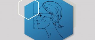
Anatomy of the nasal septum
The nasal septum divides the nasal cavity into two parts, usually unequal in size and configuration due to its curvature. The nasal septum consists of bone and cartilaginous parts. The bony part is formed by the perpendicular plate of the ethmoid bone and the vomer. The perpendicular plate from above in front adjoins the frontal bone and the inner surface of the nasal bones, and behind and below it connects with the upper edge of the vomer. Between the anterior edge of the perpendicular plate and the anterior third of the vomer there is a quadrangular cartilage. The upper edge of the cartilage forms the anterior part of the dorsum of the nose. Adjacent to the anterior edge of the quadrangular cartilage is the medial peduncle of the greater alar cartilage. The cutaneous-cartilaginous, anterior part of the nasal septum, unlike the bone part, is mobile.
The air flow passing through both halves of the nose is asymmetrical. Most healthy people experience a cyclic change in resistance to air flow passing through the left and right halves of the nose (the so-called nasal cycle), but the total resistance remains constant. Alternating changes in air flow in both halves of the nose can be explained by the need for rest to restore the nasal mucosa from microtrauma and functional overload.
The physiological nasal cycle is possible only if the nasal septum does not have a pronounced deformation and is located in the midline. In the case of anomalies in the development of intranasal structures leading to asymmetry of the lumen of both halves of the nose (for example, curvature of the nasal septum), a high degree of resistance to the air flow is constantly created on the side of the narrowing, and the speed of the air stream increases. In this case, the bulk of the air goes through the wider half of the nose. Cyclic changes in resistance are disrupted. Due to constant functional overload, after several years, chronic rhinitis develops in the wider half of the nose, leading to a constant increase in resistance to air flow in the two halves of the nasal cavity.
Thus, the septum, dividing the nasal cavity into two halves, creates a pairing of the organ. Regulated by the nasal cycle, these organs (halves of the nose) function alternately at full load, periodically resting. Complete rest is possible only with the correct position of the partition.
Types of deviated nasal septum
S-shaped curvature of the nasal septum
C-shaped deviated nasal septum
S-shaped (antero-posterior) curvature of the nasal septum
Deviation of the nasal septum in relation to the bone crest of the upper jaw
Deviation of the nasal septum and bone crest of the upper jaw
Diagnostics
The diagnosis is made by an ENT doctor when examining the nasal cavity using a nasal speculum or endoscope. It is important to assess the role and degree of participation of the deviated nasal septum in the difficulty of nasal breathing, since there are other structural features of the nasal cavity (hypertrophy of the mucous membrane, nasal polyps, vasomotor disorders, etc.) that can interfere with normal breathing, and the doctor must decide on which structures and to what extent surgical treatment should be performed.
It should be noted that a deviated nasal septum occurs to a greater or lesser extent in most people, but not all people with a deviated septum need to straighten it.
Possible unpleasant consequences
Complications are possible in three cases: violation of contraindications, non-compliance with the postoperative regimen and medical error. The best way to avoid trouble is to carefully choose your treating specialist and scrupulously adhere to the rules of rehabilitation.
Complication options:
- bleeding - with severe damage to the septum, premature removal of turundas or as a result of a blood clotting disorder (congenital or acquired as a result of taking anticoagulants);
- perforation of the nasal septum - during surgery or during the healing process. Manifested by bleeding, suppuration, mucus discharge from the nasal passages, difficult wheezing;
- hematomas - as a rule, are not dangerous, but increase the recovery period;
- damage to nerve endings - impaired sensitivity of the nose, loss of smell, in more rare cases, changes in hearing/voice, severe pain. Over time, innervation can be restored; in case of severe damage, it disappears forever;
- change in the shape of the nose for the worse (with proper plastic surgery, the shape of the nose should improve);
- infection of tissues with the formation of purulent abscesses. There is a risk of the formation of adhesions that re-block the respiratory passages.
Complications can be pronounced or barely noticeable.
In what cases should you contact the clinic immediately:
- continuous bleeding;
- the pain syndrome is not relieved by standard painkillers;
- severe increase in body temperature;
- complete absence of nasal breathing.
Even with successful execution and the absence of complications, the result may be disappointing - there is a 10% risk that the operated area will restore its original shape due to “tissue memory”. But in most cases, a deviated septum retains its shape until the end of life.
Septoplasty - surgical correction of the nasal septum
Septoplasty usually involves the most gentle surgical removal of the deviated parts of the nasal septum, previously detached from the covering mucosa and perichondrium or periosteum.
Deviated areas of the nasal septum that can be straightened are straightened in place and maintained.
Thus, by preserving our own tissues as much as possible, we ensure the preservation of the structure of the nasal septum, its strength and functionality. Incisions and sutures are made deep in the nasal cavity and are not visible from the outside.
How to prepare for septoplasty
Medical training includes a detailed diagnosis of nasopharyngeal structures with instrumental examination and laboratory tests. Among them, rhinoscopy and x-ray take first place.
There is no special preoperative preparation on the part of the patient. However, if you are scheduled for septoplasty surgery, you must follow a number of recommendations:
- report any allergies to medications;
- tell us about all the diseases you have suffered recently and the courses of treatment prescribed;
- provide a list of medications taken, including traditional medicines, vitamins, dietary supplements, traditional medicine - they may affect blood clotting, compatibility with anesthesia or drugs for the rehabilitation period;
- 2-3 weeks before surgery, avoid taking medications, especially non-steroidal anti-inflammatory drugs and aspirin (if possible);
- in 2 weeks, adjust your diet and give up alcohol and any alcohol-containing products, try to halve your daily intake of cigarettes (ideally, stop smoking altogether);
- The last meal before surgery should be 12 hours before, the last fluid intake should be 6 hours before, the last cigarette should be no later than 2-3 hours before.
Attention, women! The operation should be scheduled no later than a week before and no earlier than a week after menstruation.
Correction of the nasal septum: history of development
The most commonly used operation to correct the nasal septum was developed back in 1904, but is still used with minor modifications. The surgeon peels away the mucous membrane of the nasal septum on both sides and then completely removes the deviated nasal septum cartilage. The operation is simple to perform, but has a number of disadvantages. First of all, this is due to the risk of damage to both layers of the mucous membrane. In this case, a persistent perforation of the nasal septum is formed, which impairs breathing through the nose and which often cannot be closed even with repeated surgery. In addition, since most of the cartilage of the nasal septum is removed, the support of the nasal bridge disappears, which, even with minor trauma, can lead to its failure. However, despite the obvious disadvantages, this operation remains to this day the main type of surgical intervention, which is used in most ENT departments.
Since the middle of the last century, an operation has been developed in which the above disadvantages are minimized. Its peculiarity lies in the fact that the mucous membrane is peeled off on only one side, the cartilage is mobilized and installed in the middle position without removing it entirely.
If the curvature is complex, large fragments of cartilage must still be removed. In this case, everything that has a more or less regular shape is cut into strips and placed again between the sheets of the mucous membrane.
Although this operation is difficult to perform, it is the one that is performed in various modifications in the world's leading clinics.
Preparation for rhinoplasty
Before the operation, it is necessary to conduct a number of studies. In addition to the standard set (blood test, urine test, chest X-ray (fluorography), ECG), radiography of the paranasal sinuses or X-ray computed tomography of the paranasal sinuses is required.
Photos before rhinoplasty:
1 Rhinoplasty: BEFORE
2 Rhinoplasty: BEFORE
3 Rhinoplasty: BEFORE
Photos after the operation:
1 Rhinoplasty: AFTER
2 Rhinoplasty: AFTER
3 Rhinoplasty: AFTER
Modern septoplasty
Currently, gentle methods for correcting the nasal septum have been developed, such as cristotomy, endoscopy, thermoplasty, etc. using a laser, radio frequency system, etc. These methods are used only for minor deformities and can be performed on an outpatient basis.
In modern clinics, the choice of surgical method is determined individually, taking into account the anatomical features and condition of the patient.
The duration of septoplasty, as a rule, is no more than 1 hour, although there are complex deformities that require more time for correction.
The operation ends with the installation of nasal tampons to stop bleeding, as well as to fix the nasal septum in the desired position.
Doctors providing this service
Types of dental anesthetics
Anesthesia means absence or loss of sensation. With light anesthesia, the person is conscious. In severe cases, the patient is put to sleep. The doctor at the Nika Dentistry clinic uses medications separately or in combination. Selects medications for a safe procedure. The type of anesthetic used also depends on the person's age, health, length of the procedure, and previous negative reactions to anesthetics.
Short-term medications are applied directly to the tooth area. Long-acting is used when complex jaw surgery is performed.
The success of dental anesthesia depends on:
- drug;
- areas of anesthesia administration;
- type of procedure;
- time of the operation;
- severity of inflammation.
Local anesthesia in the lower jaw is not as strong as in the upper jaw.
Doctors give three types of anesthesia: local, sedative and general. The choice depends on the location of the manipulation, the severity of the problem and the combination with other medications.
Local anesthesia
Local anesthesia is used for simple procedures such as tooth filling, which require a short time to complete and are less complex. The person is conscious and talking when local anesthesia is given. The area is numb so that the patient does not feel pain. A popular local anesthetic is lidocaine.
Local anesthetics will numb the pain within 10 minutes, and the best anesthesia for dental treatment wears off between 30 and 60 minutes. Sometimes a vasopressor such as epinephrine is added to enhance the effect and prevent the anesthetic effect from spreading to other parts of the body.
Local anesthetics are available in the form of gel, ointment, cream, spray, patch, liquid and ampoules. Use topically (apply directly to the area to numb it) or inject into the area being treated.
Sometimes sedatives are added to anesthetics to help you relax. The patient remains fully conscious and responds to commands after slight sedation. If the drug is moderate, the person is semi-conscious or almost unconscious if the sedation is deep.
Sedatives are administered orally (tablet or liquid), inhaler, intramuscularly, or intravenously. During moderate to deep sedation, the doctor monitors your heart rate, blood pressure, and breathing.
General anesthesia
General anesthesia is used for long procedures or when the client is nervous and interferes with treatment. The person is unconscious, does not feel pain, the muscles are relaxed, and does not remember how the operation took place. The medicine is administered by putting a mask on the face or intravenously. The dose depends on the procedure and the patient's condition.
What are the side effects
The side effects of dental anesthesia depend on the type of anesthetic used. General anesthesia has more risks than local anesthesia. Reactions also vary:
- nausea or vomiting;
- headache;
- sweating or shaking;
- hallucinations, delusions, or confusion;
- slurred speech;
- dry mouth or throat;
- pain at the injection site;
- dizziness;
- fatigue;
- numbness;
- trismus caused by trauma from surgery.
Vasoconstrictors such as epinephrine added to anesthetics also cause heart and blood pressure problems. These are some side effects of anesthetics. Ask your dentist about the medicine and any problems that may arise after use.
Precautions when prescribing
Pregnancy
Special Needs
Aged people
Liver, kidney, lung, or heart problems
Neurological diseases
Other conditions
Tell your dentist if you have a hiatal hernia, acid reflux, infections or open sores in your mouth, allergies, severe nausea and vomiting, or are taking medications that cause drowsiness.
The risks are higher for those who:
- sleep apnea;
- epilepsy;
- obesity;
- hypertension;
- heart problems;
- attention or behavior disorder;
- chronic obstructive pulmonary disease;
- gastric bypass;
- Substance abuse.
What are the risks of dental anesthesia
Most people do not experience adverse reactions with local anesthesia. There is a higher risk with general anesthesia, especially in older people and people with medical complications. There is an increased risk of abnormal bleeding while taking blood thinning medications such as aspirin. If you are taking painkillers or nerve medications, tell your dentist or surgeon so they can select an anesthetic.
Anesthesia risks:
- seizures;
- coma;
- respiratory arrest;
- heart failure;
- heart attack;
- stroke;
- hypotension;
- hyperthermia;
- muscle stiffness;
- breathing problems;
- tachycardia.
Allergic reaction
Tell your dentist about any allergies, including reactions to dyes or other substances: rash, itching, swelling of the tongue, lips, mouth, or throat, and difficulty breathing; The anesthetics articaine and prilocaine at a concentration of 4% damage nerves and cause paresthesia;
After septoplasty
Sometimes after nasal septum surgery, antibiotics are prescribed to prevent infectious complications. In addition, if necessary, the patient receives painkillers. Nasal swabs remain in place for 24 hours after surgery. At this time, it is advisable to observe bed rest.
After removing the tampons, nasal breathing will be difficult at first due to swelling of the nasal mucosa and will improve within a few days. Most patients can return to work 7-10 days after surgery. Physical activity should be limited for 2-4 weeks.
How to remove a tooth
There are two extraction methods used in dentistry: simple and complex. Their choice depends on which teeth are being removed - premolars and molars with tangled branched roots are removed using a complex method. It is very difficult to pull out such elements entirely due to the fact that the tooth socket is penetrated by retaining ligaments and alveolar processes. Errors during the procedure or insufficient experience of the specialist lead to serious complications. Therefore, even despite the acute condition, always find out in advance where you can have a tooth removed from a good doctor with positive recommendations.
Factors complicating the operation:
- complete destruction of the coronal part;
- high fragility;
- acute inflammatory diseases;
- Unerupted or misaligned wisdom teeth.
The technology of the procedure depends on which teeth are removed. In some cases, tissue incision and suturing are performed.
Whether it is painful to remove a tooth or not depends largely on the condition of the element. Anesthesia is performed in all cases, with the exception of severe allergic reactions to all types of painkillers. With the development of extensive purulent lesions, the effect of the drug may be reduced.
How long it takes to remove a tooth depends on the complexity of the operation. On average, this takes no more than 5-10 minutes (including waiting for the anesthesia to take effect). In general, the procedure includes the following steps:
- anesthesia;
- if necessary, an incision is made into the mucous membrane to access the cervical area, or the doctor lowers the gum with an instrument;
- Use forceps to fix the tooth at the lowest point without excessive pressure;
- rocking and extraction from the hole is performed;
- returning the gum flap to its place.
If inflammatory processes are diagnosed in the oral cavity, a course of antibiotics is prescribed. In some cases, extraction is performed under general anesthesia.
In case of multiple lesions, the doctor determines how many teeth can be removed in your case during one visit, but more often it is 1–2 elements. This is because extraction is a traumatic operation that will take time to recover from. Too large areas of damage increase the likelihood of complications several times and take much longer to heal.
If after surgery the extracted tooth, or rather the hole left after it, hurts, this is a reason to consult a doctor. After the procedure, minor pain is allowed during the first 24 hours. Visit the dentist if your temperature rises, swelling increases, bleeding occurs, or pain spreads to the lymph nodes. These are symptoms of a wound infection. Untimely treatment can lead to alveolitis.