What is pityriasis versicolor
Pityriasis versicolor, or pityriasis versicolor, is a chronic skin disease of a fungal nature. It appears in the form of specifically pigmented spots of various sizes and shades, without signs of inflammation. Most often, the areas affected by pityriasis versicolor are located on the body (back, abdomen, neck, shoulders) and scalp. Representatives of both sexes are most susceptible to the disease at a young age, and children under 7 years of age are the least susceptible.
External manifestations of the disease usually do not cause concern, so they are often perceived as harmless cosmetic defects. For this reason, the pathology becomes chronic: spots periodically appear, then disappear, and after some time, under the influence of provoking factors, the disease worsens again.
Symptoms of the disease
When infected with pityriasis versicolor, the pathogens multiply in the superficial layers of the skin, disrupting the normal functioning of cells. First of all, melanocytes, which are responsible for the production of the pigment melanin, are affected, thanks to which the skin has one color or another. This means that under the influence of pathogenic microorganisms, the affected areas acquire an uncharacteristic, painful shade. Hence the second name for pityriasis versicolor - versicolor.
The main symptoms of pityriasis versicolor include:
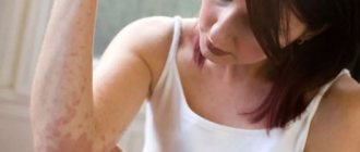
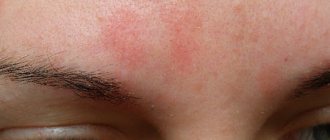
- Spots of yellow, coffee, scarlet or dark brown shades, located mainly on the back, head and neck, shoulders and stomach. In children, they also appear on the limbs, armpits and scalp. The affected areas can be of different sizes, but usually their size does not exceed 1 cm. Subsequently, the spots merge with each other, forming significant areas of the affected skin.
- No signs of inflammation in the form of redness, swelling, hyperemia and pain to the touch.
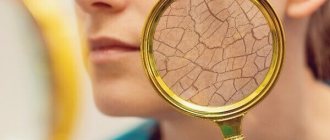
- Severe peeling of the skin in infected areas, aggravated by touch.
- No tanning on the affected areas.
Pityriasis versicolor is not characterized by itching and discomfort, so patients often mistake the manifestations of the disease for minor cosmetic defects, postponing a visit to the doctor and full treatment.
Such spots begin to itch and scratch only if a secondary infection occurs.
Reasons for development
Versicolor versicolor is a type of fungal skin infection that affects the stratum corneum of the epidermis and hair follicles. Its causative agents are two types of fungi, and infection is possible only through prolonged and close contact with the patient. And in this case, provoking factors play a big role. These include:
- weakened immunity;
- hyperhidrosis;
- disruption of the sebaceous glands;
- diseases of the endocrine system (obesity, diabetes, Itsenko-Cushing syndrome, etc.);
- hormonal imbalance due to pregnancy, menopause or taking hormone-containing medications;
- vegetative-vascular dystonia;
- abuse of antibacterial personal hygiene products;
- excessive exposure to ultraviolet rays (intense tanning, frequent visits to the solarium) and regular overheating of the body.
It is noteworthy that patients with pityriasis versicolor over 60 years of age are extremely rare. This is due to natural age-related changes in the skin, which make it less susceptible to pathogens.
In children under 10 years of age, the main causes of pityriasis versicolor infection are neglect of personal hygiene rules or improper skin care. At this age, with the protective functions of the skin intact, the body independently copes with pathogenic microorganisms attacking it, so the development of the disease does not occur. But closer to adolescence, when hormonal changes begin, the body’s susceptibility to bacteria, viruses and fungi increases, so children over 10 years old become infected with pityriasis versicolor just like adults.
Treatment of pityriasis versicolor
The diagnosis of pityriasis versicolor is made to the patient after examination by a dermatologist and dermatoscopy. Additionally, an iodine test and laboratory testing of scrapings can be used.
Treatment of pityriasis versicolor is carried out on an outpatient basis until the symptoms of the disease completely disappear. If measures were taken on time, the patient is prescribed local therapy using antifungal ointments and special agents for exfoliating dead cells.
Additionally, immunomodulators, vitamin complexes, antifungal shampoos and antihistamines are used if the patient is bothered by itching. In the most advanced cases, antimycotic agents are prescribed for oral administration. In addition, in order to avoid relapse of the disease and infection of others, the patient’s clothing and bedding are treated with disinfectant compounds.
Disease prevention also plays an important role. The following will help prevent the development of the disease:
- timely solution to the problem of hyperhidrosis (use of medicinal deodorants, creams, powders, compliance with personal hygiene rules, frequent changes of underwear, etc.);
- use of high-quality soaps and skin care products;
- regular water procedures;
- wearing clothes made from natural, hypoallergenic materials;
- avoiding stress;
- balanced diet rich in vitamins and microelements.
Experts also recommend avoiding overheating of the body and promptly seeking advice from cosmetologists and dermatologists in order to identify the problem and begin proper treatment.
Can I go to work or school with ringworm?
The presence of this diagnosis requires isolation of the patient from the team for at least two weeks. In schools and other children's institutions, it is mandatory to notify parents for timely detection of the disease in other children.
The disappearance of ringworm symptoms and 3 negative tests for the presence of fungus are a reason to return to school or work.
If a dermatomycosis infection has been detected in a team, then to prevent pathology, you can use antifungal shampoos for some time, carry out wet cleaning with the addition of antiseptic solutions, and strictly observe personal hygiene.
- Treatment of lichen in Kharkov;
- treatment of lichen in Uzhgorod;
- treatment of deprivation in Sumy;
- treatment of lichen in Poltava;
- treatment of lichen in Odessa;
- treatment of deprivation in Nikolaev;
- treatment of lichen in Mariupol;
- treatment of deprivation in the Dnieper.
3
1
1
Article rating:
3.8 out of 5 based on 5 ratings
Author: Mangusheva Victoria Yurievna
Dermatovenerologist, trichologist. Candidate of Medical Sciences, doctor of the highest category. Work experience more than 10 years.
What is erythrasma
Erythrasma is a chronic bacterial disease affecting the epidermis layer in the deep folds of the skin. It is characterized by a long course - in some cases, symptoms develop for at least 10 years, without causing significant discomfort to the patient. The clinical picture of erythrasma is similar to a fungal infection of the skin, but modern dermatology classifies it as a group of pseudomycoses.
The following main stages are distinguished in the development of the disease:
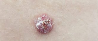
- Progression. The first characteristic spots appear on the skin, their size slowly increases, and additional symptoms develop. In some cases, secondary infections occur. The spots gradually merge with each other, forming large areas of damage.
- Stabilization. New spots do not appear, and existing ones stop growing. Peeling of the skin begins. This stage is usually associated with a change in external conditions, for example, cold weather, during which the intensity of sweating decreases and the skin condition stabilizes.
- Exacerbation or relapse. Usually associated with the beginning of the warm season. But in the case of prolonged erythrasma, the disease constantly develops in waves, and after a slight decline its symptoms again actively appear.
- Remission. Occurs with a favorable microclimate, compliance with preventive measures and proper skin care. The color of the affected areas gradually returns to normal, itching and flaking disappear, and the skin is restored.
Without timely, well-chosen treatment, erythrasma can lead to the development of serious complications.
For example, it can provoke eczema and secondary infection in patients with diabetes or obesity. Also, the course of the disease is aggravated by increased humidity and contamination of the affected areas. As a result, its typical symptoms are complicated by burning, itching and pain.
Signs of the disease
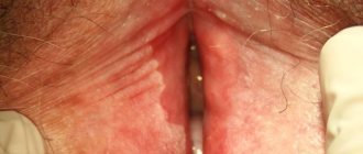
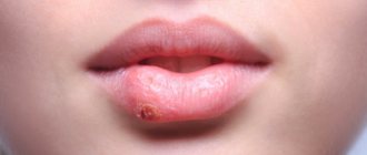
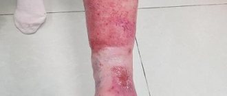
Externally, erythrasma manifests itself in the form of light brown, brick-red, brown or yellow-brown spots on the skin, most often round in shape and without signs of inflammation. The diameter of the lesions can reach several centimeters, and they tend to merge, forming large affected areas. First of all, erythrasma spots appear in the folds of the skin, where there is a favorable environment for the proliferation of bacteria.
In addition to spots, erythrasma is characterized by:
- Peeling of the skin on the affected areas, aggravated by touch. Usually this is where the development of the disease begins.
- Mild, irregular itching. It intensifies and begins to cause significant discomfort only if a secondary infection is added to the primary disease.
- Absence of fever, wounds, ulcers and ulcers with copious discharge. This distinguishes erythrasma from most bacterial skin pathologies.
- Getting wet. An optional symptom, the manifestation of which depends on the amount of sweating and the quality of skin care.
It is noteworthy that in children, symptoms of erythrasma appear extremely rarely. The risk group includes adults, primarily men, who are predominantly overweight and prone to excessive sweating. In this case, in men, the skin in the groin, navel and inner thighs is usually affected, and in women, the entire abdomen, armpits and areas under the breasts are affected.
Hygiene measures when in contact with a sick person
If you or your child have touched a sick animal or interacted with a sick person, then you need to take immediate action.
- The sooner you wash away particles containing fungus from your skin, the less likely you are to become infected.
- Wash your hands several times with antifungal soap. The simplest remedy, which is available in almost every store, is cinnamon laundry soap, or better yet, soap with birch tar.
- Wash your entire body with this soap. Suddenly, particles of the patient’s skin got under his clothes. Do not use a hard washcloth. It leaves micro-scratches on the skin, into which fungus easily penetrates.
- To wash your hair, you must use an antifungal shampoo. For example, Nizoral. You can also use it as a shower gel.
- A modern remedy with a powerful antifungal effect is Citeal. Dilute it in a small container five times. You will end up with a foaming liquid that can be used to wash your hands and entire body.
- Lavender oil, tea tree oil and turpentine have an antifungal effect. They can be used to treat small areas of skin.
Also, five days after contact, it is advisable to consult a dermatologist. He examines the body with a Wood's lamp. If you do become infected, the disease can be detected in the early stages. This will help you quickly treat her at home and avoid going to the hospital.
Causes of pathology
Corynebacteria, which are the causative agents of the disease, are normally always present on human skin. Moreover, they provoke the development of pathology only under certain, favorable conditions. Corynebacteria do not penetrate deeper than the epidermis, and also do not affect nails and hair. Since the appearance of erythrasma is directly related to increased sweating, the disease most often manifests itself in the hot summer season.
Among the main reasons for the development of erythrasma are:
- hyperhidrosis;
- deviation of the normal pH of the skin to the alkaline side;
- diaper rash, constant friction and mechanical damage to the skin;
- dermatitis and other skin diseases;
- neglect of personal hygiene rules;
- wearing synthetic, overly warm clothing;
- the use of low-quality care products or the abuse of soap with an antibacterial effect, which destroys the natural protection of the skin.
Erythrasma is transmitted by contact, most often after the use of clothing, bedding and personal hygiene products of the patient. You can also become infected during sexual intercourse, when visiting a pool or bathhouse, and when walking barefoot on the ground or beach. At the same time, it is not always possible to accurately determine the source of infection, because the carrier may not have obvious external manifestations of the disease in the form of characteristic spots and peeling.
Prevention
The main measures to prevent ringworm are careful adherence to hygiene procedures:
- Using only your own towels and combs, preventing the child or adult from sharing them and using other people’s hygiene items.
- After visiting swimming pools, bathhouses, and water parks, you should immediately wash with soap, wash and wash the items you used.
- If someone in your home shows signs of ringworm, further infestation should be prevented. His bedding and underwear must be washed separately. All items are thoroughly washed and treated with disinfectants.
- Preventing contact with stray animals, especially cats. According to statistics, about 50% of them are infected with microsporia or are its carriers.
- The patient should be isolated from the team until complete recovery.
- If there is a sick animal in the house, all surfaces must be treated with disinfectant solutions.
With careful prevention, ringworm may not appear in other household members.
Treatment of erythrasma
To diagnose a patient with erythrasma, a dermatologist first uses a visual examination. This is especially true for rashes in the groin area, which have characteristic distinctive features in the form of pronounced protrusions and bubbles along the edges. Also, the affected areas of the skin are illuminated with a Wood's lamp and a microscopic examination of the scraping is performed to exclude other diagnoses: pityriasis versicolor or pityriasis rosea, candidiasis, dermatitis or eczema.
Treatment of erythrasma is primarily based on the use of antibacterial ointments that are used to treat the affected areas of the skin.
Under their influence, corynebacteria die, and the spots gradually lighten, decrease in diameter and disappear. On average, such therapy takes at least 7-10 days. Used in parallel:
- Antiseptics. Treatment with them is carried out before applying antibacterial ointment, as well as after it, to maintain dryness of the affected areas and prevent re-infection.
- Antifungal drugs. They are prescribed together with antibacterial drugs, since corynebacteria are similar in structure to fungal micelles.
- Exfoliating ointments. They help cleanse the skin of a layer of dead cells, activating its regeneration.
- Ultraviolet irradiation. Promotes skin disinfection and restoration. Patients benefit from both natural sunbathing and physiotherapeutic UV irradiation.
If the disease has not reached an advanced stage, the use of external medications is sufficient to solve the problem. But in some cases, with multiple skin lesions, to obtain the desired result, patients are prescribed systemic antibacterial therapy.
Diagnosis of microsporia
Diagnosis of microsporia is based on the clinical picture of the disease, epidemic history (contact with cats, dogs or sick people), microscopic and cultural data, and the presence of an emerald glow under a Wood's lamp.
Microscopic examination
Microscopic examination reveals the presence of fungal mycelium and spores. It is impossible to differentiate microsporia and trichophytosis by microscopy. Identification of pathogens is carried out using a culture method followed by microscopy.
The material for microscopy is the patient's scales and hair taken from the peripheral zone of the lesion. Hair breaks off 3 weeks after the onset of the disease.
With microsporia, under a microscope, the affected hair has a characteristic appearance:
- It is surrounded by numerous round spores, tightly adjacent to each other in chains in the form of a mosaic.
- The peripheral internal part is filled with septate mycelium.
- At the end of the hair shaft, a fringe consisting of mycelium threads is sometimes visible.
- In the scales taken for research, branching mycelium with clearly defined sparse partitions can be found.
- In the affected nails, fungal elements are similar to those in the scales.
Rice. 23. Microscopy of the affected hair with ringworm.
Rice. 24. In the photo on the left is a hair affected by Microsporum canis, on the right - Microsporum
Rice. 25. Microscopy of the affected hair with trichophytosis. Pathogen spores envelop the hair like a muff (photo on the left). The inside is filled with spores (photo on the right).
Diagnosis of ringworm using a Wood's fluorescent lamp
At the base of the hair, 10 - 12 days from the moment of infection, an emerald-colored glow appears in the rays of a Wood's lamp. The study should be repeated after 3 days.
Wood's lamp is a quartz lamp. Ultraviolet radiation passes through glass saturated with potassium salts. The examination is carried out in the dark. Hair should be washed with soap before the examination. This technique is widely used by medical professionals to identify sick people and animals.
Rice. 26. The emerald glow when using a Wood's lamp is characteristic only of microsporia.
Rice. 27. Wood's lamp is used to identify ringworm and monitor the effectiveness of treatment. An emerald-colored glow in a person (photo on the left) and an animal (photo on the right).
Differential diagnosis of ringworm
Differential diagnosis of ringworm is carried out with seborrheic dermatitis, a limited form of neurodermatitis, psoriasis, alopecia areata, discoid lupus erythematosus; in the suppurative form of microsporia - with trichophytosis, boil, carbuncle, favus.