Hirsutism in women is a pathology of the endocrine system, in which the patient begins to actively grow hair according to the male type.
Unfortunately, the disease is not limited to excess hair alone: a woman also has to deal with other hormonal disorders. First of all, the menstrual cycle is disrupted, uterine bleeding and anemia may also occur, and often such women become infertile.
According to medical statistics, approximately 8 out of a hundred women experience this type of disorder. These figures are somewhat lower than the statistics on the incidence of diffuse toxic goiter, but this is largely due to the fact that the disease is unique to women.
Sometimes the manifestations of the disease are so obvious that a woman has to look for ways to mechanically combat unwanted hair. Moreover, as can be seen in the photo of hirsutism in women, this hair is hyperpigmented, that is, noticeably different in color from the rest of the hair on the body.
Unfortunately, this disease is not only a cosmetic problem: it is a serious pathology that requires correction by several specialists, primarily a gynecologist and endocrinologist.
Causes of hirsutism
The pathology is mainly caused by an increased level of androgens (male hormones) in the blood.
Often the disease is combined with other manifestations of hormonal disorders: menstrual irregularities, inability to conceive. Depending on the concentration of androgens, an increase in muscle mass, an enlargement of the clitoris, loss of hair in the temporal region, deepening of the voice, etc. may also be observed.
Hirsutism in women can occur for the following reasons:
- premature puberty or menopause;
- taking certain medications;
- pathologies of the adrenal glands;
- disorders of ovarian function;
- disorders in the pituitary gland;
- gene mutations.
Hirsutism in girls and adolescents
Hirsutism refers to excessive hair growth in females according to the male pattern, i.e. the appearance of terminal hair on the upper lip, chin, sternum, upper back, midline of the abdomen, sacrum, buttocks, inner thighs. Hirsutism is not only a cosmetic problem, but also often a symptom of serious diseases.
Physiology of hair growth
There are about 50 million hair follicles on the human body, and only a small part (100–150 thousand) are located on the scalp. Only the palms, feet, and lips are free from hair. The main part of hair follicles is formed from ecto- and mesoderm in the prenatal period. These elements are in close contact throughout the life of the hair follicle.
The first hair appears in a person at the end of the 3rd month of intrauterine life, and at the 7th month - vellus hair (lanugo), which covers all parts of the body with the exception of the palms, soles, and breast nipples. Lanugo is soft, delicate, fine hair without medulla, about 2 mm in length, moderately pigmented. As the fetus develops, their number decreases, and by the fourth month of the child’s life they completely fall out.
Before birth, they are replaced with baby hair. Terminal hair forms at the beginning of puberty - this is thick hair on the pubis and armpits, and in men on the face, abdomen, and limbs.
Hair growth has a lot to do with gender. Men have more hair and it grows faster. This is caused by the presence of a high concentration of androgens in their body. With an increase in this hormone, more body hair appears.
There are three phases of hair growth. The active phase is anagen, followed by the involution phase (catagen), in which the hair stops growing and the hair follicle decreases in size, and finally the telogen phase, in which the old hair is replaced by a new one. The duration of catagen and telogen is on average from 2–3 weeks to 3–4 months, respectively. The ratio of anagen to telogen hairs is used to determine hair growth activity, with a higher ratio indicating active growth. The frequency and location of hair follicles in the skin is an individual feature of the body [1].
Each hair consists of a shaft and a root. The entire hair root is enclosed in a hair follicle. Cells of the papillary dermis of androgen-dependent areas have a large number of androgen receptors compared to other areas of the skin.
All androgens are C19 steroids, derivatives of androstane, and have different biological activities. The main sources of androgen production in the female body are the theca cells of the ovaries, the reticular layer of the adrenal cortex, peripheral conversion from steroid hormone precursors (androstenedione, dehydroepiandrosterone (DHEA)) in the liver, muscles, skin and adipose tissue.
In circulation, along with testosterone, 5-androstenedione, 4-androstenedione, androsterone and DHEA, which have little biological activity, are determined. They are precursors or products of testosterone metabolism.
In target cells (skin, muscle, adipose tissue), testosterone is exposed to the tissue enzyme 5α-reductase, turning into 5α-dihydrotestosterone (DHT), which has maximum androgenic activity. Free fractions of testosterone have greater biological activity. Penetrating into the target cell, they bind to the nuclear receptor. Under physiological conditions, androgens act as anabolic factors [2].
Sex hormones, mainly androgens, are transported by a specific protein - sexsteroid binding globulin (SSBG). 1–3% of androgens circulate in a free state, which have a biological effect on cells. Its level decreases under the influence of androgens, glucocorticoids, growth hormone, thyroid hormones, and excess body weight.
Inactivation of sex steroids occurs mainly in the liver due to the processes of conjugation with glucuronic acid and sulfonation. The concentration of steroids in the blood is determined by the interaction of biosynthesis and degradation activities.
Sexual hair growth depends on androgen levels. Before puberty, the hair is delicate (short length, straight, light), and the sebaceous glands of the skin are little active. In response to increased androgen levels during puberty in hormone-sensitive areas, children's hair transforms into coarse terminal hair (darker, curlier). High levels of androgens are necessary for hair growth in the axillary and pubic areas. In other areas of the skin, such as the forehead, for example, the same level of androgens increases the activity of the sebaceous glands, but does not cause differentiation of vellus hair. The reasons for this are currently not entirely clear [3].
In the periphery, 5α-reductase activity is increased by local growth factors and circulating androgens. In the body, dihydrotestosterone stimulates: the production of sebum, the differentiation of the hair follicle from vellus hair to axial hair, and prolongs the anagen phase. As a result, the hair thickens.
Ethnic origin significantly influences the growth of terminal body hair. Differences between hair levels in humans are thought to be related to the sensitivity of the hair follicle to the enzyme 5α-reductase, as well as androgen receptor polymorphism. Northern peoples have the least amount of terminal hair, while Southern Europe and dark-skinned Mediterranean women have a lot of it.
Pathogenesis of hirsutism
Hirsutism is the result of an imbalance between hormone levels and the sensitivity of hair follicles to androgens. It is an important symptom of androgen-dependent dermatopathy. The severity of this symptom depends on many factors: the ethnicity of the woman, the duration and depth of the pathological process, the spectrum and level of secreted androgens, which give a different virilizing effect.
In the population, the majority of girls whose blood androgen levels are 2 times higher than the average norm have a moderate degree of hirsutism. However, the severity of hirsutism does not always correlate with androgen levels, as androgen sensitivity varies significantly between individuals. Some women with hyperandrogenism syndrome do not have hirsutism, but have other skin manifestations such as seborrhea, acne vulgaris, and alopecia. But the appearance of terminal hair in the lower abdomen, lower back, near the nipples, on the arms and legs is normal.
The level of testosterone in the middle phase of the menstrual cycle varies greatly during the day - the maximum level is in the morning and lower before the onset of menstrual flow. The biologically active part of testosterone - the free fraction - can increase, while the level of total testosterone will be within normal limits. This reflects the relatively low level of sexsteroid-binding globulin, which determines the proportion of free and albumin-bound plasma testosterone [4].
So, the pathogenetic mechanisms of the development of hirsutism are extremely diverse and insufficiently studied.
Causes of hirsutism
Androgen-dependent forms of hirsutism are the most common (75–85%). In the female body, hirsutism, acne, seborrhea, hair loss, deepening of the voice, clitoral hypertrophy can become the first and sometimes the only early diagnostic signs of hyperandrogenemia syndrome.
Polycystic ovary syndrome
Polycystic ovary syndrome (PCOS), ovarian sclerocystosis, Stein-Leventhal syndrome. A disease based on the process of cystic ovarian degeneration. It is characterized by oligo- or amenorrhea, excess body weight, hirsutism, acne vulgaris, alopecia, enlarged, polycystic ovaries and anovulatory cycles. PCOS, including incomplete forms, is detected in 70–80% of women. The pathogenesis of the syndrome has not been fully elucidated. Occurs in 1.4–2.5% of girls examined for amenorrhea. Current evidence suggests that various factors are involved in the formation of polycystic ovary syndrome. This is a disorder of the secretion of sex steroids in the ovaries; disorders in the hypothalamus-pituitary-ovarian system, androgen synthesis in the adrenal glands, ovaries; receptor damage in effector cells for these hormones involved in the implementation of biological effects.
A certain contribution to the development of polycystic ovary syndrome is made by insulin resistance, which is associated with impaired action of insulin at both the receptor and post-receptor levels, as well as hyperprolactinemia [5].
The disease is detected during puberty due to menstrual irregularities (primary or secondary amenorrhea). At the same time, hirsutism of varying severity develops. Hair growth can be above the upper lip, around the nipples of the mammary glands, along the white line of the abdomen, on the thighs. Most patients have varying degrees of obesity. The external genitalia are formed according to the female type. Only in some patients does increased androgens cause enlargement of the clitoris.
The diagnosis is confirmed by an almost 2-fold increase in luteinizing hormone (LH), with normal or even reduced levels of follicle-stimulating hormone (FSH). The LH/FSH ratio is always increased. In half of the patients, the content of testosterone and dehydroepiandrosterone sulfate (DHEA-S) was increased, in a third - prolactin. Carrying out a test with gonadoliberin (LH-RH) causes a hyperergic reaction with a sharp increase in LH and lack of response from FSH. A dynamic study of the hormones LH, FSH, estrogen, progesterone reveals monotonous indicators, which confirms the absence of an increase in rectal temperature. This indicates anovulatory cycles [6].
Ultrasound examination (ultrasound) shows the ovaries are enlarged in size, the capsule is dense, the stroma is well defined, and numerous cysts are detected.
Aromatase deficiency
Aromatase is an enzyme necessary for the conversion of testosterone to estradiol (E2) and androstenedione to estrone (E1).
Aromatase deficiency in girls leads to the absence of estrogen-dependent signs of puberty and the appearance of androgenization symptoms. In newborn girls (XX), symptoms of virilization of the external genitalia (hypertrophy of the clitoris, fusion of the labial suture) are observed.
During puberty, girls with aromatase deficiency have no enlargement of the mammary glands and no menstrual function. Symptoms of virilization intensify. Polycystic changes are observed in the ovaries.
Laboratory tests reveal high levels of testosterone, androstenedione, dehydroepiandrosterone and its sulfate. Estrogen levels are significantly reduced. Gonadotropic hormones are increased. Genetic studies reveal a mutation in the aromatase gene CYP19. Estrogen therapy has a positive effect on breast development and the onset of menarche [7].
Hyperprolactinemia
The appearance or intensification of hirsutism, especially in patients with oligoamenorrhea or amenorrhea, may be due to hyperprolactinemia (HP). An increase in prolactin secretion directly stimulates steroidogenesis in the adrenal glands, therefore, in patients with pituitary adenoma, the content of DHEA and DHEA-S significantly increases with moderate testosteronemia [8].
The secretion of prolactin is regulated by the hypothalamus, which produces prolactoliberin and prolactostatin (dopamine). The pathogenesis of hypogonadism with hyperprolactinemia is primarily due to the suppression of impulse secretion of LH-RH by excess prolactin and a negative effect on the processes of steroidogenesis in the gonads. Violation of the hypothalamic regulation of prolactin secretion - a decrease in dopaminergic influence or an increase in the production of prolactoliberin - leads to hyperplasia of pituitary lactophores with the possible development of micro- and macroadenomas.
The earliest symptom of HP is menstrual dysfunction, which is a reason for patients to consult a doctor. The examination allows, in some cases, to identify a pituitary adenoma at the microadenomas stage. Prolactinomas, according to the children's clinic of the Research Center of the Russian Academy of Medical Sciences, account for 22% of diagnosed pituitary adenomas. More often they were detected in girls during puberty and manifested themselves in the form of primary amenorrhea syndrome.
Hyperprolactinemia occurs in tumor diseases of the hypothalamus and pituitary gland, damage to the pituitary stalk, “empty sella” syndrome, as well as injuries and inflammatory processes of the base of the skull [9].
According to A. Tuzcu et al. Elevated prolactin levels are associated with more severe manifestations of hirsutism and hyperandrogenic ovarian dysfunction, with more pronounced insulin resistance. It has not yet been clarified whether HP is the cause or consequence of hyperandrogenism in some patients [10].
Congenital adrenal dysfunction (CAD)
This is a large group of diseases that have one or another genetic enzymatic defect at various stages of the biosynthesis of steroid hormones, leading to insufficient secretion of cortisol. Cortisol deficiency stimulates the production of pituitary adrenocorticotropic hormone (ACTH) in the adenohypophysis, which causes adrenal hyperplasia. The clinical picture depends on the level of enzymatic disorder. In these diseases, androgen precursors accumulate.
The severity of the influence of androgens in 21-hydroxylase deficiency is associated with individual characteristics of the metabolism of androgen precursors and differences in the activity of peripheral androgen receptors.
If the diagnosis of CAH is not made in a timely manner and appropriate treatment is not started, then due to the anabolic effect of androgens in the first years, girls grow rapidly, their skeletal muscles actively develop, a rough voice appears, and hirsutism (male-type hair growth on the face, chest, abdomen, and limbs) , i.e. signs of masculinization. In patients, the size of the clitoris increases and its tension is noted [11].
Girls in pre- and puberty do not have secondary sexual characteristics and menstruation. Increased secretion of androgens by the adrenal glands, according to the feedback principle, blocks the release of gonadotropins in the adenohypophysis. For this reason, in patients the ovaries are reduced in size with multiple cysts, the uterus is underdeveloped. Skeletal differentiation is significantly accelerated (“bone” age is ahead of passport age). By the age of 10–12 years, the epiphyseal zones of bone growth occur, which determines the final short stature of patients. Their body proportions are disturbed: wide shoulder girdle, narrow pelvis, well-developed muscles. Girls do not develop mammary glands.
With the expansion of diagnostic capabilities, more and more variants of non-classical pubertal variants of CDCN are being identified. The first clinical symptom may be accelerated isolated pubarche. In the juvenile period, with moderate hyperandrogenemia, hirsutism develops with accompanying symptoms. Hyperandrogenic ovarian dysfunction is formed.
The non-classical variant of 21-hydroxylase deficiency with manifestations of hirsutism and hypertrichosis is very common in some ethnic groups of Yugoslavia, Spain, and Ashkenazi Jews.
Premature adrenarche
Premature adrenarche (PA) is the appearance of isolated pubic and/or armpit hair in girls under 8 years of age (usually at the age of 6–8 years). PA may be a normal variant, given that the maturation of the zona reticularis of the adrenal cortex begins at 6 years of age. While the secretion of gonadoliberins, responsible for the onset of puberty, starts later. The cause of pubertal hair growth is an increase in the production of DHEA and DHEA-S by the adrenal glands, as well as 4-androstenedione, testosterone precursors that stimulate pubic and axillary hair growth. In girls, PA may be associated with excessive peripheral conversion of testosterone to dihydrotestosterone (increased 5a-reductase activity). In the absence of other signs of androgenization of the body - accelerated growth, skeletal maturation, pre-pubertal size of the uterus and ovaries, normal testosterone levels and moderately increased DHEA-S, the prognosis is favorable and sexual development does not deviate from the norm [12].
However, in some children, PA can be triggered by excess production of ACTH (hydrocephalus, meningitis, etc.). There is increasing evidence linking PA with non-classical forms of CAH and, in particular, deficiency in the activity of the enzyme 21-hydroxylase and, less commonly, 3β-hydroxysteroid dehydrogenase. The mechanism of PA is associated with the early maturation of the zona reticularis of the adrenal cortex, evidence of this can be the increased level of DHEA-S. Secretion of adrenal androgens has been shown to be stimulated by ACTH and suppressed by dexamethasone.
In the presence of a virilizing disease, clinical signs of androgenization appear: clitoral hypertrophy, high posterior perineal commissure, hirsutism, development of the muscular system; activation of sebaceous and sweat glands. These children experience accelerated growth and bone age. It should be noted that adrenarche precedes the increase in gonadotropins by about 2 years and is not associated with either an increase in the sensitivity of gonadotrophs to LH-RH or an increase in the amplitude and frequency of nocturnal LH surges.
Girls with premature adrenarche should be at risk for developing polycystic ovary syndrome. This group of patients requires corrective therapy with glucocorticoids [13].
Hyperandrogenism in patients with thyroid dysfunction (hypothyroidism) is based on a significant decrease in the production of globulins that bind sex steroids. Due to a decrease in the level of CVD, the rate of conversion of androstenedione to testosterone increases [14].
Obesity
About half of those with hirsutism are overweight. With the progression of obesity and with increasing age of children, complications or accompanying diseases appear.
Currently, abdominal fat is considered as an active hormone-producing organ that secretes a large number of factors (adipokines) with various effects. They are involved in the regulation of energy balance, cardiovascular system, endocrine system, etc.
Obesity is accompanied by the development of serious complications, including insulin resistance, hyperglycemia, dyslipidemia, arterial hypertension, and hyperandrogenism. Excessive amount of adipose tissue can affect the timing of the child’s puberty and hormonal balance.
Hyperinsulinemia associated with obesity contributes to the development of hyperandrogenemia [5]. Insulin and insulin-like growth factor 1 have the ability to increase the androgen response of ovarian theca cells to LH stimulation. It is believed that insulin increases the sensitivity of the adrenal glands to the action of ACTH. Hyperinsulinemia that occurs in obesity inhibits the hepatic production of CVD, which leads to an increase in free plasma testosterone.
Girls with obesity and PA, adolescents with PCOS have higher values of insulin resistance, a greater decrease in CVD levels and an increase in androgen concentrations compared to their peers with exogenous constitutional obesity. Girls with PA are at risk for developing polycystic ovary syndrome.
M. Mustaqeem et al. studied ethnic differences in clinical and biochemical parameters of South Asian and European women with PCOS (47 Asian and 40 European women) and controls (11 Asian and 22 European women). They reported a significantly higher prevalence of hirsutism, acne, acanthosis nigricans, and secondary infertility in Asian women with PCOS. Insulin resistance is considered to be the central link in the pathogenesis of PCOS, but the specific causes are not yet clear and are being actively studied [15].
Androgen-secreting tumors
Androgen-secreting tumors of the adrenal glands (androsteromas) are usually classified as adrenocarcinomas. They are rare in children. In early adolescence, the frequency of adrenocarcinomas increases in children with Wiedemann-Beckwith syndrome (visceromegaly, macroglossia, hemihypertrophy) and Li-Fraumeni syndrome (multiple malignant neoplasms).
In patients with adrenocarcinomas, abnormal expression of tumor markers and decreased expression of factors that suppress tumor growth, the genes of which are localized on the long arm of chromosome 11, were revealed. Abnormalities of this chromosome are detected in most patients with adrenocarcinoma.
Girls show signs of virilization: apocrine glands (sweat, sebaceous, hair follicles) are activated, body weight increases due to muscle tissue, the clitoris hypertrophies, and growth accelerates [2].
Steroid-secreting gonadal tumors
Steroid-secreting gonadal tumors are uncommon in childhood. In older girls, arrhenoblastomas (malignant tumors) are found, located in the cortex or in the area of the ovarian hilum. Undifferentiated tumors have a more pronounced virilizing effect, while differentiated ones have both a weakly expressed masculinizing and feminizing effect [16].
Excessive growth of terminal hair with a male distribution is often found in girls with hypothalamic syndrome during puberty, manifested by menstrual irregularities (oligomenorrhea, amenorrhea, uterine bleeding).
In some patients with true precocious sexual development (PPD), the cause of the disease cannot be identified. In such cases, when organic diseases of the central nervous system are excluded, a diagnosis of the idiopathic form of PPR is made. However, the improvement of research methods (computer and magnetic resonance imaging) of the brain makes it possible to more often identify the cause of the cerebral form of hyperandrogenemia (hamartoma, glioma, hCG-secreting tumors, etc.) in girls.
Cornelia de Lange syndrome
A rare genetic syndrome with an overall prevalence of 1.6–2.2:100,000 people. It is dominantly inherited, most cases are associated with de novo mutations. The syndrome is genetically heterogeneous, half of the cases are associated with mutations in the regulator gene NIPBL and HDAC8, about 5% of cases are associated with mutations in the SMC1A gene (SMC3, RAD21), which encodes a protein in the cohesin complex. The clinical picture is very variable, typical are facial dysmorphia, microcephaly, hearing loss, vesicoureteral reflux, intrauterine growth retardation and slower postnatal growth of the child, malformations of the limbs and genital organs, hirsutism, congenital heart defects and gastrointestinal tract defects. Distinctive facial features: low hair growth, thick fused eyebrows, thick and long eyelashes. Hirsutism is observed in 78% of patients. More than half of newborns experience increased hairiness on the back, and sometimes on the entire body.
The syndrome is often accompanied by epilepsy, behavioral problems in the form of attention deficit disorder, aggression, including auto-aggression. Specific treatment and prevention have not currently been developed.
Rubinstein Taybi syndrome
Rare genetic disease. Average incidence is 1 in 10,000–300,000 births. The disease is characterized by varying degrees of intellectual impairment, specific facial features and hand structure (abnormally wide and often curved thumbs), and dysphagia. Hirsutism occurs in half of the cases. The disease is associated with a de novo gene mutation and can be inherited in an autosomal dominant manner. The most common variant is a defect in a gene located on the short arm of chromosome 16 (16p13.3). The syndrome is based on a defect in the CREBBP and EP300 genes; patients with the EP300 mutation have less severe skeletal abnormalities. On the contrary, patients with a deletion of chromosome 16, containing the EP300 and CREBBP genes, have severe multiple malformations.
Diagnosis is often made phenotypically. Features of the facial structure: anti-Mongoloid eye shape, long eyelashes, arched eyebrows, high palate, low-hanging nasal septum (columella). There are many complications associated with this syndrome, for example, heart and kidney defects, obesity, otitis media, and a high risk of neoplastic processes.
Donahue syndrome
An extremely rare genetic disease characterized by impaired insulin tolerance, growth retardation, and endocrinopathies. It may be caused by a disorder in the gene located on the short arm of chromosome 19 (19p13.2), which is responsible for the regulation of insulin receptors. Children with Donahue syndrome experience delayed postnatal growth and impaired bone maturation. Low muscle mass. Children have a peculiar facial structure: low-set, poorly developed ears, thick lips and a large mouth, a flat bridge of the nose, widely spaced eyes, and microcephaly.
In most cases, there are structural features of the skin: thickening, darkening (acanthosis nigricans), hirsutism, acanthosis nigricans. Hyperandrogenism of ovarian origin.
Girls have enlarged mammary glands and clitoromegaly. Due to persistent hyperinsulinism, severe fasting hypoglycemia. The prognosis is unfavorable.
HAIR-AN syndrome
The subphenotype of polycystic ovary syndrome, manifested by hyperandrogenism, insulin resistance, obesity and acanthosis nigricans, has been known for more than 30 years. PCOS is one of the most common causes of menstrual irregularities in girls. Statistics on visits to outpatient clinics among adolescents over a 2-year period showed that out of 1002 girls (age 10–21 years), 5% (50) were diagnosed with HAIR-AN syndrome. The average age of the patients was 15.5 years, the average weight at the time of diagnosis was 94.5 kg (BMI 33.3 kg/m2). Treatment is based on weight loss and the use of metformin. 80% of patients had a response to treatment, 95% established a regular menstrual cycle, hirsutism and acne decreased [15].
About 5% of girls with hyperandrogenism and insulin resistance have acanthosis nigricans. Patients with HAIR-AN syndrome may have other signs of virilization with normal adrenal function, amenorrhea. Patients usually have normal levels of LH and FSH, although their ratio may be disrupted.
Symptoms of hirsutism can be provoked by long-term use of hormonal drugs: anabolic steroids, androgens, glucocorticoids, estrogen-containing drugs (oral contraceptives), etc. Antidepressants and some cytostatics provoke active hair growth.
Constitutional hirsutism
Excessive hair growth is genetic. Hereditary predisposition leads to the development of the so-called constitutional form of hirsutism. In this case, hirsutism is often regarded not as a pathology, but as one of the normal variants. It is found in women of the Mediterranean and Middle East. The cause of hereditary hirsutism is considered to be increased sensitivity of hair follicles to dihydrotestosterone. Even a small amount of androgens, which is normal for women, leads to faster and more abundant hair growth. Excessive hair growth begins to appear in childhood and reaches a peak during adolescence. There are no other symptoms of androgenization. In case of excessive hair growth, it is recommended to consult a dermatologist or cosmetologist and undergo regular hair removal.
Idiopathic hirsutism
The diagnosis of “idiopathic hirsutism” is made in cases where no cause for excess hair growth can be found. According to clinical symptoms and development mechanism, it is very close to the hereditary form of excess hair growth. Patients with idiopathic hirsutism do not have other complaints associated with androgenemia. Unlike hereditary hirsutism, the disease may not appear in adolescents, but at a later age. In these cases, the mechanisms that influence the sensitivity of hair follicles to androgens are not clear.
Differential diagnosis
Hirsutism should be distinguished from hypertrichosis, a general excess hair growth that is a consequence of a person’s constitutional characteristics. With hypertrichosis, excess hair is observed in areas of the body that are not typical for women (on the limbs, chest). As a rule, these areas of the skin are covered with vellus bleached or dark-colored hair. With hypertrichosis, there is no increase in hair growth only in androgen-dependent areas and is not directly related to hyperandrogenemia, although this may aggravate the manifestation of hirsutism. Hypertrichosis can be a consequence of the use of medications such as steroids, immunosuppressants, etc.
Survey
The heterogeneity of hirsutism requires a thorough search for its cause. You should pay attention to the patient’s age, relationship with medications, family history of hirsutism, and ethnicity. The severity of hirsutism is assessed using the Ferriman-Gallway scale, which allows you to assess the prevalence of coarse hair in nine androgen-dependent areas - the upper lip, chin, shoulders, chest, upper and lower abdomen, back, lower back, hips. The assessment is made on a five-point scale, and the overall severity of hirsutism can vary from 0 to 36 points. If the indicator is 8 points or higher, then we can talk about the presence of hirsutism.
During the examination, it is necessary to measure height, weight, subcutaneous fat distribution, body mass index (BMI), waist and hip circumference, and blood pressure. Determine the stage of sexual development. Note skin changes.
Laboratory tests are carried out to confirm the diagnosis of hyperandrogenemia in the appropriate clinic and to identify the source of excess androgens: adrenal glands, ovaries, central nervous system.
- Biochemical blood test: total cholesterol, LDL, HDL, triglycerides, enzymes (ALT, AST).
- Fasting glucose, glucose tolerance test.
- Hormonal profile: insulin, C-peptide, LH, FSH, cortisol, testosterone, DHEA, DHEA-S, prolactin, 17-hydroxyprogesterone, according to TSH, T4, SSSH.
- Bone age.
- Bioimpedance measurements are used to assess fat mass.
- Ultrasound of the liver, pancreas, kidneys, adrenal glands, ovaries.
- Molecular genetic research.
- To clarify the cause of hirsutism, ultrasound or computed tomography of the pelvic organs and adrenal glands, and magnetic resonance imaging of the brain are recommended.
- To exclude tumors in the ovaries, diagnostic laparoscopy is performed.
Treatment
Treatment of hirsutism, if caused by hyperandrogenemia, is difficult. The main goal of treatment is to normalize the secretion of steroid hormones that cause excess hair growth and other symptoms of androgenization. This pathology may be caused by a disorder in the gonadostatic system at various levels, requiring an individual approach to the choice of treatment method. The use of only cosmetic measures, especially in severe forms of the disease, leads to a short-term clinical effect and extremely rarely to a cure. Diagnosis and treatment of patients with hirsutism is usually carried out by an endocrinologist, gynecologist, or dermatologist.
The main problem for teenage girls with hirsutism is psychological complexes due to a cosmetic problem. Mental changes are characterized by symptoms of depression. Teenagers are depressed and almost always see the reason for their failures in defects in appearance. They need psychological support. Carrying out psychocorrective measures under the supervision of a doctor and psychologist will allow girls with hirsutism to take responsibility for control and become accomplices in the treatment of their disease.
Correctly carried out psychocorrection helps to reduce the clinical manifestations of the disease, increase the social activation of patients, their adaptation in the family and society, and also increases the effectiveness of treatment measures.
Literature
- Skripkin Yu. K., Kubanova A. A., Akimov V. G. Skin and venereal diseases: textbook. 2011. 544 p.
- Dedov I. I., Semicheva T. V., Peterkova V. A. Sexual development of children: norm and pathology. M., 2002. 232 p.
- Ragimova Z. E., Kail-Goryachkina M. V. Interdisciplinary aspects of androgen-dependent dermopathy (literature review) // Consilium Medicum. Dermatology. 2016, 3: 56–62.
- Guide to pediatric endocrinology / Ed. Charles G. D. Brooke, Rosa Lind S. Brown: trans. from English edited by V. A. Peterkova. M.: GEOTAR-Media, 2009. 352 p.
- Gurkin Yu. A. Children's and adolescent gynecology. M.: MIA, 2009. pp. 148–180.
- Bogatyreva E. M., Novik G. A., Kutusheva G. F. Phenotypes and endotypes of hyperandrogenism syndrome in adolescent girls // Treating Physician. 2016, no. 2, p. 70.
- Wilson JD, Aiman J., MacDonald PC The Pathogenesis of Gynecomastia // Advanc. in intern. Med. 1980, 25: 1–32.
- Moskovkina A.V., Puzikova O.Z., Linde V.A., Rybinskaya N.P. Hyperprolactinemia in adolescent girls with hyperandrogenism syndrome // Children's Hospital. 2013, no. 2, p. 35–39.
- Kokolina V.F. Gynecological endocrinology of children and adolescents. M.: Medical Information Agency, 2001. 287 p.
- Zhurtova I. B. Hyperprolactinemia syndrome in children and adolescents. Optimization of diagnostic and treatment methods. Author's abstract. diss. Doctor of Medical Sciences, M., 2012.
- Tuzcu A., Bahceci M. et al. Is hyperprolactinemia associated with insulin resistance in non-obese patients with polycystic ovary syndrome // J Endocrinol Invest. 2003; 26:655–659.
- Dedov I. I., Peterkova V. A. Guide to pediatric endocrinology. M.: Universum Publishing, 2006. 600 p.
- Endocrinology. National leadership. Brief edition / Ed. I. I. Dedova, G. A. Melnichenko. M: GEOTAR-Media, 2013. 752 p.
- Dedov I. I., Peterkova V. A. Federal clinical recommendations (protocols) for the management of children with endocrine diseases. M.: Praktika, 2014. 442 p.
- Onal ED, Saglam F., Sacikara M., Ersoy R., Cakir B. Thyroid autoimmunity in patients with hyperprolactinemia: an observational study // Arq Bras Endocrinol Metabol. 2014, Feb
- Mustaqeem M., Sadullah S., Waqar W., Farooq MZ, Khan A., Fraz TR Obesity with irregular menstrual cycle in young girls // Mymensingh Med J. 2015, Jan; 24 (1): 161–167.
- Peterkova V. A., Semicheva T. V., Gorelyshev S. K., Lozovaya Yu. V. Premature sexual development. Clinic, diagnosis, treatment. A manual for doctors. M., 2013. 40 p.
V. V. Smirnov1, Doctor of Medical Sciences, Professor A. A. Nakula
Federal State Budgetary Educational Institution of Russian National Research University named after. N. I. Pirogova Ministry of Health of the Russian Federation, Moscow
1 Contact information
Diagnosis of hirsutism
To make a diagnosis and draw up an individual treatment program, the doctor will collect an anamnesis (the nature of the development of the pathology, a list of medications taken, features of menstrual function). Next, laboratory tests will be ordered to determine the level of hormones in the patient’s blood.
Instrumental studies are also prescribed (ultrasound, CT, MRI of the ovaries and adrenal glands, MRI of the brain). To exclude the presence of a tumor in the ovaries, diagnostic laparoscopy may be performed.
Causes and mechanism of development of hirsutism
There are 3 types of hair:
1. Original fluff
- thin, delicate hairs on the body that appear and disappear during the period of intrauterine development of the fetus. They can only be seen in newborns born prematurely.
2. Vellus hair
- thin blond hair, the length of which does not exceed one or two centimeters.
3. Terminal hair
- hard shaft hair with pronounced pigmentation. Short, coarse hair forms eyelashes and eyebrows, and long hair grows on the head, in the armpits and external genitalia, and in men also on the face, chin, back, and abdomen.
Vellus hair is transformed into shaft hair due to the influence of male sex hormones. In girls and adult women, androgen-dependent hair growth in the pubic and armpit areas is considered natural. Hair growth on the legs and forearms is not associated with the influence of androgens. The quantity and quality of hair on a woman’s body depends on a number of factors, such as ethnicity, levels of sex hormones, and the degree of skin resistance to androgens.
Being the direct precursor of estrogens - female sex hormones, androgens in the female body are produced in the ovaries and adrenal glands, and determine the level of development of sexual desire, hair growth of the external genitalia, muscle mass, bone growth in adolescence and the closure of growth zones. In order for the ovaries to function normally, the level of androgens plays a very important role.
Peripheral tissues are influenced by testosterone and dihydrotestosterone. It is these hormones that promote the growth of coarse pigmented hair. Androgens, in turn, lengthen the activity phase of the hair follicle, increase its size and hair diameter, and also increase the production of secretion by the sebaceous glands of the scalp and face. As a result of this influence of androgens, zonal changes in hair growth are observed, namely, hair falls out from the scalp, and in androgen-dependent areas of the body, their number increases sharply.
Increased androgen production
may be observed with:
— functional disorders of the female reproductive glands
- polycystic ovary syndrome, tumor processes in the ovaries, amenorrhea of hypothalamic nature, etc. In this case, there is a disruption in the frequency of menstrual flow, infertility and enlarged ovaries. This is the most common cause of hirsutism;
— functional disorders of the adrenal glands
as a result of congenital or acquired pathology of the cortex, or as a result of an adrenal tumor;
— functional disorders of the pituitary function
, which is observed in acromegaly, prolactinoma, Itsenko-Cushing syndrome.
Hyperandrogenism in women can develop as a result of taking a certain group of medications (corticosteroids, anabolics, interferon, streptomycin, androgens, etc.)
Hirsutism can also be hereditary, and it is more often detected in representatives of the Mediterranean and Caucasian ethnic groups, and less often in Asian and European women.
If the cause of hirsutism is not identified, they speak of idiopathic androgen excess syndrome
, which is characterized by increased sensitivity of skin and hair follicle receptors to it. This form of hirsutism is also one of the most common, occurring in a quarter of all hirsutism presentations. At the same time, the ovulatory function does not suffer, the menstrual cycle and reproductive function are preserved.
Treatment of hirsutism
Treatment of the disease is aimed at eliminating the underlying cause of the pathology. However, a mild form of the disease, which is not accompanied by menstrual irregularities, does not require special medical or surgical treatment. In some cases, to normalize hormonal levels, the gynecologist prescribes oral contraceptives (selected individually).
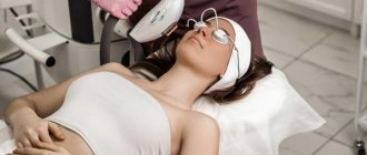
Symptomatic treatment of hirsutism consists of hair removal using one or another method. The most effective methods of getting rid of unwanted hair are photo- and electrolysis. Epilation acts directly on the hair follicle, destroying it and preventing the growth of new hair.
Many women with hirsutism experience psychological problems that interfere with their personal and social lives. However, hirsutism often indicates serious internal pathologies that require immediate treatment. At a medical clinic you can undergo a complete examination of the body and get rid of excess hair.
Hirsutism - causes, diagnosis and solutions
Hirsutism is excessive growth of coarse pigmented hair in women on the face and body according to the male pattern.
The appearance of coarse shaft hair in certain areas - above the upper lip, chin (like a mustache and beard), on the back, abdomen, hips and other androgen-dependent areas that are highly sensitive to male sex hormones, causes cosmetic problems, accompanied by development in women psychological complexes. Hirsutism occurs in 5-10% of women of childbearing age. Its severity is sometimes so great that patients resort to mechanical removal of excess hair. Hirsutism is not a problem of an exclusively cosmetic nature; on the contrary, it is often a sign of serious diseases of the endocrine system, and therefore requires careful examination, monitoring and correction by a gynecologist and endocrinologist.
It is necessary to differentiate hirsutism from hypertrichosis, which develops against the background of a number of diseases (hypothyroidism, anorexia, reaction to taking a number of medications, etc.) and is characterized by total excess hair growth, not limited to androgen-dependent areas.
Europe believes it will be better
The Western European cultural tradition is more fluid and the issue of shaving the intimate area in men is one of the burning topics on many Internet forums. According to the results of a study conducted last year in the United States, commissioned by the editors of a major men's magazine, it became known that about half of the American women surveyed prefer the complete or partial absence of hair in the groin of their partner. Women associate the shaved genitals of a man with intelligence, high culture and sexual fantasy of this individual. In addition, the editors of the American men's magazine concluded that the absence of hair in the bikini area of the stronger sex is a very advantageous point. Shaved male genitals appear larger than they are, look aesthetically pleasing and make a proper, very impressive impression on women.
Should you shave hair in the intimate area: statistics
A recent study published in JAMA Dermatology suggests that despite this stance, more and more women are choosing to have their pubic hair removed. In the US, 62% of respondents decided to completely remove hair from their intimate areas, while the rest regularly trim the hair growing there.
The largest percentage in both groups were women aged 18 to 34 years. Interestingly, this trend also applies to men. About 20% of them also shave their private areas. Most do this because of their partners, who claim to prefer "clean and tidy" men.
When asked about their preference for women's private parts (a survey conducted for AskMen.com), men admitted that they prefer women to completely remove their intimate hair (41%) or trim it (38%). Only 15% of respondents did not care, and they left it to women to decide.


