Neoplasms on the scalp and neck account for about 5% of benign tumors. They can be located in different areas and have different origins. The medical clinic in Kaluga successfully treats neoplasms on the head and facial skin using modern innovative methods. The treatment method is determined individually after examining the patient and a full diagnosis.
Reasons for development
It is still unclear why soft tissue sarcomas occur, but certain risk factors have been identified.
Genetic predisposition
- nevoid basal cell syndrome (Gorlin syndrome);
- neurofibromatosis (von Recklinghausen disease);
- tuberous sclerosis (Bourneville disease);
- Gardner's syndrome;
- Werner's syndrome.
Carcinogens
The risk of developing angiosarcomas increases significantly with previous exposure to phenoxy herbicides (2,4-dichlorophenoxyacetic acid, 2,4,5-trichlorophenoxyacetic acid) and dioxins.
Immunosuppression
The most striking example is Kaposi's sarcoma in people with AIDS, autoimmune hemolytic anemia, and organ transplant recipients.
Most patients come with a complaint of tumor growth after an injury or bruise. There is no connection between trauma and the development of soft tissue sarcomas.
Table of contents
- Dermatofibrosarcoma protuberans
- Angiosarcoma
- Merkel cell carcinoma
- Sebaceous gland carcinoma
- Diagnostic imaging of non-melanocytic skin cancer
Non-melanocytic skin cancer (non-melanoma skin cancer) are all types of skin cancers that do not develop from melanocytes. Today, dermatology identifies about 200 such neoplasms, the most common of which are:
- Basal cell carcinoma
- Squamous cell carcinoma
- Cutaneous B-cell lymphoma
- Cutaneous T-cell lymphoma
- Dermatofibrosarcoma protuberans
- Angiosarcoma
- Merkel cell carcinoma
- Sebaceous gland carcinoma
In our company you can purchase the following equipment for diagnosing non-melanocytic skin cancer:
- FotoFinder dermoscope Vexia (FotoFinder)
- FotoFinder ATBM bodystudio (FotoFinder)
- FotoFinder (FotoFinder)
- Handyscope (FotoFinder)
Classification
Classification by tissue from which tumor formation occurs:
- Fatty – liposarcoma;
- Muscular:
- striated muscle tissue - rhabdomyosarcoma,
- unstriated muscle tissue – leiomyosarcoma;
- Vessels:
- circulatory – hemangiosarcoma,
- lymphatic – linfangiosarcoma;
- Connective – fibrosarcoma;
- Synovial membrane – synovial sarcoma;
- Nervous – neurogenic sarcoma;
- Skin – fibrosarcoma.
Symptoms
Clinical manifestations and severity of symptoms depend on the primary location and size of the tumor.
Most often, soft tissue sarcomas of the head and neck are asymptomatic.
May be observed:
- pain;
- paresthesia is a disturbance of sensitivity along the course of peripheral nerves, which can manifest itself as burning, numbness, tingling;
- nerve palsies;
- trismus – spasm of the masticatory muscles, which is characterized by limitation of movements in the temporomandibular joint;
- ulceration of the mucous membrane;
- dysphonia is a phenomenon characterized by impaired vocal function;
- dysphagia – difficulty swallowing.
When soft tissue sarcomas are located near the nose, nasal congestion may occur that does not go away over a long period.
Cutaneous B-cell lymphoma
A fairly rare type of non-melanoma skin cancer in which immune system cells (B lymphocytes) attack the skin. The incidence of cutaneous lymphomas overall is 0.3 cases per 100,000 population per year, of which 10% (in the US) to 20% (in Europe) are B-cell lymphomas. Variants of the disease:
- Primary cutaneous follicular lymphoma is a hard, tumor-like nodule without ulceration, most often on the head or neck (but can occur in other places).
- Primary cutaneous marginal zone cell B-cell lymphoma is reddish, dome-shaped papules, nodules, or erythematous plaques, usually located on the trunk and extremities, less commonly on the head or neck ( Fig. 3 ).
- Primary cutaneous diffuse large B-cell lymphoma, lower extremity type - tumor-like nodules on the legs, prone to invasion into soft tissues and regional lymph nodes.
- Intravascular diffuse large B-cell lymphoma is a fairly rare variant that is mildly visible on the skin.
The development of cutaneous B-cell lymphomas is only partially understood. Today it is believed that this disease begins as a hyperreactive inflammatory process. Subsequently, inadequate regulation of cell proliferation and defective oncogene expression contribute to the transition from preneoplastic conditions to neoplasia.
The average 5-year survival rate for most patients is more than 90% if the tumor is detected early. However, the prognosis sharply worsens with late diagnosis of cutaneous B-cell lymphomas and in advanced cases.
Rice. 3. Single nodule of primary cutaneous marginal zone cell B-cell lymphoma (Medscape)
Examples of soft tissue sarcomas of the head and neck
Kaposi's sarcoma
Osteogenic sarcoma of the alveolar process of the maxilla
Osteogenic sarcoma of the mandible
Pleomorphic sarcoma of the soft tissues of the neck on the right
Diagnostics
The diagnosis is made based on histological examination of tumor tissue.
There are two methods for obtaining material for research:
- core needle biopsy;
- open biopsy.
The biopsy should be performed in the location that during the operation will be included in the area of excision of the tumor formation.
Diagnostic algorithm:
- Examination: A lumpy, rounded yellow or gray nodule may be observed. It can have different densities and consistencies. Soft nodes - with liposarcomas, dense - with fibrosarcomas, jelly-like formations - with myxomas;
- Complete blood count with calculation of leukocyte formula and platelet count;
- Biochemical blood test with determination of liver and kidney function indicators (including electrolytes);
- Coagulogram;
- MRI of the head and neck area;
- CT scan of the lungs;
- Ultrasonography;
- Radioisotope study of skeletal bones (for myxoid liposarcomas);
- CT scan of the brain (for alveolar soft tissue sarcoma and hemangiopericytoma).
Important: for the purpose of initial examination and clarification of the stage, routine PET scanning is not recommended.
Varieties
There are three main types of seborrheic dermatitis:
- dry – with reduced production of sebaceous secretions, excessive exfoliation of the skin, the formation of dense dry crusts, cracking of the skin and hair loss;
- oily – with a change in the composition of sebum and an increase in its production, the formation of red spots on the skin and yellow, easily separated crusts;
- mixed - with the appearance of oily crusts on the scalp and dry seborrhea on the face, the most severe type of disease.
A relatively mild form of seborrhea is dandruff - the formation of particles of exfoliated skin on the scalp.
Treatment
The treatment of a patient with sarcomas of the soft tissues of the head and neck requires an integrated approach with the involvement of a number of specialists: a morphologist, a surgeon, a radiation diagnostician, a chemotherapist, and a radiologist.
Surgery
Surgery is the main treatment method for patients with this pathology.
Soft tissue sarcomas grow within the capsule, which subsequently pushes away nearby tissue. This shell is called a pseudocapsule. Surgery involves removing the pseudocapsule en bloc with negative resection margins without damaging it, since violating the integrity of this formation increases the risk of tumor recurrence.
Postoperatively, radiation therapy may be administered to provide local control.
Additional treatments include chemotherapy and radiation therapy.
Radiation therapy
Preoperative radiation therapy provides significant advantages - the operating conditions are improved and the size of the tumor formation is reduced.
The negative aspect of this treatment is the high incidence of postoperative complications of an infectious nature.
Chemoradiation therapy
Treatment that combines chemotherapy with radiation therapy is called chemoradiotherapy.
Chemotherapy can improve the effectiveness of radiation therapy, so they are sometimes used together. This is a combination of systemic and local therapy.
Benign and malignant neoplasms on the skin: what are the differences?
Benign pathologies do not pose a threat to human life. If they reach large sizes, they can interfere with the adequate functioning of various body systems. In contrast, malignant ones grow quickly and aggressively, penetrate into surrounding tissues, and form metastases over time. Some damage vital organs and cause death.
Sometimes benign skin tumors change due to external or hereditary causes. They acquire the ability to degenerate into malignant pathologies. Such conditions are called borderline or precancerous. They pose a great danger to health and life, although they do not always have pronounced symptoms.
Treatment for a common process
Chemotherapy is the mainstay of treatment for advanced disease because the drugs injected enter the bloodstream and reach cancer cells throughout the body.
The most common chemotherapy drugs used for soft tissue sarcomas are doxorubicin, trabectedin, gemcitabine, docetaxel, and paclitaxel. These drugs may be prescribed alone or in combination.
Chemotherapy in patients with progressive disease should be based on doxorubicin or epirubicin, both drugs belong to the anthracyclines. In patients with angiosarcoma, paclitaxel or docetaxel may be offered instead of doxorubicin.
Adding another drug(s) to doxorubicin or epirubicin may enhance the effect of systemic chemotherapy in some patients. This choice primarily depends on the histological type of cancer.
If the first chemotherapy does not give the expected result, then another chemotherapy may be offered. The choice of one or more drugs will depend on previously used drugs, as well as on the histological type of the tumor. Drugs that may be considered include ifosfamide, trabectedin, gemcitabine, docetaxel, and paclitaxel.
Targeted therapy
This treatment works by binding to a specific protein or structure involved in tumor growth and progression. Side effects differ from traditional chemotherapy and depend on the mechanism of action of the drug. Targeted drugs approved for use in soft tissue sarcomas in Russia are:
- pazopanib – for soft tissue sarcomas other than liposarcomas;
- imatinib – for dermatofibrosarcoma, when systemic therapy is required.
Radiation therapy
Radiation therapy may be used to relieve symptoms or prevent complications, such as bone metastases.
Surgery
Surgical treatment of metastases may be considered depending on their location and medical history. For example, when lung metastases appear long after initial treatment and when, in the opinion of the surgeon, they can be completely removed.
Diagnostic imaging of non-melanocytic skin cancer
Success in treating most (if not all) types of non-melanocytic skin cancer lies in diagnosing them as early as possible. For the initial detection of malignant skin tumors, dermatoscopy is most often used. The essence of this method is to identify specific signs of a tumor when studying it with a dermatoscope.
To successfully identify these signs, you need not only high-quality equipment, but also experienced personnel. Although a doctor can determine most criteria for skin cancer after basic training in dermatoscopy courses, it is believed that high diagnostic accuracy is achieved provided that the dermatologist has more than two years of experience in treating patients with skin tumors.
Modern technologies can help the doctor determine the degree of malignancy of the detected tumor. These technologies are based on digital dermatoscopy , which allows you to photograph a tumor in high resolution and send it with a detailed description for review by a more experienced colleague. This approach significantly speeds up the diagnostic process - if previously the patient had to wait his turn to see an experienced specialist, now, thanks to the capabilities of digital dermatoscopy and telemedicine, the suspicion of a malignant neoplasm is confirmed in a matter of hours.
The newest direction in identifying skin cancer is the use of artificial intelligence - systems for analyzing images of tumors that use convolutional neural networks (CNN) to determine the likelihood of a malignant process. The leader in digital dermatoscopy today is the FotoFinder company, whose software and hardware systems allow you to examine the surface of the skin, take high-resolution images with maximum transfer of all details, use a second opinion service (when the results of dermatoscopy are instantly sent to an experienced specialist), and also use artificial intelligence, which instantly examines the image of the tumor and gives it an assessment.
Modern technologies open up wide opportunities for doctors who do not specialize in identifying malignant skin tumors - first of all, for cosmetologists. It is especially important to use digital dermatoscopy for those specialists who remove tumors using laser. A doctor inexperienced in the field of dermatoscopy may mistake a malignant tumor for a benign one and remove it, which is fraught with serious consequences for the patient, which can cost him health and even life.
Questions from our users:
- treatment of non-melanocytic cancer
- skin cancer treatment price
- laser skin cancer treatment
- skin cancer treatment with liquid nitrogen
Forecast
The prognosis for soft tissue sarcomas of the head and neck largely depends on the size of the tumor, primary location, etiology, and the presence of local or distant secondary foci of malignancy. With early diagnostic measures and adequate timely therapy, the prognosis is favorable.
Bibliography:
- NCCN Guidelines for Patients: Soft Tissue Sarcoma, 2022.
- Shah. Head and Neck Surgery and Oncology 5 ed., 2022.
- Soft tissue sarcomas: a guide for patients – Information based on ESMO Clinical Practice Guidelines – v.2016.1
- Fedenko A. A., Bokhyan A. Yu., Gorbunova V. A., Makhson A. N., Teplyakov V. V. Practical recommendations for drug treatment of soft tissue sarcomas. Malignant tumors: Practical recommendations RUSSCO #3s2, 2022 (volume 9). pp. 272–282.
Sebaceous gland carcinoma
This is a malignant tumor of the cells of the meibomian glands and Zeiss glands located on the eyelids. It is more common in older people, especially women over 70 years of age. However, this carcinoma also occurs in relatively young patients who have been exposed to the sun a lot. The average incidence is 3.2% of all malignant tumors and 0.8% of eyelid tumors. The mortality rate of patients is at 22%.
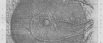
The clinical manifestations of sebaceous gland carcinoma vary greatly. This tumor can simulate various benign conditions - chalazion (inflammation of the edge of the eyelid), blepharoconjunctivitis, keratitis, etc. The classic manifestation of carcinoma is a hard, painless compacted mass or ulceration on the eyelid, in the area of which eyelashes fall out ( Fig. 8 ).
Sebaceous gland carcinoma is prone to metastasis, so timely diagnosis is of great importance to improve the prognosis of the disease.
Rice. 8. Sebaceous gland carcinoma (American Academy of Dermatology)