Photos before and after Reviews Videos Rehabilitation Prices Analyzes Questions and Answers
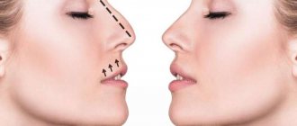
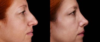
Lateral curvatures of the nasal dorsum (deviations) are often the result of injuries, including birth injuries.
There are 4 degrees of lateral curvature of the nasal dorsum:
- I degree
. Lateral displacement of the nasal bridge by no more than 1/2 of its width. - II degree
. The lateral displacement of the nasal bridge is no more than the value of its width. - III degree
. Lateral displacement of the nasal bridge by more than its width. - IV degree
. Excessive displacement of the nasal bridge to the side.
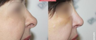
The external manifestations of a curvature of the nasal bridge may vary. With a thin and long nose, the same degree of curvature of the dorsum will be more pronounced than with a thick and short nose.
With deformities of the nasal dorsum of II-IV degrees, patients are also diagnosed with trauma to the nasal septum (straight vertical fracture, C-shaped fracture).
Correction of II – IV degrees of curvature of the nasal bridge
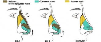
Most often, when correcting this kind of curvature of the nasal bridge, the plastic surgeon is faced with the task of eliminating the deformation at two levels: cephalic and caudal.
Cephalic deformity is located in the area of the fracture of the base of the nasal bones. The apex of the cephalic deformity is located in the area of the bridge of the nose and is often partially masked by soft tissues.
The apex of the caudal deformity is mainly located at the level of the osteochondral junction and/or the cartilaginous part of the nasal dorsum. In this case, as a rule, there is damage to the cartilaginous part of the back of the nose, deformation of the nasal septum, which causes disturbances in nasal breathing.
When eliminating lateral curvatures of the nasal bridge, the plastic surgeon faces the following list of tasks:
:
- eliminate cephalic deformity;
- eliminate caudal deformation;
- correction of the location of the nasal septum.
Surgical correction often involves performing a specific sequence of actions
:
- application of open access;
- submucosal excision of deformed parts of the nasal septum and vomer;
- submucosal separation of the elements of the cartilaginous part of the nasal dorsum;
- osteotomy with comparison of displaced areas of the nasal bones;
- elimination of deformation of the cartilaginous part of the nasal dorsum;
- final manipulations on the back of the nose.
First of all, correction of the nasal septum is performed, which subsequently creates conditions for successful comparison of displaced sections of the nasal bones. At the same time, the elements of the cartilaginous part of the nasal dorsum are separated. Afterwards, the position of the bones and cartilage of the nasal dorsum is corrected.
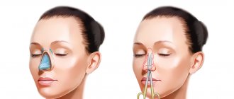
Correcting the position of the nasal bones involves performing an osteotomy.
Special cases of rhinoplasty
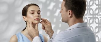
Indications for surgery
Rhinoplasty is performed for aesthetic or medical reasons.
Aesthetic rhinoplasty is used in cases where the nose is disproportionate and unattractive based on generally accepted standards or subjective views. For the most part, this plastic procedure is aimed at eliminating snub and hump noses, as well as changes in the tip of the nose. In other words, such an operation allows you to achieve harmony in appearance. Medical rhinoplasty is more aimed at eliminating obstacles to the normal functioning of the nose that were caused by various injuries or congenital pathologies.
There are cases when rhinoplasty is performed simultaneously for both medical and aesthetic reasons. However, it is worth noting that not a single plastic surgeon will correct the shape and size of the nose to the detriment of its natural functions, just as he will not perform an operation whose result will be disharmonious.
Contraindications for rhinoplasty
In addition to age restrictions (up to 18 years), nose modeling surgery cannot be performed on people who suffer from:
- diabetes mellitus;
- high blood pressure;
- acute viral diseases;
- exacerbations of chronic pathologies;
- nervous system disorders;
- various skin lesions in the surgical area.
Methods of carrying out
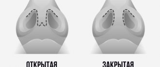
Depending on the desired result and the individual characteristics of the patient, rhinoplasty is performed in two ways: open and closed.
Closed rhinoplasty is used for a small amount of correction that can be performed on the side of the nasal passages. The advantage of this method is the complete absence of seams on the outside.
An open operation is used in situations where it is not possible to achieve the goal using a closed method. The plastic surgeon makes an incision along the lines of the natural folds of the skin; therefore, traces of stitches after restoration are practically invisible.
Operation options
Before going to a plastic surgeon, you should understand that any correction has certain limits, beyond which it is extremely undesirable, and in some cases even dangerous to health. The nose is a bone-cartilaginous structure that is hidden behind soft tissue. Rhinoplasty affects almost all tissues, so during the consultation you should listen to the recommendations of the plastic surgeon, and not just focus on your “want”. After all, for example, in the case of disproportionate shortening of the nose, its natural shell will be too large and, of course, this fact will be reflected in the harmony of the face. In addition, no aesthetic operation should go against the functionality of the organ. In other words, normal breathing through the nose is a prerequisite for rhinoplasty, and not a single surgeon will put a beautiful nose above this.
Correction of deformities of the cartilaginous part of the nasal dorsum
Often, when correcting the cartilaginous part of the nasal dorsum, the plastic surgeon is faced with the following tasks: eliminating deformation of the cartilage of the nasal dorsum; correction of the position of the lateral cartilaginous walls of the nose after osteotomy; change in the height of the nasal bridge; in case of deformation of the caudal part of the nasal septum, it is eliminated.
Elimination of curvature of the cartilage of the nasal dorsum
can be achieved by straightening it in the opposite direction. If this is not enough to eliminate the deformation, then additional manipulations are performed.
Correction of the position of nasal cartilage
. After an injury, as a result of displacement of the lateral walls of the nasal pyramid, a significant difference in their height may occur. This is most typical for the consequences of perinatal trauma.
With severe curvature, moving the lateral walls of the nose into the desired position leads to the emergence of counteracting forces, which are eliminated after submucosal separation of the superolateral cartilages and nasal septum. In this case, after comparing the displaced areas, one of the cartilages is located above the level of the nasal septum, the other below. Subsequent partial excision of the protruding sections of cartilage makes it possible to obtain the nasal dorsum of the required height. Such manipulations are effective both in eliminating lateral displacement and in reducing the height of the nasal bridge.
Change in the height of the cartilage of the nasal dorsum
. In rare cases, due to crushing of the cartilage, depression of the nasal bridge is observed at the level of its cartilaginous part. To eliminate such deformation, a plastic surgeon installs cartilage grafts, thereby restoring the damaged relief of the nasal dorsum.
Recovery period
After fixing the new tissue, the incision site is closed with stitches, a plaster cast is applied, and gauze turundas are inserted into the nostrils. After 1-2 days they are removed, after which the patient breathes normally through his nose.
The plaster is removed after 1-2 weeks. While wearing the bandage, it is recommended to refrain from eating solid food so as not to cause pain. Swelling and bruising completely heal by the end of the second week. For a month, it is recommended to refrain from physical activity, visiting baths, solariums, and swimming pools.
At Dr. Panov's Clinic, various nasal defects are eliminated, including saddle-shaped deformities of varying degrees of severity. The professional work of our specialists will allow you to enjoy your appearance for the rest of your life!
Closed rhinoplasty
more aesthetic than open, as it does not leave scars on the columella (the skin partition on the outside between the nostrils).
Open
rhinoplasty
is more traumatic: damage to a larger number of vessels supplying the back and especially the tip of the nose causes a longer rehabilitation process (resolution of swelling). It is believed that with open rhinoplasty a greater view of the nasal structures is possible, but these structures are also visible with endonasal (intranasal) incisions, although in the first case it is better. However, with a good knowledge of anatomy and clear planning of the operation, this is of little relevance. As for correcting the curvature of the nasal dorsum, open rhinoplasty has no advantage over closed rhinoplasty, since the curvature is eliminated by eliminating the curvature of the nasal septum and clear positioning of the nasal bones after osteotomy. Both are performed from independent approaches to the nasal mucosa and wide exposure of supporting structures is not required for this.
Open rhinoplasty can be more convenient if various materials are placed on the bridge of the nose (cartilage, implants) and fixed.
1.
The patient is on the table before surgery. To prevent infection, the operating area (face) is treated with a disinfectant solution. The eyes are sealed to prevent the cornea from drying out. A tube fixed in the mouth is visible: during rhinoplasty, general anesthesia is most often performed - anesthesia with tracheal intubation or, at best, using a laryngeal mask (the tube is not passed through the larynx with the vocal cords, but covers it from the outside, so there are problems with hoarseness, sore throat throat, etc. after such anesthesia there is no). Usually, all drugs are administered through a vein (intravenous anesthesia), since they are quickly eliminated from the body and allow the anesthesiologist to do the job so that the patient is at least comfortable, and at most, pleasant after surgery. Local anesthesia can be used, but this may worsen the outcome of the operation, since to achieve adequate pain relief it is necessary to inject a large amount of anesthetic, which makes it difficult to assess the shape of the nose during the operation.
2.
Infiltration is in progress. Infiltration is intended to facilitate the detachment of skin from the osteocartilaginous frame of the nose. Composition of the solution: adrenaline helps reduce bleeding, ice caine helps to achieve pain relief (despite the fact that the patient is under anesthesia, the body still hurts, i.e. the nerve endings continue to signal pain to the brain, even though the brain is asleep).
3.
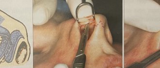
Access to the nasal septum. The blood is removed using an aspirator - a tube attached to a machine that creates a vacuum.
4.
Extraction of the bony part of the nasal septum.
5.
The bony (in blue) and cartilaginous (in purple) portions of the nasal septum. The curved bone part of the septum is removed (from a functional point of view this is not significant). The deviated cartilaginous part of the septum can be straightened, so it is removed temporarily (from a functional point of view, this part of the septum is desirable). The cartilaginous part, being returned back, always takes root.
6.
Access to the septal cartilage: incision, detachment and cartilage isolation. The scissors are passed between the septal cartilage and the medial crura of the alar cartilages. This is done anatomically, that is, bypassing structurally significant formations (the lower edge of the septum and the medial legs of the alar cartilages).
7.
Shortening of the nose due to excision of the septal cartilage. With this manipulation, the nose is shortened in length: the tip of the nose is raised, which leads to an increase in the nasolabial angle - visually this is perceived as a shortening of the nose.
8.
Shortening the nose due to skin excision. The skin covering the excised area of cartilage is removed. If the skin is not excised, but sewn as is, the length of the nose will not change.
9.
Additional infiltration: in the area of the marginal incisions used to treat the tip and dorsum of the nose.
10.
Edge accesses. This manipulation is the threshold to a new stage - the reduction of wing cartilage.
11.
Skin detachment at the tip of the nose. The alar cartilage is visible in the left nostril.
12.
Subcutaneous detachment in the dorsum of the nose.
13.
The wing cartilage is isolated.
14.
Excision of the cephaloid (cephalo - head: upper, close to the head) edge of the wing cartilage.
15.
Removal of the perichondrium - the less tissue we leave in the area of the tip of the nose, the more narrowing we can achieve. In addition, the perichondrium is an additional source for the growth of scar tissue.
16.
The treated cartilage is turned outward.
17.
Narrowing of the tip of the nose by suturing the arches of the alar cartilages.
18.
After shortening and narrowing the tip of the nose.
19.
Infiltration was carried out in the area of the nasal dorsum. The alar cartilages are highlighted in blue: the lateral crus is visible in the right nostril, and the medial crus is visible in the left nostril.
20.
Removal of the bone part of the hump. It is removed with an osteotome - a chisel with two stops that are felt under the skin as it is carried out, which allows the second hand to control the position of the cutting part of the osteotome.
21.
Removed bone.
22.
Removal of the cartilaginous part of the hump. The cartilaginous part is cut with a scalpel at the level of the removed bone hump. It is cut with one confident movement, which allows you to get a clear contour of the bridge of the nose.
23.
Straightening the bridge of the nose by grinding the bony part of the hump with a tool called a rasp. Resurfacing is not a method of removing a hump, but rather a way to smooth out the sharp edges after an osteotomy. If a surgeon says that he is removing a hump by grinding, this means that he is afraid of an osteotome, in which case the operation will take too long, since grinding is not a very effective way to remove a large amount of bone matter. Increasing the operation time leads to increased blood loss, increased morbidity of the operation, and increased anesthetic risk.
24.
Assessment of the relief of the nasal dorsum.
25.
Suturing of intranasal accesses.
26.
Lateral osteotomy to narrow the dorsum of the nose (lateral osteotomy of the nasal bones). The nasal bones break at the base and move closer to each other.
27.
Osteotomy. The stage of breaking the nasal bones and bringing them closer together.
28.
Assessment of the profile of the nasal dorsum.
29.
Packing of the nasal vestibule. Tampons protect wounds from infection and absorb wound discharge (blood, ichor, mucus).
30.
Plaster application. The purpose of the cast is to distribute pressure evenly over the surface of the skin and determine its fixation (“adhesion”) to the newly created osteochondral frame of the nose.
To communicate with patients who have undergone rhinoplasty, come to our forum, Rhinoplasty