To the reference book Operations in otolaryngology are usually aimed at restoring hearing and may involve the installation of an auditory prosthesis.
Author:
- Oganesyan Tigran Sergeevich
ENT pathology expert
5.00 (Votes: 1)
- Operations
- Preparation
- stapedoplasty using modern prostheses;
- myringoplasty;
- tympanoplasty (hearing-improving surgery) using modern prostheses;
- general cavity (radical) surgery on the middle ear;
- mastoidoplasty;
- shunting and drainage of the tympanic cavity for exudative otitis media;
- removal of exostoses of the external auditory canal;
- revision of the tympanic cavity;
- reoperations: tympanoplasty, stapedoplasty;
- operations for tinnitus;
- operations for ear injuries;
- formation of the external auditory canal in congenital and acquired atresia.
Operations in otolaryngology are usually associated with diseases accompanied by hearing loss and requiring surgical treatment.
How does the ear work?
The ear is divided into three sections: the outer ear, the middle and the inner.
The external ear includes the pinna, the external auditory canal and the eardrum, which is the boundary between the outer and middle ear. The middle ear is represented by the tympanic cavity, the mastoid cavity and their contents - primarily the auditory ossicles, as well as the auditory (Eustachian) tube connecting the tympanic cavity with the pharyngeal cavity.
Figure 1. Sections of the ear and how they are viewed with an otoscope. Source: openpediatrics.org
The inner ear is represented by the vestibule, cochlea, semicircular canals and is located in the pyramid of the temporal bone.
The most painful thing for humans is otitis media - swelling and inflammation of the mucous membrane of the tympanic cavity.
Radical middle ear surgery
Sanitation surgery of the middle ear is performed for chronic inflammation of the middle ear (chronic purulent otitis media). It is carried out with the aim of ridding the patient of a chronic source of infection and preventing the development of severe and life-threatening ear (otogenic) complications (such as meningitis, brain abscess, arachnoiditis, etc.) and with the aim of preventing secondary cochlear neuritis (sensorineural hearing loss) that develops as a result of penetration toxic products of inflammation of the middle ear into the inner ear. In recent decades, sanitizing operations have also begun to be performed with the aim of “preparing” the middle ear (first stage) for hearing-improving surgery (second stage).
Many methods of sanitizing operations have been developed, differing in both the surgical approach (through a postauricular or intraauricular incision) and the volume of bone removed. A variety of surgical techniques allows you to choose the most suitable one for a particular patient, depending on the characteristics of inflammatory changes, the degree of auditory disorders, the condition of the auditory tube, the general condition of the patient, etc. It should be borne in mind that the longer the course of chronic otitis media (for example, several decades ), the greater the volume of the operation and the less chance there is for the patient to completely get rid of discharge from the ear and for the possibility of performing hearing-improving surgery.
Main indications for surgery
The main indication for sanitizing surgery is persistent discharge from the ear or that temporarily stops under the influence of treatment. They can be abundant or scanty; purulent, mucous or mucopurulent, less often watery. Sometimes the discharge stops, but when you wipe the ear with a cotton wool wrapped around a probe or a match, the fetid crust is removed or the cotton wool simply has an unpleasant odor. This indicates that despite the apparent absence of discharge, inflammation continues, but the discharge is so small that it has time to dry out, turning into a crust.
Relative readings
Another indication for sanitizing surgery is dull pain in the ear, dizziness and hearing loss. It should be noted that in some cases, such as, for example, with a small perforation of the eardrum, when it is localized in the upper part of the membrane, while the auditory ossicles are preserved, hearing may be normal. However, this does not mean that sanitizing surgery is not necessary. Often, with almost normal hearing, cholesteatoma develops, which always poses a threat to health, and therefore must be completely removed during surgery. Sometimes during a sanitizing operation in such patients it is possible to preserve hearing. If the operation is postponed for a long time, then cholesteatoma can so destroy the structures of the middle ear and impair hearing that it is no longer possible to perform a hearing-improving operation.
How is the operation performed?
Radical sanitizing surgery consists of creating a common bone cavity with smooth walls, communicating with the ear canal. In other words, a single cavity of the middle ear is formed, as if wide open into the ear canal. During the operation, all “affected” elements of the middle ear are removed, including altered auditory ossicles, even if they are partially destroyed by the inflammatory process. Typically, radical surgery is performed behind the ear.
With minor destruction of the elements of the middle ear, as, for example, in the initial stage of development of epitympanitis, you can limit yourself to a small operation called atticotomy. It is performed through the external auditory canal. In this operation, a small area of bone tissue is removed and a small cavity is created. As a rule, after such an operation, hearing-improving surgery can be performed in the future.
There is another operation called a separate atticoanthrotomy, which includes elements of radical surgery and atticotomy. It is usually performed for chronic purulent mesotympanitis, when the mucous membrane of the middle ear is mainly affected by inflammatory changes and there is no cholesteatoma. At the same time, the hearing-improving stage of the operation (tympanoplasty) is performed. During this operation, as with atticotomy, an operating microscope is used.
For some types of surgery, a repeat operation (revision surgery) may be required after 8-14 months to prevent recurrence of cholesteatoma. Indications for it are determined by the operating surgeon.
In addition to the indicated main 3 types of sanitizing operations, there are many modifications.
The success of the operation largely depends on the general condition of the patient and on the implementation of the necessary regimen after the operation.
Preparing for surgery
Before the operation, they find out whether there are any contraindications to the operation or any pathology in other organs that may affect the outcome of the operation. First of all, there should be no carious teeth or purulent sinusitis. If necessary, teeth should be sanitized. Pathology of the pharynx (chronic tonsillitis), nose and near the sinuses is subject to treatment. Infection in the nose and throat may contribute to poor postoperative healing.
A very important place is occupied by the preparation of the ear for surgery, which is carried out both in the clinic and in the hospital. In the clinic, local treatment (drops) is carried out, sometimes, according to indications, they are lubricated with ointment or insufflated with powder; sometimes they give injections of vitamins, etc. Before the operation in a hospital, the ear is carefully examined under a microscope, washed, the ear canal is cleaned of crusts and, sometimes, drug injections or tablet medications are prescribed.
On the eve of the operation, the patient must take a shower, since after the operation he will be deprived of this pleasure for an average of 7-10 days; cleanse the intestines, take a sedative or sleeping pill at night. On the day of surgery, you should not take food or liquids in the morning. The hair around the ear should be cut short and 2-3 cm shaved. The operation usually lasts 1-3 hours in the supine position; It is usually performed under local anesthesia or general anesthesia (the type of anesthesia will be determined individually). As a rule, patients tolerate the operation easily and painlessly.
Postoperative regimen
The postoperative regimen, both in the hospital and after discharge, is prescribed by the otosurgeon individually, depending on the nature and characteristics of the operation. Usually the next day patients are allowed to get up. Restriction of walking and sudden movements lasts 3-7 days. A more free regimen with walks along the corridor, and in the summer - in the hospital yard, is usually prescribed after complete dressing, that is, after all tampons have been completely removed from the ear. During the hospital stay, the patient is prescribed treatment aimed at accelerating the healing of the postoperative cavity, and daily dressings are performed. Physiotherapeutic procedures are often prescribed. Treatment in a hospital lasts from 3 to 7 days; with sluggish healing, this period is sometimes extended. Dressings consist of changing tampons in the first 7-10 days, removing wound discharge and introducing medications into the ear.
Upon discharge from the hospital, the otosurgeon in the epicrisis notes the nature of the operation, the recommended postoperative regimen and, if necessary, indicates drug treatment, which is carried out in the clinic at the place of residence. Taking into account the specifics of the operation and healing, the certificate also notes the approximate period of home stay with the right to extend sick leave. It usually lasts 1-2 weeks after discharge.
Rehabilitation
Ear infections should be avoided for the first 6 months. You should not overcool, swim in open pools, and when using a bath or shower, especially when washing your hair, you should tightly close the ear canal with cotton wool moistened with sterile Vaseline.
Usually, discharge from the ear stops after 4 weeks, so during this period the patient is observed by a doctor who cleans the ear. Sometimes the discharge continues for a longer period; in this case, the patient himself can, as directed by the doctor, apply ointment or instill medications. With a sudden change in weather or changes in atmospheric pressure, dull pain in the operated ear may occur for many years. In this case, you should massage the skin in front of the tragus in the area behind the ear.
As necessary, the doctor prescribes frequent or infrequent visits to him; In the first 6 months, the patient should usually be examined by a doctor once a month, and then less often - once every 2-3 months until 1 year after surgery. During this entire period, you should avoid not only hypothermia, but also contact with people with influenza and colds. In case of acute inflammation of the respiratory tract and influenza, you should consult a doctor.
Of great importance both before and after surgery are general strengthening procedures aimed at increasing defenses, improving general blood circulation and oxygen enrichment. Proper alternation of work and rest, regular physical exercise and spending time in the fresh air help to harden the body.
It is advisable to include in a set of physical exercises those that increase breathing volume. Running or brisk walking, light physical labor, deep breathing, turns and rotations of the head and neck are useful.
The diet should be complete and varied, including protein products (milk, cottage cheese, unfried meat) and fortified foods (vegetables, fruits, juices). Under no circumstances should you drink alcoholic beverages, including beer, as they increase the secretion of mucus and discharge from the ear. In any case, this prohibition is categorical for two years, even with good healing.
Compliance with the listed recommendations and timely visits to the doctor contribute to reliable healing and complete relief of the patient from ear discharge. For those patients who are prescribed a second hearing-improving operation, compliance with the noted rules increases the chances of its effectiveness.
What is myringotomy, paracentesis, tympanotomy?
All these are different names for a surgical operation, the essence of which is to puncture the eardrum, removing harmful contents from the tympanic cavity that has formed there due to inflammation. A through incision ensures free release of fluid accumulated in the middle ear, after which the person feels much better and the pain subsides.
The intervention is associated with certain difficulties. A long-term inflammatory process thickens the membrane, and it can be difficult to make a small through puncture. The operation is usually prescribed for children, because they suffer from otitis media more often than adults due to age-related characteristics of the anatomy of the auditory tube.
Chronic otitis media with cholesteatoma (epitympanitis)
With epitympanitis, the inflammatory process is localized mainly in the supratympanic space - the attic and in the antrum of the mastoid process; The perforation is usually located in the loose part of the eardrum; it often spreads to other parts of the eardrum. Epitympanitis is characterized by a more severe course compared to mesotympanitis: epitympanitis is characterized not only by all the morphological processes that occur with mesotympanitis, but also by caries damage to the bone walls of the middle ear, most often in the attic, aditus, antrum and mastoid cells. In addition, with epitympanitis, in most cases, cholesteatoma (epidermal formation) is formed. Gradually increasing, it fills the attic and antrum and destroys the surrounding bone. This process is facilitated by constant pressure on the bone walls of the cholesteatoma mass and the ingrowth of matrix cells into bone cells. As a result, cholesteatoma can destroy the labyrinthine bone capsule, the wall of the facial nerve canal, the mastoid process, exposing the membranes of the temporal lobe of the brain, the cerebellum and the wall of the sigmoid sinus. Chronic purulent epitympanitis, complicated by cholesteatoma, can last a long time without pronounced symptoms.
Indications
A puncture of the eardrum is indicated in the following cases:
- severe pain syndrome with confirmed otitis media;
- fluid in the middle ear, identified through a special examination - tympanometry;
- there is no positive dynamics with long-term use of antibiotics and other medications;
- Eustachian tube dysfunction in adults;
- hearing impairment due to fluid accumulation in the tympanic cavity;
- the need for analysis to identify the causative agent of a possible infection and conduct adequate drug therapy.
Photo: asier_relampagoestudio / freepik.com
If exudate from the middle ear cavity is removed on time, without waiting for complications, otitis media will not progress to the chronic stage.
Important! Myringotomy of the tympanic membrane does not replace antibiotic therapy, does not reduce the incidence of otitis media, and does not reduce the number of relapses.
Where can I watch a video about ear otoplasty?
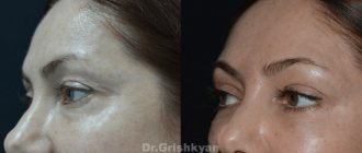
You can find videos about otoplasty on the websites of various plastic surgery clinics. On our website of Dr. Maxim Osin there is a section dedicated to videos about ear plastic surgery. By watching the video you will be able to get a closer look at the work of our plastic surgeon. In addition, a video about otoplasty surgery in our clinic allows you to compare the results “before” and “after” the operation performed by Maxim Osin. The video also presents comments from our patients, given both immediately after surgery and after rehabilitation.
How is the operation performed?
The procedure for performing the operation is determined by the ENT doctor based on a comprehensive examination of the patient, a confirmed diagnosis and the presence of indications for intervention.
To get the best view of the middle ear cavity, a microscope is used. A small incision is then made in the eardrum and the fluid in the middle ear is removed. In most cases, a small tube may be needed to remain in the incision site to allow drainage to continue.
No stitches are required to close the incision - it will heal on its own.
Otosclerosis
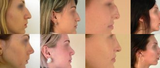
Otosclerosis is a process based on focal damage to the bone capsule of the ear labyrinth. The pathological essence of the disease is that healthy bone in the lesion is replaced by newly formed spongy bone, rich in blood vessels. Therefore, the name otospongiosis is more correct. Typically, the otosclerotic lesion is located in the region of the vestibular window. While the changes are localized only in the bone, the process does not manifest itself clinically. With the transition of the process to the annular ligament of the stapes, the mobility of the stapes is limited and the transmission of sounds through the middle ear gradually deteriorates. There is progressive hearing loss and a feeling of tinnitus. Otosclerosis is classified as a hereditary disease with an autosomal dominant mode of inheritance. Stapedoplasty is an operation performed for otosclerosis, which involves removing a fixed bone - the stapes - and replacing it with a prosthesis. There are different types of prostheses: made of titanium, autocartilage, Teflon. The efficiency of these operations is very high - 95%.
Diagnostics
Photo: kuprevich/freepik.com
Acute otitis media is determined by a combination of signs:
- anamnesis data;
- otoscopy indicators - examination of the outer ear and eardrum using a special device;
- characteristic symptoms of the presence of fluid behind the eardrum: severe ear pain, hearing loss and intoxication: pallor, weakness, lack of appetite, headache and fever.
In some cases, the doctor prescribes additional diagnostic procedures: bacterial culture of the exudate and x-ray of the temporal bone.
Operation process
Myringotomy.
Photo: Dr Paulose FRCS - ENT SURGEON / YouTube The puncture of the eardrum is carried out in several stages:
- removal of wax from the ear canal;
- rinsing the ear with an antiseptic solution;
- administration of anesthetic;
- a linear incision of the eardrum with a sterile paracentesis needle;
- antiseptic treatment;
- installing a thick gauze swab (turunda) with medicine.
Tympanoplasty with revision of the trepanation cavity
The purpose of this operation is to try to stop the flow of suppuration from the trepanation cavity and to improve hearing in patients who have undergone general ear surgery in the past.
The operation is performed under local or general anesthesia using a behind-the-ear approach. After removal of pathological formations, the mastoid cavity can be filled with muscle and fatty tissue from the postauricular area or bone. Over time, the ear canal can be repaired using cartilage or bone. The eardrum is restored and, if possible, the transmission mechanism is also restored. However, in most cases, a second operation to restore hearing is necessary (see: Tympanoplasty: a planned second stage).
The patient is usually hospitalized for a few days and can return to work 1-3 weeks after discharge. Complete healing of the cavity inside the ear occurs after 4 months.
Postoperative period and rehabilitation
In the postoperative period, the ear canal is washed several times a day with furatsilin or boric alcohol to quickly relieve inflammation and improve the patient's condition. In order for the harmful contents of the tympanic cavity to flow out more quickly, it is recommended to lie down with the operated ear down on a pillow.
Typically, complete recovery after surgery occurs within 4 weeks, but after about 5 days after surgery, the hole in the eardrum will heal. For 3-5 days after surgery, pus may be discharged from the puncture site; this is normal. The recovery period requires compliance with the following recommendations:
- the ear canal should be cleaned daily;
- if the doctor has prescribed ear drops, you need to instill them strictly in accordance with the prescription;
- it is necessary to regularly change cotton swabs in the ear;
- Check with your doctor about the rules for performing water procedures.
Photo: ronstik/Depositphotos
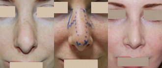
Types of cuts
Postauricular tissue is used to close the eardrum defect.
Taking the fascia
Plastic perforation of the tympanic membrane
The patient is hospitalized for several days and can begin work 1-2 weeks after discharge. Complete healing and improvement in hearing in most cases occurs after 2-3 months.