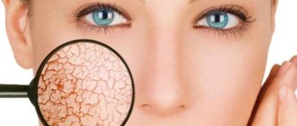
Skin as an organ
Skin is the dense outer covering of a person.
It is an independent organ that performs specific functions. First of all, the skin is necessary to protect internal organs from physical, chemical and microbiological influences. In addition, the skin contains a large number of receptors that provide tactile, temperature and pain sensitivity. Damage to the integumentary tissue always increases the risk of developing an infection or inflammatory process. Diseases of internal organs can also cause skin lesions. Cover departments:
- The epidermis is the outermost part of the skin, formed by five layers of cells. The upper layer of the epidermis is represented by dead (keratinized) cells necessary for the formation of a biological barrier. The lower layers ensure the renewal of the cellular composition of the epidermis.
- The dermis is the middle layer of the skin. This is the area where smooth muscle fibers, blood vessels, nerves, glands, hair follicles and other structures are located.
- Adipose tissue is the deepest layer of the skin, mainly consisting of adipose tissue. This section of the organ provides protection to underlying tissues from temperature changes and external physical influences.
Scientists include external respiration, water-salt regulation, vitamin D formation and blood deposition as additional functions of the skin. The structure of the skin differs in different parts of the body. Thus, the thicker skin of the soles of the feet does not contain hair follicles. Nails, which are derivatives of the skin, form at the tips of the fingers. Also, different areas of the skin differ in the number of sweat and sebaceous glands.
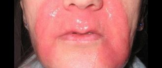
Clinical guidelines for the treatment of rosacea
Rosacea is a chronic disease accompanied by inflammation of the skin. The capillary network stands out sharply, and purulent rashes appear. Blood rushes to the site of inflammation, the walls of blood vessels stretch, and excess fluid gradually accumulates in adjacent tissues.
There are four subtypes of the disease and one variant, granulomatous rosacea. Subtypes of rosacea are: erythematotelangiectatic (predominant), papulopustular, phymatous (hypertrophic), ocular (ophthalmic rosacea).
Classification
Pyoderma is not a separate disease. In medicine, this term is used to refer to any pathologies manifested by purulent skin lesions. Different types of pyoderma differ in the depth of damage to the skin and the causative agent of infection. The most common type of pyoderma is a boil, which is a purulent inflammation of the hair follicle.
Other types of pathology:
- Streptoderma is superficial pyoderma, in which multiple ulcers appear on the surface of the patient’s face, torso and limbs. A bullous rash occurs due to the invasion of streptococci into the skin.
- Ecthyma vulgaris is a streptococcal infection of the deep layers of the skin. Blisters with pus and deep ulcers form on the surface of the skin.
- Folliculitis is a pustular inflammation of the hair follicles caused by fungal or bacterial invasion. Whitish-yellow ulcers appear in the hair growth area. The release of exudate when the abscess membrane ruptures leads to the appearance of an ulcer.
- Intertrigo is a lesion of skin folds, characterized by swelling, redness and tissue deformation. Also, blisters filled with pus appear on the surface of the skin.
- Carbuncle is an infectious lesion of the skin. A carbuncle is a purulent-necrotic ulcer that quickly spreads in the peripheral direction. The inflammatory process reaches the subcutaneous fatty tissue.
- Furunculosis is a purulent lesion of several hair follicles at once. This form of pyoderma can occur against the background of chronic infection.
- Sycosis is a staphylococcal lesion of the scalp due to bacterial invasion of the hair follicles. Often, multiple ulcers appear in the area of beard growth in adult men.
- Gangrenous pyoderma is the appearance of a deep purulent-necrotic ulcer reaching the subcutaneous tissue. Large ulcers can reach 20 cm in diameter. Hemorrhages may form in the area of skin damage. This pathology is usually not associated with infection.
Only an experienced dermatologist can determine the type of disease by external signs. Some pathologies manifest themselves simultaneously in several forms of skin lesions.
Pathogenesis and stages of development
During the development process, the boil goes through three successive stages:
- initial (infiltration): the inflammatory process begins at the mouth of the follicle and gradually spreads;
- necrotic (maturation): inflammation engulfs the entire cavity of the follicle, causing necrosis and purulent melting of tissue; the process lasts from several days to 2 weeks and ends with a rupture of the skin over the center of the boil;
- healing stage: after the pus has drained and the cavity of the lesion has been cleaned, it gradually scars with the formation of a scar.
Causes
Neutral, opportunistic and pathogenic microorganisms are constantly present on human skin. These are fungi and bacteria that do not penetrate deep tissues and do not provoke the development of infection. The constancy of the microflora of the skin and the inhibition of the vital activity of microorganisms allows maintaining the integrity of tissues. Weakening of the body's defense systems can provoke the proliferation of pathogenic and opportunistic microorganisms with damage to various layers of the skin. In some cases, ulcers are not associated with infection.
The main mechanism of pyoderma formation is due to the infectious process. Pathogenic bacteria begin to actively multiply and produce toxins that damage the skin. The immune system immediately begins to fight the infection by activating inflammatory processes and protective cells. The release of inflammatory mediators leads to even greater damage to the skin. Destroyed cells and dead microorganisms form purulent exudate that accumulates in various cavities.
Possible causes of the disease:
- Damage to integumentary tissues due to cuts, combing, as well as chemical and thermal exposure. Violation of the integrity of the epidermis causes the penetration of pathogenic microorganisms into less protected areas of the skin. In addition, cuts and burns themselves cause an inflammatory response.
- Disruption of the body's defense systems. In some cases, immune cells begin to attack healthy tissue. The most severe form of autoimmune damage to integumentary tissues is pyoderma gangrenosum. In addition, allergic and autoimmune processes increase the risk of tissue infection.
- Penetration of parasites into the skin. This is a rare variant of the etiology of the disease. Parasitic invasion can also lead to a purulent-inflammatory process.
Clarifying the cause of the disease is necessary to select a treatment method and prevent relapses.
Symptoms of a boil in a child
The main symptoms of a boil in a one-year-old child, preschool and school-age children are the same and depend on the stage of development of the disease.
At the initial stage, an area of redness appears on the skin, which gradually increases, but practically does not cause discomfort. As the pathology progresses, a purulent area appears in the center of the lesion, and the area of inflammation increases in size and becomes sharply painful. Then a necrotic rod is formed inside the cavity, which from the outside looks like a dark dot in the very center of the boil. After a few days, the abscess breaks through, its contents come out, after which the healing process begins. A small boil, especially if it is located on a child's hand, foot or finger, rarely causes general symptoms. With multiple inflammatory foci, as well as when localized in an area with a large number of blood and lymphatic vessels (face, neck, groin, armpits), the baby may have a fever, weakness, malaise and other signs of general intoxication.
Risk factors
The etiology of pyoderma is not limited to the mechanisms of formation of the purulent-inflammatory process. Dermatologists know the forms of predisposition to this disease associated with lifestyle, heredity and the patient’s individual history.
Known risk factors:
- Failure to comply with personal hygiene rules. Constant skin contamination increases the risk of infection.
- Overheating or hypothermia of integumentary tissues. Temperature changes disrupt the functions of local immunity. Often, after hypothermia, the skin becomes a gateway to infection.
- Age. In newborns and the elderly, pyoderma most often occurs due to insufficient effectiveness of local immunity.
- The presence of a large number of allergic reactions and autoimmune diseases (especially in children.)
- Pathologies of the endocrine system. The condition of the skin largely depends on hormonal influences.
- Metabolic diseases: diabetes and obesity.
- Congenital and acquired immunodeficiency conditions.
- Poor nutrition leading to a deficiency of certain vitamins and microelements in the body.
- Unfavorable family heredity. Skin diseases may be associated with the transmission of genetic mutations.
The causative agents of pyoderma dermatitis are found in the environment and on the surface of the integumentary tissue. These are staphylococci, streptococci and some pathogenic fungi. People often become infected with pyogenic bacteria in a hospital setting.
Treatment of boils in children
If the child’s boil is small in size and is not located on the face, neck or knee, treatment at home is allowed. In case of extensive lesions or multiple purulent foci, hospitalization is required.
With a relatively mild course of the disease, conservative treatment is sufficient, which depends on the stage of development of the disease:
- at the initial stage, alcohol solutions and dry heat are used to accelerate ripening;
- at the necrotic stage, antibacterial agents, proteolytic drugs and regular wound washing are necessary;
- at the healing stage, agents are used that accelerate regeneration in combination with antibacterial treatment.
In more severe cases, local treatment is supplemented with systemic drugs: antibiotics, anti-inflammatory drugs, immunostimulants, etc. If the infection spreads to adjacent tissues with the development of abscesses or phlegmons, surgical assistance is required: opening and draining the abscess.
Symptoms
The external manifestations of pyoderma depend on the depth of damage to the skin, the causative agent of the infection and other factors. Many dermatitis manifests itself in several types of skin formations at once. Additional symptoms may be due to the underlying cause of the pathology and the effect of bacterial toxins on the body.
Symptoms of body intoxication:
- dizziness and weakness;
- nausea and vomiting;
- sleep disturbance;
- increased body temperature;
- decreased appetite;
- headache;
- chills.
If the symptoms listed above appear, you should immediately consult a doctor. The infection that causes pyoderma can spread through the blood and damage internal organs. Self-removal of skin lesions increases the risk of sepsis.
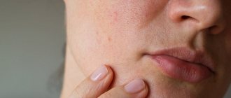
Symptoms of the disease
Most often, acne affects the skin of the face, since this area contains the most sebaceous glands. Acne can occur in the following forms:
- Open comedones (“blackheads”). It occurs as a result of expansion of the sebaceous duct and disruption of keratinization of its walls.
- Closed comedones (“whiteheads”) are formed as a result of blockage of the sebaceous gland. At the same time, the whitish contents of the cavity are visible under the skin. In closed comedones, favorable conditions are created for the proliferation of propionobacteria - these microorganisms love an oxygen-free environment.
- Painful lump.
- Pustule. It is the result of infection of a closed comedon.
Other types of acne:
- Globular acne. They are compactions under the skin of quite large sizes. They can merge with each other, resulting in formations of quite large sizes. Complications in the form of suppuration are common. This type of acne is a serious disease. They often leave disfiguring scars on the skin.
- Lightning acne. A serious disease that most often affects young men. A large number of pustules and ulcerations appear on the skin of the body (but not the face). Body temperature rises to 38?C. There is a general deterioration in health and loss of appetite.
- Inverse acne forms in the armpits and groins, where there is friction between the skin and clothing. Lumpy elevations appear, which then open with the release of pus.
Diagnostics
To undergo the examination, you must make an appointment with a dermatologist. The doctor will ask the patient about complaints and study medical history to identify risk factors for the disease. The next stage of diagnosis is an initial examination of damaged skin. The dermatologist evaluates the shape and size of the ulcers, and also determines the depth of tissue damage. To clarify the cause of pyoderma and the severity of the patient’s condition, instrumental and laboratory examinations are prescribed.
Additional diagnostic methods:
- Dermatoscopy is a method of visual examination of areas of tissue damage. To determine the type of disease, the doctor uses an optical or digital device that allows you to repeatedly enlarge the image (photo). Dermatoscopy is used to search for specific signs of different types of pyoderma and differential diagnosis.
- Microbiological examination of the contents of blisters, boils and other pathological structures. The doctor carefully punctures the membrane of the bladder and collects the exudate in a sterile container. In the laboratory, specialists inoculate the material on various nutrient media to identify the causative agent of the disease. After specifying the type of causative agent of dermatitis, specialists conduct a test for the sensitivity of microorganisms to antibiotics. Bacterial culture is the most reliable way to diagnose the infectious form of the disease.
- Blood analysis. In the treatment room, a nurse collects venous blood and sends the material to the laboratory. First of all, specialists evaluate the ratio and quantity of blood cells. An immunological test can detect signs of an autoimmune disease. Also, if necessary, serological diagnostics are carried out: specialists look in the blood for specific antibodies produced by the body in response to infection. It is often necessary to exclude a sexually transmitted infection.
In case of nonspecific symptoms, the doctor must exclude the presence of other diseases with similar symptoms. For this purpose, differential diagnosis of toxicerma, epidermolysis bullosa, pemphigus vegetans and fungal skin lesions is carried out. The presence of HIV infection is excluded. If necessary, a consultation with an immunologist, allergist, rheumatologist and infectious disease specialist is scheduled.
Diagnosis of boils in children
Diagnosis and treatment of boils in children is carried out by a dermatologist, often requiring the help of a surgeon and/or otolaryngologist. Making a diagnosis is not difficult, since the formation is clearly visible during a routine examination. If boils appear in a child too often and/or in large numbers, a more detailed examination is required to clarify the cause of this condition. Depending on the situation, the following is prescribed:
- microscopy and bacterial culture of the abscess contents;
- general and biochemical blood test;
- glucose tolerance test for suspected diabetes mellitus;
- study of the state of immunity;
- Ultrasound of internal organs, examination of the nasal cavity and sinuses, chest x-ray and other studies aimed at searching for chronic foci of inflammation.
Pediatric doctors of other specialties often join the examination: endocrinologists, pulmonologists, gastroenterologists and immunologists.
In addition, if you suspect the development of a boil, it is imperative to involve a pediatric surgeon in the diagnosis. His intervention will help to promptly notice the formation of an abscess and take the necessary measures (opening the abscess, washing the cavity, bandaging).
Treatment
The method of treatment depends on the depth of damage to the integumentary tissue, the identified cause of pyoderma, the extent of the process and the complications that arise. Most often, local treatment is sufficient. The doctor opens the ulcers, removes the exudate and treats the affected skin with antiseptic solutions. If necessary, topical antibiotics are used. A serious condition, accompanied by intoxication of the body and damage to internal organs, requires hospitalization.
Additional treatments:
- Prescribing antibiotics in the form of tablets, intramuscular or intravenous injections. Typically, such treatment is necessary for systemic infection. Before receiving the results of a drug sensitivity test, the doctor prescribes broad-spectrum antimicrobial agents.
- Use of corticosteroids in the form of topicals or tablets. These medications are necessary to suppress the autoimmune process and relieve inflammation. If necessary, immunomodulators and cytostatic medications are prescribed.
- Physiotherapy. The effect of ultra-high frequency electric current on the skin improves the condition of the tissues.
- Prescribing additional medications, such as antihistamines and painkillers.
During treatment, your doctor may prescribe a special diet and vitamin supplements. When prescribing a course of antibiotics, the specialist constantly monitors the patient’s condition.
Ulcers* on the face: treatment
If you have acne, it is important to consult a dermatologist in a timely manner. If you notice that you often have a rash, even in the form of comedones, you need to visit a doctor. Acne is a chronic disease prone to recurrence18, do not allow it to worsen, do not wait until pustules appear next to the comedones.
Topical antibiotics can be used to treat mild to moderate acne. One of them is Clindovit®18.6 gel. It must be applied twice a day to dry, previously cleansed skin6.
Due to the presence of clindamycin, Clindovit® gel exhibits antibacterial activity against Propionibacterium acnes6. The drug reduces the level of free fatty acids6.
Clindovit® gel is recommended to be used together with azelaic acid (for example, Azelik® gel) or benzoyl peroxide, which helps reduce the risk of developing antibiotic resistance28.
*acne
Prevention
Preventing infectious and inflammatory skin diseases on your own is not a difficult task.
Basic methods of prevention:
- hygienic skin care;
- timely treatment of cuts and burns;
- treatment of chronic infections;
- regular examinations by a dermatologist for chronic skin diseases;
- control blood sugar levels in diabetes.
Consultation with a dermatologist and immunologist will help the patient learn more about methods of prevention and treatment of pyoderma.