The aesthetic perception of the face is largely determined by the state of the periorbital zone.
It is around the eyes that signs of aging often appear earlier than in other areas of the face. This is due both to anatomical features and to active functional and emotional stress. The aesthetic features of the periorbital zone depend largely on ethnicity.
The shape of the eyes and eyelids is determined by the structure of the orbit. The width of the palpebral fissure in Caucasians is on average 10–13 mm, length – 30–33 mm, while the outer corner of the eye is located slightly higher than the inner corner, forming an angle of 7–10 degrees with the horizontal line passing through the middle of the pupil.
The clinical picture of dark circles under the eyes is quite polymorphic and is formed under the influence of a number of endogenous (internal) and exogenous (external) factors.
Aesthetic problems that arise in the periorbital area can be very diverse: thinning, dryness, decreased skin turgor and elasticity, dehydration, atrophy of subcutaneous fat. This causes the appearance of many periorbital wrinkles, dark circles under the eyes, and the formation of hernias.
Previously, it was believed that soft tissue hypertrophy is the underlying cause of the formation of the characteristic clinical picture of worsening furrows. Studies have revealed significant age-related atrophy of the bone structures of the skull in the orbital area, which inevitably entails a change in the points of fixation of soft tissues. Due to a change in the location of the orbital ligament from horizontal to vertical, a redistribution of SOOF fatty tissue occurs, which is manifested by a worsening of the palpebral (infraorbital) groove. Weakening of the ligaments and grooves in the infraorbital region contributes to the formation of a tired face even in young patients and aggravates it in older patients.
Tired face
Characteristic features of a tired face are sunken eyes, dark circles under the eyes, and worsening of the lacrimal, palpebral and nasozygomatic grooves. Most often these are signs of involutional changes, accompanied by atrophy of bone structures, soft tissues and skin. Fragmentary correction of wrinkles will not bring results; it is necessary to restore the lost regional volume.
The upper edge of the cheek fat pad is spaced from the lower edge of the orbit and is interrupted at the level of the midpupillary line. The subcutaneous fatty tissue of the lower eyelid is loose and practically devoid of fat. On the cheeks there is a more pronounced subcutaneous layer, which is penetrated by multiple connective tissue bridges and fixed to the skin. Such a significant difference in the thickness of the subcutaneous fat causes the heterogeneity of the relief and contributes to the formation of depression in the infraorbital region.
Age-related atrophy and tissue involution at the morphological level lead to a significant decrease in the number and functional activity of cells, and a decrease in the synthesis of extracellular matrix components. Along with this, there is a decrease in the number of active vessels, their dilatation (expansion), which leads to a slowdown in blood flow. These changes further emphasize the existing difference in skin at the eyelid-cheek border.
The orbicularis oculi muscle, which is visible through the skin, gives the area under the lower eyelid a bluish tint, which we call dark circles under the eyes.
Treatment with medications
Drug therapy is prescribed for any degree of retinal damage: at the initial stage it is the basis of therapy, and at an advanced stage it serves as an auxiliary method that accelerates recovery.
What medications are prescribed:
- antiplatelet agents (Clopidogrel, Ticlopidine and their analogues) in the form of tablets and injections;
- angioprotectors (“Ascorutin”, “Complamin” and their analogues) in the form of tablets or injections;
- vasodilators or antispasmodics (“No-Shpa”, “Papaverine”);
- hypolipidemics (“Atorvastatin”, “Simvastatin” and their analogues) in tablets;
- microcirculation stimulants (“Pentoxifylline”);
- polypeptides (“Retinolamine”) in the form of intrabulbar injections.
Additionally, vitamin and mineral complexes in tablets (Blueberry-Forte, Okuvate-plus, Ophthalm-Katachrome and others), B vitamins, artificial tears, vitamin complexes enriched with antioxidants (Aevit and its analogues) are used. .
Monthly courses of medication are repeated at least twice a year. After therapy, diagnostics are carried out to record any changes. If treatment in this way is ineffective, they move on to radical methods.
Prerequisites for the formation of age-related changes in the periorbital region
Anatomical and morphological features
Bones
Due to the resorption of bone structures, we observe sunken temples, smoothed cheekbones, sunken eye sockets, deep nasolacrimal and nasozygomatic grooves. Age-related hypoplasia (reduction in size) of the upper jaw and zygomatic bone aggravates the pattern of grooves in the infraorbital region and leads to displacement of the zygomatic fat and cheek. A decrease in the volume of the bone frame leads to a situation where the area of the integumentary soft tissue exceeds the supporting capabilities of the underlying structures.
Leather
Changes also occur at the level of the skin, which loses its turgor and elasticity and becomes thinner. The skin sags, wrinkles and folds worsen. In people with constitutionally thin skin in the periorbital area, it becomes almost transparent over the years. In this zone, the thickness of the epidermis is smaller than in other zones, especially the stratum corneum (9 microns, and in other areas of the face - 15 microns), fewer rows of the spinous layer (2-3, in other areas of the skin - 3-15) . Already at an early age, blood vessels can be visualized, and blue or brown circles under the eyes can be observed. An age-related decrease in elastic properties contributes to the early formation of wrinkles in areas of facial activity. In many patients, the first wrinkles in the lateral orbital region can be detected as early as 25 years of age.
Muscles
During waking hours, the eyes are constantly in motion. There is simultaneous and complex work of both oculomotor and facial muscles. Facial muscles are actively involved in emotional expression (smile, surprise, anger, etc.). In addition, constant blinking movements are necessary to moisturize and cleanse the cornea and conjunctiva.
No area of the face experiences such facial stress. The orbicularis oculi muscle is a sphincter and is in good shape all the time. The fibers of the orbicularis oculi muscle are closely woven into the lower layer of the dermis, thereby causing significant mobility of the skin around the eyes, and any increase in tone can manifest itself in the form of wrinkles. Over time, the orbicularis oculi muscle partially atrophies and stretches.
SFA (subcutaneous fat)
The periorbital region is practically devoid of the subcutaneous fat layer, however, in the deeper layers there is periorbital fatty tissue, intended to form the eye bed. The peculiarities of the skin and the location of the pancreas in the periorbital zone cause the appearance of edema under the eyes, although all adipose tissue accumulates fluid. The periorbital tissue is located in the bony cavity of the orbit and limits the possibilities of its expansion, except in the anterior direction, which leads to compression of the venous and lymphatic vessels, as a result of which the swelling is aggravated. The fascia that limits the anterior protrusion of the swollen tissue weakens, which ultimately leads to the formation of a fatty hernia. The skin over the hernia stretches even more and loses its tone. The pancreas atrophies with age, contributing to the formation of recesses in the area of the nasolacrimal groove and gives the face a haggard appearance.
Causes
Retinal thinning can develop for many reasons. Since the mechanism of occurrence of the disease is associated with various factors, experts identify several groups of phenomena that provoke it:
- Vascular changes
. Even a well-formed and adequately loaded retina undergoes thinning due to a lack of nutrients and oxygen. They reach it through the choroid of the fundus of the eye. If the local circulatory network does not function well enough, over time the rate of death of retina cells exceeds the rate of their renewal. The result of the process is thinning of the retina. - Metabolic changes
. The framework of the retina is collagen, which is also the connecting element between the choroid of the eyeball and the retina. When metabolic processes are disrupted or collagen synthesis pathologies in the body, the base of the retina becomes thinner. - Systemic diseases
- hypertension, atherosclerosis and diabetes mellitus. These ailments lead to a gradual weakening and destruction of the vascular walls, impaired patency, as a result of which the retina does not receive enough nutrients and becomes thinner. - Ophthalmic infections
. Not the most common cause of retinal thinning, as it might seem at first glance. Disturbances in retinal nutrition due to infectious and post-infectious changes are possible only when the pathological process moves to the intraocular membranes. Most often this happens with tuberculosis or herpes infection, as well as with the penetration of pathogenic flora (streptococci, staphylococci, Pseudomonas aeruginosa) through open wounds on the cornea. - Complications of viral infections
(influenza), which affect the vascular system of the eyes. - Kidney disease
, when the body increases the level of compounds that negatively affect the condition of the smallest vessels, including in the inner lining of the eye. - Mechanical damage to the inner membranes of the eye
due to injury or surgery.
Ophthalmologists also identify a congenital form of thinning of the retina. In this case, the fetus initially develops a retina of insufficient thickness. Visual impairment in this case is observed from the very birth of the child, and as the child grows, vision is completely lost.
Important! The only way to recognize a vision problem in a timely manner is to have your vision checked regularly. If a person is at risk, it is recommended that he visit an ophthalmologist twice a year, and for others - annually.
Correction of the lacrimal and palpebral grooves
Aesthetic correction of dark circles is carried out using a wide arsenal of therapy methods and often requires an integrated approach.
The choice of correction method depends on the etiology and pathogenesis of the problem. Identifying cause-and-effect relationships is important in the correction of the periorbital region.
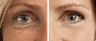
The definition of “young look” includes a number of criteria: uniform color and texture of the skin around the eyes, absence of grooves (tear, palpebral, cheek-zygomatic) and fatty hernias of the lower and upper eyelids.
Fillers based on hyaluronic acid have proven their safety and effectiveness. Rare adverse events are associated exclusively with the technical nuances of the procedure.
The procedure for correcting the nasolacrimal groove is carried out with biodegradable fillers based on hyaluronic acid; the preparation for correcting such a delicate area must be quite soft and plastic.
The goal is to eliminate the volume deficit in the periorbital region. The choice of drug depends on the thickness of the skin and soft tissues. The drug is administered in small portions - 0.05 bonewise along the anterior (facial) surface of the zygomatic process of the maxillary bone and the lower orbital edge of the zygomatic bone.
When the filler is injected, the relief in the infraorbital area is leveled, the eyes do not look sunken, light reflection improves and dark circles under the eyes disappear.
Correction of this area requires the doctor to understand the mechanisms of formation of certain cosmetic defects, good knowledge of anatomy, and the ability to assess the individual and ethnic characteristics of the patient.
The procedure can be performed with either a needle or a cannula. The point for inserting the cannula is 1.5 cm below the temporal edge of the orbit. The depth of drug administration is strictly under the muscle. With intradermal administration, the risk of contouring increases. The filler is injected slowly and evenly into the projection of the palpebromalar groove. Subperiosteal administration guarantees a natural result, no Tyndall effect (change in skin color). The elevation test allows you to determine the level of the cannula.
You can increase the effectiveness of contouring of the periorbital area and prolong the effect by combining contouring with procedures aimed at tightening and toning the skin:
- fractional laser resurfacing;
- needle RF lifting;
- phototherapy;
- biorevitalization;
- plasma lifting;
- mesotherapy.
Methods for diagnosing retinal dystrophy
More information about the clinical picture of the disease is provided by an integrated approach, which the specialists of Dr. Trubilin’s clinic use in their work. A complete diagnosis includes the use of the following methods:
- Visual acuity study.
- Carrying out measurements of intraocular pressure.
- Checking the fundus with a dilated pupil.
- Non-contact examination using optical coherence tomography (OCT). The technique is used to identify the causes and consequences of retinal diseases. Profile equipment allows the diagnostician to see the smallest sections of the central part of the organ being examined. Based on the information received, it is easier to assess real visual acuity. Screening is carried out with a laser beam or IR illumination. At the end of the diagnostic procedure, the patient receives a two-dimensional or three-dimensional image of the fundus.
Modern technology makes it possible to detect the earliest manifestations of the disease, which opens up good prospects for preventing the progression of disorders. Patients are encouraged to use simple self-diagnosis methods. You need to pay attention to the following signs:
- deterioration in the quality of vision when trying to look at any object at a distance or at close range;
- decreased visibility when reading or writing texts, if the same glasses that were used earlier are used for this;
- the desire to turn on the light brighter, to make the lighting more intense in a room where previously, under exactly the same conditions, this was not required;
- the appearance of a feeling that there is an obstacle in front of one of the eyes, impairing visibility, the spot has a fuzzy outline and is transparent to light, it cannot be removed with your hands or washed with water.
Any changes in the perception of the surrounding space should alert you: distortion of contours, colors, brightness. These signs are a good reason to contact an ophthalmologist. Patients are also recommended to use the Amsler mesh in accordance with the recommendations of the attending physician. This diagnosis is carried out for each eye separately.
Periorbital hyperpigmentation
The formation of hyperpigmentation can be due to various reasons:
- pathology of the endocrine system (hyperfunction of the thyroid gland, Addison's disease, pathology of ovarian function);
- chronic sinusitis, diseases of the gastrointestinal tract and liver (hepatitis, cirrhosis, cholelithiasis, chronic colitis);
- taking medications that stimulate melanin synthesis (tetracycline antibiotics, antidepressants, hormonal contraceptives, etc.);
- chronic dermatoses (allergic rhinoconjunctivitis, atopic dermatitis, eczema).
The intensity of pigment spots is determined by the amount of melanin, and the color is determined by the depth of deposition. Shades of brown indicate a superficial epidermal accumulation of pigment, but shades of black, blue and gray indicate a deep intradermal location of the pigment.
Medications that block melanogenesis are used as therapy. Externally used: arbutin, mulberry extract, kojic and azelaic acids, hydroquinone.
Injectable mesotherapy drugs that regulate melanogenesis have proven themselves well: vitamin C, taurine, glutathione, sodium pyruvate.
Hardware methods for correcting hyperpigmentation have become very popular: BBL and IPL phototherapy, laser systems for melanin destruction.
Treatment
Successful treatment of the disease is possible only with early detection. Advanced forms of retinal thinning can be stopped, but vision cannot be restored in such cases. The sooner the diagnosis is made, the less effort it will be possible to restore the structure of the retina. At the initial stage of retinal dystrophy, conservative methods are used: medications, physiotherapy, diet correction and taking vitamin and mineral complexes. In case of extensive damage to the tissues of the inner lining of the eye, surgical or minimally invasive intervention is indicated.
Circles under the eyes of vascular origin and swelling
An abundant, superficially located vascular network in the eyelid area contributes to the formation of bluish circles under the eyes. Since the skin here is very thin, even minor hemodynamic disturbances are immediately reflected in the appearance.
A simple test helps determine the presence of a vascular component in the formation of dark circles. Pressing the soft tissue with the fingers against the bony edge of the orbit causes blanching, which suggests a predominance of the vascular component in the formation of dark circles.
Swelling of the eyelid area is associated with a pathological increase in the amount of interstitial (intercellular) fluid as a result of transudation (exit) of the liquid part from the vascular bed. A large number of vascular anastomoses contribute to fluid retention in the tissues. Fatty tissue is loose and highly hydrophilic compared to other zones. An age-related decrease in the tone of the orbicularis oculi muscle also contributes to increased swelling. Interstitial pressure decreases and lymph drainage is disrupted.
Endogenous (internal) causes of edema formation:
- diseases of the excretory system;
- cardiovascular pathology;
- venous and lymphatic insufficiency;
- hormonal disorders (thyroid gland, adrenal glands).
- Exogenous (external) causes:
- violation of the drinking regime (lots of fluids at night);
- alcohol;
- salty food;
- taking hormonal drugs.
Methods of treating swelling (it is necessary to strengthen the drainage activity of the lymphatic and vascular network):
- microcurrent therapy;
- manual lymphatic drainage massage;
- phonophoresis;
- mesotherapy with drugs that have a drainage effect (caffeine, L-carnitine, artichoke extracts, ginkgo biloba, Asian centella and green tea, rutin).
Skin tightening procedures also have a good effect: fractional photothermolysis, RF needle lifting, photorejuvenation, etc.
Symptoms
The clinical manifestations of retinal thinning are very diverse, but boil down to one thing - a gradual loss of visual acuity, the ability to distinguish colors and navigate in space in low light. The specific set of symptoms depends on the form of the disease and the degree of its progression.
Congenital thinning develops slowly. There is no manifestation, that is, a sudden onset of symptoms, with this form. Signs of the disease depend on the type of retinal damage:
- with pigment thinning, the patient ceases to see at dusk, the disease first appears in adulthood;
- with dotted white, whitish, blurred spots appear in the field of vision, which grow and merge over time. The disease appears in childhood and progresses slowly.
Acquired types of pathology are more diverse, but have similar manifestations. In most patients, visual acuity suddenly drops, the ability to adapt to poor lighting deteriorates, and entire sectors disappear from the field of vision. The set of symptoms depends on the location of the thinned area and the presence of exudate underneath it.
Based on these characteristics, acquired retinal thinning is divided into four subgroups:
- With damage to the peripheral part of the retina. The disease causes a deterioration in the quality of vision - the appearance of “blind” spots, the inability to see and recognize objects with peripheral vision, spots and black dots in front of the eyes.
- With damage to the central part of the retina. Unlike other varieties, this one manifests itself as a sudden clouding of the visual image in the center. The spot may appear as a dark or whitish mist. Peripheral vision is preserved. Contact with the outside world is difficult due to the inability to perceive the environment as a three-dimensional picture.
- With the release of exudate. The initially thin retina with this type of pathology is subject to increased stress due to the pressure of the fluid accumulated behind it. Patients with this form of the disease complain of distortion of the visual image, loss of multiple areas in the field of vision.
- With the accumulation of semi-solid deposits under the retina. This form of the disease is accompanied by slow death of retinal tissue from the center to the periphery. It manifests itself as a gradual loss of fragments in the field of vision, deterioration in light sensitivity, and the inability to determine the distance to an object or focus the gaze on it.
The sooner you pay attention to the listed symptoms and begin treatment, the higher the chance of at least partially preserving your vision.
Prevention
Currently, there are no guaranteed working measures to prevent the thinning of the retina, especially if the pathology is traced in the family history. The only way to avoid serious vision problems is to regularly visit an ophthalmologist and undergo fundus and retinal examinations.
Following the general principles of a healthy lifestyle will help increase the chances of maintaining a healthy retina: a balanced diet, giving up bad habits, moderate physical activity. These measures will be enough to maintain the vascular system in good shape. In addition, eye injury should be avoided, be it mechanical particles, bruises, or prolonged viewing of TV.
Important points of rehabilitation
The most common questions from patients: will there be pain or bruising after eyelid blepharoplasty? When can I go to work and are the stitches noticeable after eyelid surgery?
Painful sensations
We are dealing with surgery, so you will experience some discomfort in the early postoperative period.
How to reduce?
Analgesics in tablets can easily cope with this.
Hematomas and edema
Hematomas (bruises) and swelling are inevitable. The manifestation reaches its peak within two to three days after correction.
How to reduce?
During the first days after eyelid blepharoplasty, apply ice to your cheeks. Try to sleep on a higher pillow to minimize swelling. Do not bend over or lift heavy objects.
Two weeks after the procedure, swelling disappears almost completely.
When can you put on makeup and go out in public?
2 weeks after blepharoplasty, you can use cosmetics and resume an active social life. But heavy physical activity and active sports are not yet allowed.
How to care for seams
Scar healing takes place in several stages. The color of the scar evens out with the skin tone after about 3 months.
Price
The cost of the operation is affected by the complexity of the manipulations. Details can only be clarified during a face-to-face consultation with surgeon Vladimir Kosinets. To register, please contact the administrator by phone.
| Blepharoplasty | Price |
| Upper eyelid surgery | 60 000 |
| Lower classic blepharoplasty | 70 000 |
| Inferior transconjunctival surgery | 80 000 |
| Lower correction with fat redistribution and myopexy | 90 000 |
| Circular blepharoplasty | 130 000 |
| Volumetric plastic surgery of the upper and lower eyelids | 150 000 |