Services Doctors Results Reviews
Expert tested
Zadiran Alina Valerievna Dermatovenerologist, cosmetologist. Candidate of Medical Sciences
Publication date: October 10, 2022
Review date: November 14, 2022
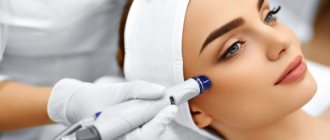
The laser is a real magician. It helps to improve skin health, restore youth, get rid of wrinkles, scars and stretch marks, age spots, and excess hair. With its help, blepharoplasty is now also performed - excess skin on the eyelids is removed. Although quite recently this was only possible through surgery.
Laser procedures are good because they do not cause discomfort to patients, are easily tolerated and do not require serious rehabilitation when you need to give up your usual lifestyle. I did a laser procedure and after a few hours there will be no trace left of the intervention. The only condition: they need to be done during the period of least solar radiation, that is, from mid-autumn to early spring.
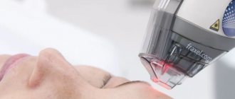
There are many laser procedures today, but there are several of the most popular and in demand. They are carried out using the Fraxel
(USA) and
Dermal radio wave optical thermolysis - DROT
(Italy). These lasers are recognized as the safest and most effective.
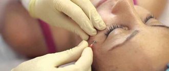
Non-surgical blepharoplasty DROT (DROT)
This technology was developed by Italian scientists. It is based on a combination of RF radiation and fractional CO2 laser.
For eyelid lifting S-Pulse and D-Pulse pulses are used, as well as the Stack mode, with which you can vary the depth of penetration of the laser beam. The use of different types of laser pulses in combination with RF radiation allows doctors to work at different depths of the skin depending on the task at hand. As a result, the eyebrows are raised, sagging tissue on the upper and lower eyelids is removed, the skin is noticeably thickened, and wrinkles are smoothed out.
Laser eyelid lift provides a very noticeable skin tightening effect after the procedure, comparable to surgical circumferential blepharoplasty. It removes up to 2 mm of excess skin. And the facial expression becomes 10 years younger.
The technology can be used to correct existing age-related changes in the periorbital area, as well as for preventive purposes for all skin phototypes.
Types of skin neoplasms
Laser removal of skin tumors is effective for the following pathologies: atheroma, basal cell carcinoma, keratoma, botryomycomoma, cutaneous horn, warts, mole, fibroma, papilloma, condyloma, xanthelasma, molluscum contagiosum, nevus, etc.
What are the benefits of non-surgical laser blepharoplasty?
- There are no bruises or swelling. Due to the exposure to high temperatures during laser blepharoplasty, blood clotting in small vessels occurs without the formation of swelling and bruising.
- Fast recovery period. Patients who have undergone laser blepharoplasty note the fastest possible recovery period.
- Infection is excluded. Due to the high temperature during laser blepharoplasty, the possibility of microbes getting into the skin is eliminated, which means that the risk of infectious diseases and various complications disappears.
- No scars. The coagulation process, which is characteristic of laser blepharoplasty, allows wounds to heal without scar formation.
- There is a general rejuvenation of appearance. As a result of laser exposure, not only excess eyelid skin is eliminated, but also a general facelift occurs. In addition, the skin becomes dense, uniform in color, smooth and soft, especially in the lower eyelid area. As a result of laser exposure, the look becomes more expressive and attractive.
The results obtained from laser eyelid rejuvenation last for 3-8 years.
Laser stretch mark removal
At A Clinic, stretch marks (that is, rupture of the dermis due to stretching of the skin) are removed using the American Fraxel laser and the Italian technology of Dermal Radio Wave Optical Thermolysis (DROT-therapy).
Fraxel
is capable of destroying scar tissue at a depth of up to 1.5 mm and stimulating the growth of normal tissue at this site. In this case, the surface of the skin is not affected. The Fraxel laser safely removes stretch marks from any area of the body, including the chest and inner thighs.
DROT
generates microthermal damage zones that destroy scar tissue. In areas subjected to fractional exposure, regeneration processes are launched, and new healthy tissue is formed without aesthetic defects. The number of sessions required to improve skin texture is determined individually depending on the patient’s skin condition.
Application of laser in dermatology and cosmetology
The search for new means and methods of treating dermatoses is due to intolerance to many drugs, the development of allergic reactions of varying severity, side effects of drugs, the low therapeutic effectiveness of generally accepted methods of treatment, and the need to improve and optimize existing methods. In this regard, studying the capabilities of various physical factors - ultrasound, cryotherapy, phototherapy, magnetic and laser radiation - is an important practical task of modern dermatology. This article describes the main physical and therapeutic properties of laser radiation, as well as the range of its applications in dermatology and cosmetology.
The term "laser" is an abbreviation for the English Light Amplification by Simulated Emission of Radiation - amplification of light using induced radiation.
A laser (or optical quantum generator) is a technical device that produces electromagnetic radiation in the form of a directed, focused, highly coherent monochromatic beam.
Physical properties of laser radiation
The coherence of laser radiation determines the constancy of the phase and frequency (wavelength) throughout the operation of the laser, i.e., this is a property that determines the exceptional ability to concentrate light energy in various parameters: in the spectrum - a very narrow spectral line of radiation; in time - the possibility of obtaining ultrashort light pulses; in space and direction - the possibility of obtaining a directed beam with minimal divergence and focusing of all radiation in a small area with dimensions on the order of the wavelength. All these parameters make it possible to carry out local effects, down to the cellular level, as well as to effectively transmit radiation through optical fibers for remote effects.
The divergence of laser radiation is a plane or solid angle that characterizes the width of the radiation pattern in the far field at a given level of energy distribution or power of laser radiation, determined in relation to its maximum value.
Monochromaticity is the spectral width of the radiation and the characteristic wavelength for each radiation source.
Polarization is a manifestation of the transversality of an electromagnetic wave, i.e., maintaining a constant orthogonal position of mutually perpendicular vectors of electric and magnetic field strength in relation to the speed of propagation of the wave front.
The high intensity of laser radiation allows significant energy to be concentrated in a small volume, which causes multiphoton and other nonlinear processes in the biological environment, local thermal heating, rapid evaporation, and hydrodynamic explosion.
The energy parameters of lasers include: radiation power, measured in watts (W); radiation energy, measured in joules (J); wavelength, measured in micrometers (µm); radiation dose (or energy density) - J/cm².
Laser radiation differs in its properties from other types of electromagnetic radiation (X-ray and high-frequency γ-radiation) used in medicine. Most laser sources emit in the ultraviolet or infrared ranges of electromagnetic waves, and the main difference between laser radiation and the light of conventional thermal sources is its spatial and temporal coherence. Thanks to this, laser radiation energy is relatively easy to transmit over a considerable distance and concentrate in small volumes or in short time intervals.
Laser radiation affecting a biological object for therapeutic purposes is an external physical factor. When laser radiation energy is absorbed by a biological object, all processes occurring during this process are subject to physical laws (reflection, absorption, dispersion). The degree of reflection, scattering and absorption depends on the condition of the skin: moisture, pigmentation, blood supply and swelling of the skin and underlying tissues.
The penetration depth of laser radiation depends on the wavelength, decreasing from long-wave to short-wave radiation. Thus, infrared (0.76–1.5 μm) and visible radiation have the greatest penetrating power (3–5–7 cm), while ultraviolet and other long-wave radiation are strongly absorbed by the epidermis and therefore penetrate into tissues to a shallow depth (1– 1.5 cm).
Application of laser in medicine:
- destructive effects on biological structures and processes - coagulation (in ophthalmology, oncology, dermatovenereology) and tissue dissection (in surgery);
- biostimulation (in physiotherapy);
- diagnostics - study of biological structures and processes (Doppler spectroscopy, flow cytophotometry, holography, laser microscopy, etc.).
Application of lasers in dermatology
In dermatology, two types of laser radiation are used: low-intensity - as laser therapy and high-intensity - in laser surgery.
Lasers are divided according to the type of active medium:
- to solid-state (ruby, neodymium);
- gas - HE-NE (helium-neon), CO2;
- semiconductor (or diode);
- liquid (based on inorganic or organic dyes);
- metal vapor lasers (the most common are copper or gold vapor).
Depending on the type of radiation, there are ultraviolet, visible and infrared lasers. At the same time, both semiconductor lasers and metal vapor lasers can be both low-intensity (for therapy) and high-intensity (for surgery).
Low-intensity laser radiation (LILR) is used for laser therapy of skin diseases. The effect of LILI is to activate cell membrane enzymes, increase the electrical charge of proteins and phospholipids, stabilize membrane and free lipids, increase oxyhemoglobin in the body, activate tissue respiration processes, increase cAMP synthesis, stabilize oxidative phosphorylation of lipids (reduce free radical complexes).
When exposed to LILI on biological tissue, the following main effects are observed:
- anti-inflammatory,
- antioxidant,
- anesthetic,
- immunomodulatory.
The pronounced therapeutic effect in the treatment of human diseases of various etiologies and pathogenesis suggests the existence of a biostimulating mechanism of action of low-power laser radiation. Researchers consider the immune system's response to laser radiation to be one of the most important factors in the mechanism of laser therapy, which, in their opinion, is the trigger point in the reaction of the whole organism.
Anti-inflammatory effect
When exposed to LILI on the skin, an anti-inflammatory effect is observed: microcirculation in tissues is activated, blood vessels dilate, the number of functioning capillaries increases and collaterals are formed, blood flow in tissues increases, the permeability of cell membranes and osmotic pressure in cells is normalized, and the synthesis of cAMP increases. All these processes lead to a decrease in interstitial edema, hyperemia, peeling, itching, delimitation of the pathological process (focus) is observed, and acute inflammatory manifestations subside within 2–3 days. The effect of LILI on the area of inflammation in the skin, in addition to the anti-inflammatory effect, provides an antibacterial and fungicidal effect. According to literature data, the number of bacteria and fungal flora is reduced by 50% within 3–5 minutes of laser irradiation of the pathological area.
Taking into account the anti-inflammatory and antibacterial effect of LILI when applied locally to the skin, lasers are used in the treatment of diseases such as pyoderma (folliculitis, boils, impetigo, acne, streptostaphyloderma, chancriform pyoderma), trophic ulcers, allergic dermatoses (true eczema, microbial eczema, atopic dermatitis , urticaria). LILI is also used for dermatitis, burns, psoriasis, lichen planus, scleroderma, vitiligo, diseases of the oral mucosa and red border of the lips (bullous pemphigoid, exudative erythema multiforme, cheilitis, stomatitis, etc.).
Antioxidant effect
When exposed to LILI, an antioxidant effect is observed, which is ensured by reducing the production of free radical complexes, when cellular and subcellular components are protected from damage, as well as ensuring the integrity of organelles. This effect is associated with the pathogenesis of a significant number of skin diseases and the mechanism of skin aging. As studies by G. E. Brill and co-authors have shown, LILI activates the enzymatic component of antioxidant protection in erythrocytes and somewhat weakens the stimulating effect of stress on lipid peroxidation in erythrocytes.
The antioxidant effect of LILI is used in the treatment of allergic dermatoses, chronic skin diseases and during anti-aging procedures.
Analgesic effect
The analgesic effect of LILI is achieved due to the blockade of pain sensitivity along the nerve fibers. At the same time, a slight sedative effect is observed. Also, the analgesic effect is provided by reducing the sensitivity of the skin receptor apparatus, increasing the threshold of pain sensitivity, and stimulating the activity of opiate receptors.
The combination of analgesic and mild sedative effects plays an important role, since in various skin diseases itching (as a perverted manifestation of pain) is the main symptom that disrupts the patient’s quality of life. These effects make it possible to use LILI for allergic dermatoses, itchy dermatoses, and lichen planus.
Immunomodulatory effect
Recently, it has been proven that in various skin diseases there is an imbalance of the immune system. Both with local irradiation of the skin and with intravenous irradiation of the blood, LILI has an immunomodulatory effect - dysglobulinemia is eliminated, the activity of phagocytosis increases, apoptosis is normalized and the neuroendocrine system is activated.
Some techniques using LILI
Allergic dermatoses (atopic dermatitis, chronic eczema, recurrent urticaria). LILI irradiation of venous blood is carried out using an invasive or non-invasive method, as well as local laser therapy.
The invasive method consists of venipuncture (venesection) in the area of the radial vein, collecting blood in an amount of 500–750 ml, which is passed through a laser beam, followed by reinfusion of irradiated blood. The procedure is carried out once, once every six months with an exposure of 30 minutes.
The non-invasive method involves applying a laser beam to the projection of the radial vein. At this time, the patient clenches and unclenches his fist. As a result, 70% of the blood is irradiated within 30 minutes. The method is painless, does not require special conditions, and involves the use of both continuous and pulsed laser radiation - from 5 to 10,000 Hz. It has been established that vibrations of 10,000 Hz correspond to vibrations on the surface of cell membranes.
Blood irradiation is performed only with a helium-neon laser, wavelength 633 nm, power 60.0 mW and semiconductor lasers with a wavelength of 0.63 microns.
S. R. Utz et al used laser heads with a reflective surface to treat severe forms of atopic dermatitis in children using a non-invasive method; Immersion oil was applied to the skin at the irradiation site, and compression was created with the head. The irradiation zone was the great saphenous vein at the level of the medial malleolus.
The listed methods are supplemented with local laser therapy. Recommended maximum area sizes for laser therapy during one session: for the skin of the face and mucous membranes of the nasal cavity, mouth and lips - 10 cm², for other areas of the skin - 20 cm². For symmetrical lesions, it is advisable to sequentially work on two contralateral zones during one session with an equal division of the recommended area.
When working on the skin of the face, it is strictly forbidden to direct the beam at the eyes and eyelids. It follows that helium-neon laser radiation should not be used to treat eyelid skin diseases.
Helium-neon laser radiation is used mainly in remote mode. To treat skin diseases with a lesion area greater than 1–2 cm², the laser beam spot is moved at a speed of 1 cm/s over the entire area selected for the session so that it is all evenly irradiated. A spiral scanning vector is advisable - from the center to the periphery.
In atopic dermatitis, irradiation is carried out across fields, covering the entire affected surface of the skin according to the configuration of the pathological area from the periphery to the center, with irradiation of healthy tissue within 1–1.5 cm or scanning with a laser beam at a speed of 1 cm/s. The radiation dose per session is 1–30 J/cm², session duration is up to 25 minutes, course of 5–15 sessions. Treatment can be carried out against the background of antioxidant therapy and vitamin therapy.
When irradiating venous blood using LILI in patients with allergic dermatoses, we achieve all the above-mentioned effects of laser radiation, which contributes to a faster recovery and a reduction in relapses.
Psoriasis. For psoriasis, blood irradiation is used, laser inductothermy of the adrenal glands is used, as well as local effects on plaques. It is usually performed with infrared (0.89 nm, 3–5 W) or helium-neon lasers (633 nm, 60 mW).
Laser inductothermy of the adrenal glands is carried out by contact on the skin in the projection of the adrenal glands, from 2 to 5 minutes, depending on the patient’s weight, the course is 15–25 sessions. Laser irradiation is carried out in the stationary and regressing stages of psoriasis, ensuring the production of endogenous cortisol by the patient's body, which leads to the resolution of psoriatic elements and allows achieving a pronounced anti-inflammatory effect.
The effectiveness of laser therapy for psoriatic arthritis has been shown. During treatment, the affected joints are irradiated, sometimes local therapy is combined with irradiation of the adrenal glands. After two sessions, an exacerbation is noted, which becomes less intense by the 5th session, and by the 7th–10th sessions the condition stabilizes. A course of laser therapy consists of 14–15 sessions.
A fundamentally new direction in the treatment of psoriasis and vitiligo is the development and clinical use of an excimer laser based on xenon chloride, which is a source of narrow-band ultraviolet (UVB) radiation with a length of 308 nm. Since the energy is directed only to the area of the plaque and healthy skin is not affected, the lesions can be irradiated using radiation with a high energy density (from 100 mJ/cm² and above), which enhances the antipsoriatic effect. Short pulses of up to 30 ns allow you to avoid vaporization and thermal damage. A narrow monochromatic radiation spectrum with a length of 308 nm acts only on one chromophore, causing the death of mutagenic keratinocyte nuclei and activating T-cell apoptosis. The introduction of excimer laser systems into widespread clinical practice is limited by their high cost, lack of methodological support, insufficient knowledge of long-term results, and difficulties associated with calculating the depth of exposure as plaques thin out during therapy.
Lichen planus (LP). In case of LLP, the technique of local irradiation of rashes by contact method, sliding movements from the periphery to the center is usually used. Exposure - from 2 to 5 minutes, depending on the affected area. The total dose should not exceed 60 J/cm². Such procedures provide an anti-inflammatory and antipruritic effect. To resolve plaques, the exposure is increased to 15 minutes.
When LLP is localized on the scalp, laser irradiation is carried out with an exposure time of up to 5 minutes. In addition to the above-mentioned effects, stimulation of hair growth in the irradiation zone is achieved.
When applying these methods, infrared, helium-neon and copper vapor laser radiation is used. In case of LP, irradiation of venous blood can also be performed.
Pyoderma. For pustular skin diseases, the technique of LILI irradiation of venous blood and the technique of local irradiation by contact method, sliding movements with an exposure of up to 5 minutes are also used.
These techniques make it possible to achieve anti-inflammatory, antibacterial (bacteriostatic and bacteriocidal) effects, as well as stimulation of reparative processes.
For erysipelas, LILI is used contact, remotely and intravenously. When using laser therapy, body temperature normalizes 2–4 days earlier, regression of local manifestations occurs 4–7 days faster, cleansing and all repair processes occur 2–5 days faster. An increase in fibrinolytic activity, the content of T- and B-lymphocytes and their functional activity, and an improvement in microcirculation were revealed. Relapses with traditional treatment are 43%, with LILI - 2.7%.
Vasculitis. For the treatment of skin vasculitis, V.V. Kulaga and co-authors propose the invasive LILI method. 3–5 ml of blood is taken from the patient’s vein, placed in a cuvette and irradiated with a 25 mW helium-neon laser for 2–3 minutes, after which 1–2 ml of irradiated blood is injected into the lesions. 2–4 injections are given in one session, 2–3 sessions per week, the course of treatment consists of 10–12 sessions. Other authors recommend intravascular irradiation of blood with helium-neon laser energy with a power of 1–2 mW for 10–30 minutes, sessions are carried out daily or every other day, the course consists of 10–30 sessions.
Scleroderma. J. J. Rapoport and co-authors propose to conduct laser therapy sessions using a helium-neon laser through a light guide inserted through a needle at the border of healthy and affected skin. The session lasts 10 minutes, the dose is 4 J/cm³. Another technique involves external irradiation of lesions with radiation at a power of 3–4 mW/cm² with an exposure of 5–10 minutes, a course of 30 sessions.
Viral dermatoses. Laser therapy has been used quite successfully for herpes zoster. A. A. Kalamkaryan and co-authors proposed remote segmental irradiation of lesions with a helium-neon laser with a power of 20–25 mW, in which the laser beam moves along the nerve trunks and to the sites of rashes. Sessions are held daily and last from 3 to 20 days.
Vitiligo. To treat vitiligo, helium-neon laser radiation and external photosensitizers, such as aniline dyes, are used. Immediately before the procedure, a dye solution (diamond green, methylene blue, fucorcin) is applied to the lesions, after which local irradiation is carried out with a defocused laser beam with a power of 1–1.5 mW/cm². The duration of the session is 3–5 minutes, daily, the course is 15–20 sessions, repeated courses are possible after 3–4 weeks.
Baldness. The use of a copper vapor laser in an experiment carried out on the skin, according to electron microscopy, revealed a marked increase in proliferative and metabolic activity in epidermocytes, including hair follicles. An expansion of the microvessels of the papillary dermis was noted. In connective tissue, in particular in fibroblasts, a relative increase in the volume of intracellular structures associated with collagen synthesis was detected. An increase in activity was recorded in neutrophils, eosinophils, macrophages and mast cells. The listed changes are the basis for the treatment of baldness. Already after the 4th–5th session of laser therapy, growth of vellus hair on the head is noted.
The vitiligo treatment technique described above is also used to treat patchy baldness.
Scarring. Using light and electron microscopy, changes that occur in skin scars as a result of the use of laser radiation in humans were studied. Thus, the use of ultraviolet and helium-neon LILI did not cause significant changes due to the shallow penetration of laser energy. After using infrared laser radiation, the number of collagen-resorbing fibroblasts increases, while the collagen fibers become thinner, the number of mast cells and the release of secretory granules slightly decrease. The relative volume fraction of microvessels increases to some extent.
When using LILI to prevent severe scarring of skin surgical wounds, a decrease in the content of active fibroblasts and, consequently, collagen was revealed.
Use of high-intensity laser radiation (HILI)
VILI is obtained using CO2, Er:YAG laser and argon laser. CO2 laser is mainly used for laser removal (destruction) of papillomas, warts, condylomas, scars and dermabrasion; Er:YAG laser - for laser skin rejuvenation. There are also combined CO2 and Er:YAG laser systems.
Laser destruction. VILI is used in dermatology and cosmetology for the destruction of tumors, removal of nail plates, as well as for laser vaporization of papillomas, condylomas, nevi and warts. In this case, the radiation power can range from 1.0 to 10.0 W.
Neodymium and CO2 lasers are used in clinical practice. When using a CO2 laser, surrounding tissues are less damaged, and a neodymium laser has a better hemostatic effect. In addition to the laser physically removing lesions, studies have shown the toxic effects of laser radiation on human papillomavirus (HPV). By varying the laser power, spot size and exposure time, the depth of coagulation can be controlled. Well-trained personnel are required to perform the procedures. Lasers require anesthesia, but topical or topical anesthesia is sufficient, allowing procedures to be performed on an outpatient basis. However, 85% of patients still report mild pain. The method has approximately the same effectiveness as electrocoagulation, but is less painful, causes fewer postoperative side effects, including less pronounced scarring, and provides a good cosmetic effect. The effectiveness of the method reaches 80–90% in the treatment of genital warts.
Laser therapy can be successfully used to treat common warts that are resistant to other treatments. In this case, several courses of treatment are carried out, which allows increasing the cure rate from 55 (after 1 course) to 85%. However, in special cases with many years of ineffective treatment with various methods, the effectiveness of laser therapy is not so high. Even after multiple courses of treatment, it can stop recurrence in only about 40% of patients. Careful studies have shown that such a low rate is due to the fact that the CO2 laser is ineffective in eliminating the viral genome from lesions that are resistant to treatment (according to PCR, molecular biological cure occurs in 26% of patients).
Laser therapy can be used to treat genital warts in teenagers. The method has been shown to be highly effective and safe in treating this group of patients; in most cases, 1 procedure is sufficient for cure.
To reduce the number of relapses of genital warts (recurrence rate from 4 to 30%), it is recommended to use laser “cleaning” of the surrounding mucosa after the removal procedure. When using the “cleansing” technique, discomfort and pain are often observed. In the presence of large condylomas, before laser therapy, their preliminary destruction is recommended, in particular with electrocautery. This, in turn, avoids the side effects associated with electroresection. A possible cause of relapse is the persistence of the HPV genome in the skin near the treatment sites, which was identified both after laser application and after electrosurgical excision.
The most severe side effects of laser destruction are: ulceration, bleeding, and secondary wound infection. After laser excision of warts, complications develop in 12% of patients.
As with electrosurgical methods, HPV DNA is released through smoke, which requires appropriate precautions to avoid contamination of the physician's nasopharynx. At the same time, some studies have shown no difference in the incidence of warts among surgeons involved in laser therapy compared with other groups of the population. There were no significant differences in the incidence of warts between groups of doctors who used and did not use protective equipment and smoke evacuators. However, because the types of HPV that cause genital warts can infect the lining of the upper respiratory tract, laser smoke containing these viruses is dangerous for surgeons performing vaporization.
The widespread use of laser destruction methods is hampered by the high cost of high-quality equipment and the need to train experienced personnel.
Laser hair removal. Laser hair removal (thermal laser hair removal) is based on the principle of selective photothermolysis. A light wave with specially selected characteristics passes through the skin and, without damaging it, is selectively absorbed by melanin, which is contained in large quantities in the hair follicles. This causes heating of the hair follicles, followed by their coagulation and destruction. To destroy the follicles, the required amount of light energy must be supplied to the hair root. For hair removal, radiation with a power of 10.0 to 60.0 W is used. Since hair is in different stages of growth, complete hair removal requires several procedures. They are carried out on any part of the body, non-contact, at least 3 times with an interval of 1–3 months.
The main advantages of laser hair removal are the comfort and painlessness of the procedures, the achievement of stable and long-term results, safety, high processing speed (hundreds of follicles are removed simultaneously with one pulse), non-invasiveness, and non-contact. Thus, this method represents the most effective and most cost-effective method of hair removal today. Prolonged exposure to the sun and tanning (natural or artificial) significantly reduces the effectiveness of procedures.
Laser dermabrasion. Dermabrasion is the removal of the upper layers of the epidermis. After exposure, a fairly soft and painless laser scab remains. Within 1 month after the procedure, new young skin is formed under the scab. Laser dermabrasion is used to rejuvenate the skin of the face and neck, remove tattoos, polish scars, and also as a treatment for post-acne in patients with severe forms of acne.
Laser skin rejuvenation. The laser provides precise and superficial ablation with minimal heat damage and no bleeding, resulting in rapid healing and resolution of erythema. For this purpose, Er:YAG lasers are mainly used, which are good for superficial skin rejuvenation (including in dark-skinned patients). The devices allow for quick and uniform scanning of the skin, as well as even out color boundaries after CO2 laser treatment.
Contraindications to the use of laser therapy
Laser therapy is used with caution in patients with cancer, diabetes mellitus, hypertension and thyrotoxicosis in the stage of decompensation, severe heart rhythm disturbances, angina pectoris of the 3-4th functional classes and circulatory failure of the 2nd-3rd stage, blood diseases, threat bleeding, active form of tuberculosis, mental illness, as well as individual intolerance.
Thus, laser radiation is a powerful adjuvant in the treatment of patients with various dermatological diseases and the method of choice in surgical dermatology and cosmetology.
Literature
- Bogdanov S. L. et al. Laser therapy in cosmetology: Method. recommendations. - St. Petersburg, 1995.
- Brill G.E. et al. Physical medicine. - 1994. - No. 4, 2. - P. 14-15.
- Grafchikova L.V. et al. Physical medicine. -1994. - No. 4, 2. - P. 62.
- Egorov BE and others. Proceedings of the International Conference Clinical and experimental application of new laser technologies. Kazan. - 1995. - P.181-182.
- Kalamkaryan A.L. et al. Vestn. dermatol. and venerol. - 1990. - No. 8. - P. 4-11.
- Kapkaev P.A., Ibragimov A.F. Current issues in laser medicine and surgical endoscopy: Proceedings of the 3rd International Conference. - Vidnoe, 1994. - pp. 93-94.
- Korepanov V.I., Fedorov S.M., Shulga V.A. Application of low-intensity laser radiation in dermatology: A practical guide. - M., 1996.
- Kulaga V.V., Shvareva T.I. Vestn. dermatol. and venerol. - 1991. - No. 6. - P. 42-46.
- Mandel A. N. The effectiveness of laser therapy in patients with focal scleroderma and its effect on the parameters of serotonin, dopamine, norepinephrine and urocanic acid: Abstract of thesis. dis. ...cand. honey. Sci. -M., 1982.
- Mandel A. N. Efficacy of laser photochemotherapy in patients with chronic dermatoses: Dis. ... doc. honey. Sci. - M. 1989. - P. 364.
- Mikhailova I. V., Rakcheev A. P. Vestn. dermatol. - 1994. - No. 4. - P. 50.
- Petrischeva N. N., Sokolovsky E. V. Application of semiconductor lasers in dermatology and cosmetology: A manual for doctors. - St. Petersburg: St. Petersburg State Medical University, 2001.
- Pletnev S. D. Lasers in clinical medicine; Guide for doctors. - M.: Medicine, 1996.
- Rakcheev A.P. Prospects for the use of lasers in dermatology // All-Union Conference on the Use of Lasers in Medicine. - M., 1984.
- Rapoport Zh. Zh. et al. Application of lasers in surgery and medicine. - Samarkand, 1988. - Part 1. - P. 91-93.
- Rodionov V. G. The influence of laser radiation on capillary toxic factors in the blood of patients with allergic vasculitis of the skin // All-Union Conference on the Application of Lasers in Medicine. - M., 1984.
- Utz S.R. et al. Vestn. dermatol. and venerol. - 1991. - No. 11. - P. 11.
- Khalmuratov AM Current issues of laser medicine and surgical endoscopy // Materials of the 3rd International Conference. - Vidnoe, 1994. - P. 482-483.
- Shulga V. A., Fedorov CM Information sheet on the problem “Dermatology and venereology”. - M.: TsNIKVI, 1993.
- Bergbrant I.M., Samuelsson L., Olofsson S. et al. Acta Derm Venerol. 1994; 74(5): 393-395.
- Bonis B., Kemeny L., Dobozy A. et al. 308 nm eximer laser for psoriasis. Lancet. 1997; 3509:1522.
- Damianov N., Mincheva A., de Villiers E.M. Khirurgia. 1993; 46(4): 24-27.
- Handley JM, Dinsmore WJ Eur Acad Dermatol Venerol. 1994; 3(3): 251-265.
- Gerber W., Arheilger B., Ha T. A. et al. Ultraviolet B 308-nm eximer laser treatment of psoriasis: a new phototherapeutic approach. British J of Dermatol. 2003; 149: 1250 -1258.
- Gloster HM, Roenigk RK J Amer Acad Dermatol. 1995; 32(3): 436 – 441.
- Lassus J., Happonen HP, Niemi KM et al. Sex Transm Dis. 1994; 21(6): 297-302.
- Novak Z., Bonis B., Baltas E. et al. Xenon chloride ultraviolet B laser is more effective in treating psoriasis and in including T cell apoptosis than a narrow-band ultraviolet B. J Photochem and Photobiol. 2002; 67: 32-38.
- Petersen CS, Menne T. Acta Derm Venerol. 1993; 73(6): 465-466.
- Schneede P., Muschter R. Urology. 1999; 33(4): 299-302.
- Schoenfeld A., Ziv E., Levavi. H. et al. Gynecol & Obstet Invest. 1995; 40(1): 46-51.
- Smyczek-Garsya B., Menton M., Oettling G. et al. Zentralbl Gynakol. 1993; 115(9): 400-403.
- Townsend DE, Smith LH, Kinney WK J Reprod Med. 1993; 38(5): 362-364.
- Vasileva P., Ignatov V., Kiriazov E. Akush Ginekol. 1994; 33(2): 23-24.
- Wozniak J., Szczepanska M., Opala T. et al. Gin Pol. 1995; 66(2): 103-107.
A. M. Solovyov, Candidate of Medical Sciences, Associate Professor K. B. Olkhovskaya, Candidate of Medical Sciences
MGMSU, Moscow
What are the benefits of laser stretch mark removal?
- Almost any stretch marks can be removed. The laser acts at a certain depth, which is set individually, depending on the age of the stretch marks.
- During the procedure, healthy tissues are not affected and burns are avoided.
- Suitable for sensitive areas. The laser can be used in the most sensitive areas - on the chest, abdomen, inner surfaces of the arms and thighs.
- There is no rehabilitation period. The maximum that may appear after laser exposure is slight redness. It goes away within a day. The skin recovers quickly.
- In most cases, the procedure is carried out immediately after the initial consultation.
How is it removed?
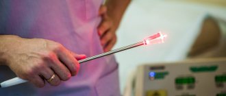
The process of removing formations with a laser is based on the effect of carbon dioxide on the cells of angiomas, plantar warts, and moles, which causes a loss of fluid in them and triggers a destruction mechanism. At this moment, the tissue temperature reaches 100 ̊ C, the surrounding skin heats up to 70 ̊ C, which causes the death of bacteria and determines the bactericidal properties of the laser.
Before surgery, a local anesthetic is applied to the affected area, after which it is evaporated and removed. The laser beam prevents bleeding and does not harm healthy tissue.
The number and time of sessions depends on the severity of the disease and the affected area. It will take 1-2 sessions of 5-20 minutes. The healing period is 7-10 days.
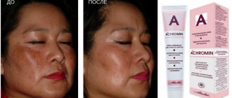
Laser removal of pigment spots
The most effective and safest method of removing pigment spots of any complexity is treatment with fractional laser Fraxel.
The principle of the effect of this laser on pigment spots and areas of hyperpigmentation differs from other common ones. Fraxel does not act on melanin, but on the cells that synthesize and accumulate this pigment. Where the laser penetrates the skin, these cells die, and the melanin they contain is processed by surrounding “utilizer cells.” It does not matter at what depth in the skin the pigment is located: Fraxel effectively treats any form of hyperpigmentation. Subsequently, in place of the destroyed micro-area of the skin, young cells appear, which synthesize and accumulate an amount of pigment adequate to external solar exposure. Thus, old cells in which the formation of melanin is impaired are replaced with new ones: normally colored skin is formed in place of the pigment spot.
ABCD principle
In dermatology, a simple rule is used to determine the signs of malignancy called ABCD. Each letter of the word corresponds to a specific characteristic of a mole:
A - asymmetry:
symmetrical (good) asymmetrical (bad).
B - random coloring:
uniform coloring (good) uneven coloring (bad).
B - height:
uniform height (good) uneven height (bad).
G - boundaries:
clear boundaries (good) unclear boundaries (bad).
D - diameter:
not growing (good) growing (bad).
Important: any changes in moles or their trauma is a reason to pay attention to them! Carefully examine all your formations; if you notice any suspicious signs, contact a specialist immediately!
What are the benefits of laser pigment spot removal?
- Pigmentation can be treated with the Fraxel laser on skin of any color (even black!).
- The laser removes pigment from any part of the body. Thus, many standard techniques cannot be applied or are ineffective when used on the skin of the hands. Fraxel is recommended for the treatment of pigmentation on the hands, neck, and décolleté, and the removal of pigment is accompanied by an impressive rejuvenation result.
- General skin rejuvenation occurs. In the microzones of laser exposure, the process of regeneration of new structural proteins involved in the process of skin renewal - collagen and elastin - begins. Therefore, the skin after Fraxel will look noticeably younger - dense and elastic, as in its youth.
Is it worth removing tumors and why?
The category of skin neoplasms includes diseases with a potential risk of cancerous degeneration:
- basalioma, pigmented nevus - provokes the development of melanoma
- keratoma - squamous cell skin cancer
- papillomatosis (HPV infection) – causes dysplastic processes in the mucous membranes of the genitals
There are signs by which you can determine the changes that occur with the tumor:
- size
- form
- shade
- the appearance of pain
- the appearance of infiltrate (accumulation of blood and lymph)
To avoid relapses, it is recommended to remove the affected areas by cleansing the problem area of the skin.
Note! If the tumor formation is caused by a viral infection, the attending physician must carry out adequate treatment with antiviral drugs, and then laser removal of the skin tumor.
Skin formations are removed immediately. Malignant processes are first treated to prevent progression and metastasis.
Laser acne removal
Post-acne is a complex of skin defects remaining after acne.
Acne most often occurs on young skin. This is an inflammation in the hair follicle, which is clogged with the secretion of the skin glands and in which pathogenic bacteria multiply. The body directs all its energy to remove these bacteria. As a result, inflammation often ends in the formation of a scar - irregularly structured, rough connective tissue.
Fraxel removes microparticles of scar tissue, and normal skin begins to form at the site of exposure due to the production of new collagen. Evens out skin color and texture.
Today Fraxel is one of the most popular hardware procedures for post-acne treatment in Europe and the USA.
Gutsenko Liliya Anatolevna
Cosmetologist
You can also remove post-acne using DROT. The device operates in fractional mode, in which the beam is divided into separate beams (that is, fractions). They penetrate into the deep layers of the skin, causing targeted tissue damage and leaving undamaged areas. Due to microthermal effects, regeneration processes are launched, even in areas untouched by the laser.
A microthermal treatment zone (MTZ) is formed at the site of exposure to microrays. This is done in order to stimulate the skin cells located around the MLZ to begin producing collagen and elastin. All this allows you to tighten and renew the skin. As a result, you can get rid of post-acne: the skin texture becomes smoother and the elasticity of the skin increases.
What types of tumors are there and which ones can be removed with a laser?
There is a large list of skin diseases that can be easily corrected using laser destruction. These include:
- Angioma is a benign tumor of blood and lymphatic vessels. Affects large areas of the skin surface and is a serious cosmetic problem.
- Basal cell carcinoma (basal cell carcinoma) - affects the skin of the face, head, neck, corners of the eyes, wings of the nose, has an unknown etiology, and can occur in a malignant form.
- Warts are growths of various types and colors that are of viral origin.
- Botriomycomoma (pyogenic granuloma) - a neoplasm characterized by a benign course, is a red or bright pink nodule of various sizes, and bleeds when damaged.
- Keratoma is a benign formation that occurs as a result of keratinization of all layers of the epidermis, manifested by the formation of single or multiple plaques with a rough surface, prone to destruction. Refers to borderline tumors in patients who have a tendency to develop cancer.
- Molluscum contagiosum is a disease caused by the chickenpox virus, affecting the mucous membranes and skin.
- Nevus (mole) is a formation on the skin, characterized by an accumulation of melanocytes, and has a benign and malignant course.
- Fibroma is a neoplasm of connective tissue of a benign nature, yellowish-flesh in color, formed on the mucous membranes of the oral cavity, skin, and ovaries.
- Chalazion is a disease of the eyelid and cartilage tissue around the eyes, a chronic proliferative process.
- Condyloma, papilloma (pointed tumor) are soft tissue papillary formations on a stalk, which are caused by the human papillomavirus. Some strains are highly oncogenic and cause severe damage to the genitals and mucous membranes of the cervix (dysplasia).
Laser technology for delicate areas
Fractional laser rejuvenation with Fraxel can be used as an independent technique for neck rejuvenation, where the skin is very thin and delicate, or perhaps in combination with injections. A thin (thinner than a human hair) laser beam forms thousands of micro-zones of influence on every centimeter of skin. In these microzones, old and defective collagen and excess pigment are destroyed. At the same time, around each microzone of exposure, many viable cells remain, which are activated under the influence of heat. During this process, which can take from several hours to several days, skin free of defects appears in place of each microzone.
How to prepare for the procedure
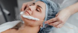
Laser removal of skin tumors does not require special preparation. All that the doctor may require is a conclusion from a histological laboratory about the benign nature of the process, a consultation with a therapist to exclude other contraindications and an oncologist.
Before the procedure, it is recommended to refrain from bad habits, avoid visiting the solarium and sunbathing.
When should a histological examination be performed? Dermatoscopy (examination under a dermatoscope) or histological examination is indicated if a tumor is suspected of malignancy.
The histological method is the examination of postoperative material for the presence of malignant neoplasms under a microscope. Laser removal of skin diseases is a minimally invasive surgical operation in which a section of the affected tissue can be obtained and sent to a histology laboratory without fail for a final diagnosis.
What are the benefits of laser rejuvenation of delicate areas?
- After the laser fractional rejuvenation procedure, the skin structure itself changes. It becomes smoother, moisturized, elastic, less susceptible to inflammation and rashes. It becomes easier to care for her at home.
- A unique feature of Fraxel is that the laser does not destroy the uppermost layer - the stratum corneum - the epidermis. Therefore, after the procedure, the skin looks natural.
- Fractional laser acts delicately on the skin, so there is no need for a rehabilitation period.
The number of procedures depends on the initial data and the problem being solved. For some, one procedure per year is enough, while others may require a course of 3-5 procedures.
The result obtained, namely young, beautiful skin, can last for a long time.
At what age can laser procedures be performed?
It is advisable to resort to laser procedures after 25 years for indications that depend on the severity of age-related changes.
The first sign that you can think about laser rejuvenation is enlarged pores. The pore is surrounded by muscle fibers, which over time lose their tone, causing it to expand. And while the muscles of the body can be trained, there are no exercises for the pores. The second sign that it is time to resort to laser procedures is the appearance of sagging skin, fine wrinkles in the area around the eyes, on the cheeks, and a decrease in its elasticity.
But when it comes to treating the consequences of acne or stretch marks, laser procedures are prescribed at an earlier age.
Dangerous degeneration of moles into melanoma!
Every year, doctors diagnose 200,000 patients with melanoma, and a third of cases are fatal. On average, after positive treatment results, patients with melanoma live about 5 years. These scary statistics are presented here so that you think about your health and pay attention to it.
How to spot melanoma?
Melanoma, in simple words, is a former mole. The harbingers are situations when a mole changes color, redness appears around the mole, grows unevenly, hurts, and bleeding appears. It is possible that new moles will appear during life, which look unusual and differ from those that previously existed on the body. Any changes or appearance of “unusual” moles require special attention and are a reason to consult a dermatologist.