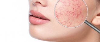
Symptoms
The appearance of the formation and anamnestic data suggest melanosis. This disease has the following characteristic clinical manifestations:
- observed with equal frequency in both sexes;
- occurs, as a rule, in old age;
- single spot;
- the size varies from 2 to 10 cm;
- blurred boundaries;
- irregular shape (the outlines sometimes resemble a “geographic map”);
- brown, bluish, gray or black;
- slight peeling;
- uneven coloring;
- preservation of skin pattern;
- surrounding skin without inflammatory changes;
- at first it is level with the skin or slightly raised;
- over time, the surface becomes uneven with nodules, papules, plaques;
- localized more often on areas of the skin exposed to sunlight (face, forearms, hands);
- sometimes found on mucous membranes (oral cavity, etc.).
Disease prevention
To prevent the development of melanosis, you should protect your skin from direct sunlight in the summer.
Aggressive ultraviolet rays damage the top layer of skin, leading to the development of excessive pigmentation. Therefore, it is important to wear a hat or Panama hat with a wide brim to protect the face. The use of sunscreen will help disperse ultraviolet rays without causing harm to the skin.
It is also necessary to regularly undergo a comprehensive medical examination in an effort to prevent the development of dangerous diseases.
Newly appeared moles and age spots should be examined to rule out the development of skin cancer. Proper nutrition and an active lifestyle will strengthen the immune system, protecting the body from viruses and bacteria. Doctors recommend including foods containing vitamins and beneficial microelements in your daily diet.
A sufficient amount of fresh vegetables and fruits will enrich the body with essential substances and improve health. Timely diagnosis and seeking medical advice can identify melanosis at an early stage. With proper systematic treatment prescribed by the doctor, the disease recedes.
Diagnostics
A competent specialist can diagnose Dubreuil's melanosis only after seeing the patient. But further examination may be required to confirm this diagnosis:
- dermatoscopy (examination under magnification allows a more detailed assessment of the boundaries and structure of the pigment formation);
- mandatory morphological examination of the material obtained during excision of the formation (reveals an increase in the number and accumulation of melanocytes at the border of the dermis and epidermis, inflammatory and dystrophic changes in the dermis);
- scintigraphy with radioactive phosphorus (to exclude degeneration into melanoma);
- determination of the level of melanoma-specific tumor markers.
The first signs of melanoma
Melanoma is the acquisition by cells of unfavorable signs of malignancy (malignancy properties), expressed by various symptoms.
To make it easier to remember the signs of melanoma, use the “FIGARO” rule:
Shape – swollen above the surface;
Changes – accelerated growth;
Borders are openwork, irregular, jagged;
Asymmetry - the absence of mirror similarity of the two halves of the formation;
Size – a formation with a diameter of more than 6 mm is considered a critical value;
Coloring – uneven color, inclusion of random spots of black, blue, pink, red.
In widespread practice, the English version is also popular, summarizing the main, most typical features - the “ABCDE rule”:
Asymmetry - asymmetry in which, if you draw an imaginary line dividing the formation in half, one half will not be similar to the other.
Border irregularity - the edge is uneven, scalloped.
Color – a color that is different from other pigment formations. Interspersed areas of blue, white, and red colors are possible.
Diameter – diameter. Any lesion larger than 6 mm requires additional observation.
Evolution – variability, development: density, structure, size.
Without special studies, it is difficult to determine the type of nevus, but timely changes in the nature of the spot will help detect malignancy.
Treatment
Due to the high risk of malignancy, patients with Dubreuil's melanosis must be treated, and not just observed by an oncologist for years. Depending on the location and size of the formation, concomitant pathology, they undergo:
- surgical intervention (wide excision of the formation and 1 - 2 cm of unchanged skin, with a significant area of removed tissue, plastic surgery is then performed);
- close-focus X-ray therapy (if melanosis of significant size is located on the face, if surgical treatment is impossible).
General therapeutic recommendations
- From an analysis of the literature, it follows that micrographic surgery according to Mohs gives the lowest degree of relapse, although the number of studies is limited and the series of patients are few.
- Other treatments that have a similar relapse rate of 7-10% include traditional surgery, cryosurgery and radiation therapy. Despite the disadvantages of cryosurgery and radiation therapy, there is no significant difference in recurrence rates when comparing these three methods. Therefore, all three modalities can be recommended equally as the primary treatment for LM, if the doctor has the appropriate training. With destructive therapeutic techniques, careful follow-up is extremely important, since a complete histological assessment of the lesion is not possible. The use of destructive techniques is especially indicated for elderly and frail patients who do not agree to surgery or for whom it is not suitable.
- Treatment should not be carried out without prior histological confirmation, since the differential diagnosis includes both benign lesions, such as seborrheic keratosis, solar lentigo, and malignant changes, such as MLM or superficial spreading melanoma.
Recommended Treatments
Surgical treatment
Surgical excision is widely considered the most reliable method of adequate removal. Most surgeons and dermatologists argue that the treatment of choice is extensive excision, since destructive techniques are performed blindly, without providing a biopsy for a complete diagnosis and a histological basis for adequate treatment. In support of this opinion, the cure rate is 91% or higher. It was shown that out of every 85 pigmented spots diagnosed with LM before treatment, 45 had lesions of LM. These studies are used to demonstrate the need for surgical treatment and obtaining a complete biopsy to establish a histological diagnosis. Other studies suggest, however, that a single incisional biopsy is an adequate method for obtaining a representative biopsy if nonoperative treatment is considered. Surgical treatment of the disease has been available for many years. For the eventual acceptance of wide-margin excision as a permanent primary treatment, a larger number of patients is needed in a prospective study with a comparison group and long-term follow-up.
Micrographic surgery according to Mohs The advantage of micrographic surgery according to Mohs over traditional surgery and destructive methods is almost 100% assessment of the tumor boundaries, determination of the spread of the disease beyond the clinical border of the lesion and maximum preservation of tissue. The latter is especially important since most lesions are located in the head and neck area. The main disadvantage is the difficulty in distinguishing between normal and abnormal melanocytes in frozen sections. To overcome this difficulty, rapid permanent sections and monoclonal antibodies HMB-45 are used. The main problem with the use of NMV-45 is the lack of sensitivity in detecting melanoma cells. From a review of the literature, it follows that Mohs micrographic surgery is a logical surgical method for treating both LM and MLM. The most likely source of progression to metastasis is the presence of satellite foci of abnormal melanocytes distant from the microscopic border of the lesion. Series studies with a larger number of patients are needed to evaluate this treatment modality for both primary and recurrent lesions. When using this method, one should take into account both its technical and financial components, as well as the general health of the patient. Studies with long-term follow-up could also answer the question (in the presence of many relapses) whether LM/MLM is a pathological field defect. In this case, this would be an indication for the use of non-operative methods.
Non-operative methods
Cryosurgery
Cryosurgery has long been used in the treatment of LM, and in some cases of early stage LM, as an alternative to surgical excision. In standard dermatology textbooks, it is recommended for elderly and frail patients who are not suitable for surgical methods. It is known that melanocytes die at temperatures from -4° to 0°C. These are not as low temperatures as are required for the death of flat epithelial cells. Cryosurgery techniques vary in different clinics. However, it is considered necessary to carry out aggressive treatment using a double freeze-thaw cycle with a margin of 1 mm at temperatures from -40 to -50 ° C at the base of the lesion. There are many reports of the effectiveness of cryosurgery in the treatment of LM, as well as the lack of success, with relapse rates ranging from 0 to 50%.
The total number of patients in all reports on the use of cryosurgery for LM over the past 15 years was 169, of which 16 cases had relapses, thus the relapse rate was 9.5%. It has been observed that relapses, if they occur at all, occur within the first 3 years after treatment. The most common side effects include hypopigmentation, atrophy, hypertrophic scars, and post-inflammatory transient peripheral hyperpigmentation. Reactive lentiginous hyperpigmentation may occur. In this case, a biopsy is necessary to exclude relapses if pigmentation persists for longer than 2-3 months. after treatment.
A review of the literature on cryosurgery shows that a problem in assessing the value of cryosurgery is often the lack of standardization in treatment, both within individual patient groups (freezing time depends on the size of the lesion) and between different medical centers. In addition, these studies, as well as studies on surgical methods, did not have a large enough number of patients.
The main argument against cryosurgery is the lack of histological confirmation of cure. However, follow-up studies show that cryosurgery is not inferior to traditional surgery in terms of relapse rates (except in cases of MLM).
It is important to emphasize that cold is a powerful stimulus for lymphocyte-mediated immunity. It is therefore possible that cryosurgery reduces tumor burden compared to other treatment methods if MLM occurs.
Radiation therapy
Radiation therapy is recommended, in particular, for elderly patients, as well as for lesions in difficult-to-surgery areas. In Europe, the Miescher technique or its variants are often used for treatment. It includes:
- the use of an X-ray tube with a beryllium window generating 12 kV: 15 mA;
- distance to target on skin - 20 cm;
- 50% dose is achieved in the skin at a depth of 1-1.3 mm;
- the dose is 20 Gy per session for 5 sessions at intervals of 3-4 days;
- The irradiation field includes a border area of healthy skin 5 mm wide.
The main disadvantage of this treatment method is the rapid attenuation of the radiation and the limited penetration depth of the X-rays. We believe that radiation therapy is an entirely acceptable treatment option when administered by experienced professionals. Experience is also required in subsequent monitoring of the condition of the wound and the expected degree of hyperpigmentation to distinguish these conditions from relapses of the disease.
Alternative and experimental treatments
Azelaic acid
Azelaic acid is a competitive, concentration-dependent inhibitor of various oxidoreductases. It has been shown to be effective in the treatment of melasma and post-inflammatory melasma. The effect of azelaic acid on the hyperfunction and abnormal proliferation of melanocytes is achieved through the reversible blocking of tyrosinase and inhibition of mitochondrial enzymes. On this basis, 20% azelaic acid in the form of a cream and 15-35% azelaic acid in the form of an ointment have been used by a number of researchers for treatment for several months up to 1 year. However, results varied across studies. This is mainly due to the use of different formulations and concentrations of azelaic acid, different treatment durations, and the selection of lesions that had previously been treated with other methods, or the presence of MLM lesions that were not diagnosed on biopsy before treatment. B There are reports in the literature of 81 patients treated with azelaic acid. Of these, 58 patients had complete resolution of the lesions, 17 patients had no improvement, 3 patients had partial improvement, 2 patients developed lesions into MLM, and 1 patient did not undergo follow-up.
Further modification of the molecular structure of azelaic acid may improve treatment outcomes. Relapses at the beginning of treatment and a relatively long time until complete resolution of the lesions require very careful follow-up monitoring to exclude the development of lesions in the MLM. We believe that the relapse rate of 22% is too high and the response time is too long for this method to be considered as a primary choice for the treatment of LM.
5-fluorouracil (5-FU)
Topical use of 5-FU for the treatment of LM was reported in 1975. However, there is no clear evidence of significant benefit of 5-FU for the treatment, and therefore it is not recommended as a therapeutic option for this disease.
Laser methods
An argon laser, a blue-green light laser with 80% output (recoil) at a wavelength of 488 and 514.4 nm, which is absorbed by hemoglobin and melanin and very weakly by tissues that do not contain a chromophore, were used. Only 3 patients are reported in the literature, so it is not possible to draw an adequate conclusion about the effectiveness of this treatment regimen. There are currently no reports on long-term follow-up of laser-treated LM and MLM, so it is not possible to compare this treatment modality with other modalities.
The carbon dioxide laser emits infrared light at a wavelength of 10,600 nm. This light energy is absorbed by water and converted into heat. Corega used this laser on 4 patients with LM. Follow-up was carried out for an average of 15 months, with no relapses. However, as with the argon laser, although these results are promising, the study was conducted on only 4 patients and the follow-up was short-term.
Topical tretinoin
Retinoids have been shown to block proliferation and lead to differentiation of cultured mouse melanoma cells. Topical retinoids have been tested for invasive melanoma, dysplastic nevi and lentigo melanosis. This method is not recommended for routine clinical practice.
Forecast
.Transformation into malignant melanoma is observed in 25-30% of patients (in some studies the figure is up to 50%). Lesions in a preinvasive form can exist from 1.5 to 30 years. The causes of malignancy have not been established
Stages
The course of melanoma is determined by the specific stage to which the patient’s condition corresponds at a particular moment; there are five of them in total: stage zero, stages I, II, III and IV. Stage zero allows you to identify tumor cells exclusively within the outer cell layer; their germination to deep-lying tissues does not occur at this stage.
- Melanoma in the early stages. Treatment involves local excision of the tumor within normal, healthy tissue. The total amount of healthy skin that must be removed depends on the depth of disease penetration. Removing lymph nodes near the melanoma does not increase the survival rate of people with stage I melanoma;
- Stage 2. In addition to excision of the formation, a biopsy of regional lymph nodes is performed. If a malignant process is confirmed during sample analysis, the entire group of lymph nodes in this area is removed. Additionally, alpha interferons can be prescribed for prevention purposes.
- Stage 3. In addition to the tumor, all lymph nodes that are located nearby are excised. If there are several melanomas, all of them must be removed. Radiation therapy is performed in the affected area, immunotherapy and chemotherapy are also prescribed. As we have already noted, relapses of the disease cannot be ruled out even with correctly defined and administered treatment. A pathological process can return either to an area that was previously damaged or to form in a part of the body that was not related to the previous course of the process.
- Stage 4. At this stage, melanoma patients cannot be completely cured. With the help of surgical operations, large tumors that cause extremely unpleasant symptoms are removed. It is extremely rare that metastases are removed from organs, but this directly depends on their location and symptoms. Chemotherapy and immunotherapy are often used in this case. Forecasts at this stage of the disease are extremely disappointing and on average amount to up to six months of life for people who develop melanoma and reach this stage. In rare cases, people diagnosed with stage 4 melanoma live several more years.
The main complication of melanoma is the spread of the pathological process through metastases.
Postoperative complications include the appearance of signs of infection, changes in the postoperative incision (swelling, bleeding, discharge) and pain. At the site of the removed melanoma or on healthy skin, a new mole may develop or discoloration of the skin may occur.
Causes
Melanoma develops from melanocyte cells that produce the pigment melanin. Melanocytes are responsible for the color of eyes, hair, skin, and for protecting the body from ultraviolet rays. Most often, a malignant neoplasm is localized on the skin, less often in the mucous membranes of the rectum, vagina, or oral cavity; a tumor can also form on the retina of the eye.
The exact causes of this type of cancer are unknown. The study of melanoma is also complicated by the fact that it is usually asymptomatic, especially in the early stages
It attracts attention after a mole changes color to a darker color or due to tumor growth. Malignancy of moles and birthmarks occurs due to the following factors:
- excessive ultraviolet irradiation - sunburn, trips to the solarium - all this negatively affects not only the general condition of the skin, but also increases the risk of malignancy of moles;
- congenital nevi;
- hereditary predisposition (cases of melanoma in the family);
- thyroid diseases;
- diseases of the endocrine system;
- injury to the skin, birthmarks and moles;
- increased sensitivity to ultraviolet radiation;
- skin cancer (even in remission);
- age factor;
- Skin phenotypes 1 and 2 – people with fair skin, blond or red hair, blue or gray eyes and freckles are most susceptible to melanoma due to genetic predisposition.
It is important to understand that if you have at least 3 risk factors listed above, you should undergo regular examinations with a dermatologist, checking all newly formed moles and freckles. Timely removal of malignant tumors increases the chances of stable long-term remission and relieves the patient from secondary complications
Oncologists and dermatologists at the Yusupov Hospital closely monitor the health status of their patients, regularly prescribing the necessary examinations to avoid progression or relapse.
Types of melanomas
neviClinical forms of melanoma During melanoma there are two phases:
- Radial growth: melanoma grows on the surface of the skin, spreading horizontally
- Vertical growth: the tumor grows into the deep layers of the skin
Signs of skin melanoma
Melanoma of the eye
However, there are signs that should alert you:
- One or more spots appear on the iris of the eye
- Visual acuity does not suffer for a long time, but gradually it worsens on the side of the diseased eye
- Over time, peripheral vision decreases (objects located to the side are difficult to see)
- Flashes, spots or glare appear in the eyes
- Initially, there is pain in the affected eye (due to increased eye pressure), then it subsides - a sign of the tumor spreading beyond the eyeball
- Redness (inflammation) occurs on the eyeball, and blood vessels become visible
- A dark spot may appear on the white of the eyeball
Therapy methods
A doctor of appropriate qualifications can determine the type of melasma and draw up a treatment regimen. Having identified the provocateurs of the disease, the doctor selects medications to eliminate the root cause of the pathology and age spots from epithelial tissues.
https://youtube.com/watch?v=QBSOfq6ykdI
Drug therapy
To treat melanosis use:
- vitamins C, A, E and PP;
- enterosorbents - Polysorb;
- hepatoprotectors with antioxidant properties - Hepatosan, Sirepar, Heptral, Gepabene, Karsil.
With the development of erythema (excessive redness of the skin resulting from dilation of blood capillaries), antihistamines are used:
- Suprastin;
- Tavegil;
- Zyrtec.
To treat melasma that has progressed to severe forms, corticosteroids are used:
- Hydrocortisone;
- Prednisolone;
- Prednisone.
In case of toxic melasma, the patient is isolated from the source of poisoning. Medicines are prescribed that can restore the functioning of internal organs affected by the effects of poisons. First of all, endocrine organs, kidneys and liver begin to be treated.
Reticular pigmentation is eliminated with local drugs. Coffee stains disappear under the influence of:
- hydrogen peroxide - a liquid with bleaching properties;
- ointments and creams with retinol - preparations containing vitamin A that can moisturize the skin and restore epithelial cells (Retinoic ointment);
- Salicylic ointment - a product that has an exfoliating and brightening effect;
- cosmetic creams with photoprotection that prevent the occurrence and progression of pigmentation (Shiseido Urban, Bioderma Photoderm, anti-sun cream from Biocon);
- 3% citric acid solution, which has a whitening effect.
Cosmetological methods
Skin defects are removed in beauty salons.
Cosmetologists remove darkened areas of skin using the following procedures:
- Laser resurfacing. The specialist carefully treats damaged epithelial tissue without affecting healthy areas. The skin in problem areas is smoothed and brightened.
- Chemical peeling. During the procedure, damaged, keratinized skin is exfoliated.
- Photorejuvenation. The method allows you to cleanse the skin of the dead layer and improve the condition of the epithelium.
- Ultrasonic peeling. Pigment spots are cleaned with an ultrasonic scrubber.
- Enzyme peeling. Special enzymes are used to remove pigment. The procedure brightens and rejuvenates the skin.
- Biorevitalization or mesotherapy. Injection solutions that can whiten the skin are injected into the lesions.