Rhinoplasty is the most spectacular operation that can change and beautify your face! Why put off solving the problem “for later” if you can become beautiful here and now.
Nose surgery is one of the most common aesthetic surgeries in the world. From year to year, rhinoplasty in Moscow ranks first or second in popularity among all plastic surgeries. Correction may be required after injury or illness to restore the nose to its original shape and breathing function. However, rhinoplasty is most often performed simply to become more attractive.
People of different ages, genders and nationalities resort to surgical correction of the nose. Many of them specially come to Ekaterina Sergeevna Kudinova from other cities and countries to get their perfect nose done by the best plastic surgeon, in a good clinic, and at a very reasonable price.
What is rhinoplasty and what problems does it solve?
| Rhinoplasty is a nose job. With the help of rhinoplasty, you can not only change the size and shape of the nose, improve breathing, but also harmonize the proportions of the face, make it more feminine, achieve the effect of visual enlargement of the eyes, and create an additional emphasis on the lips. Removing a hump in the nose, adjusting its length, or correcting a wide nasal tip are the most popular problems that rhinoplasty solves. |
Beauty is often difficult to define objectively.
This can be a combination of factors such as symmetry, aesthetically pleasing proportions and relationships. In the modern world, in conditions of tough professional and social competition, the importance of the appearance factor has become extremely important. The nose, as the most prominent part of the face, plays an important aesthetic role. Its shape largely determines the individuality and attractiveness of a person.
The presence of asymmetry, disproportion and incorrect relationships of the nose are a common cause of dissatisfaction with one's appearance.
But no need to worry!
The development of plastic surgery has made it possible to adjust the shape of the nose, which can make your appearance more impressive and harmonious, and you more successful.
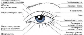
What is a snub nose?
The main sign of this defect is that the tip is directed upward. The ideal nasolabial angle is different for men and women. For men it should not exceed 95º, and for women from 95 to 115º.
An upturned nose is when this angle is higher than the specified norm. At the same time, the nostrils are very noticeable and the nose appears short. It happens that the problem is aggravated by the fact that the wings and its tip are significantly thickened. In this case, it visually appears not only wide, but also large. Outwardly, it may resemble a “heel”.
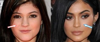
Rhinoplasty before and after photos
What does the nose look like after rhinoplasty? Are the seams visible?
Look at the photos before and after rhinoplasty. The nose looks great, there is no longer any hump, and there are no visible seams either. The patient is very satisfied.
The photo shows the result of removing Galina Khrushch's nasal hump using open rhinoplasty. The following was performed: removal of the nasal hump, a columella strut graft was installed in the area of the medial crura of the lower lateral cartilages, the domes of the alar cartilages were brought closer and fixed to the anterior edge of the septal cartilage, median and lateral osteotomy, augmentation of the nasal dorsum with a platelet automembrane with cartilage shavings. Photos were taken 1.5 months after rhinoplasty surgery.
View more photos before and after rhinoplasty
Possible complications
Complications usually arise due to the surgeon’s unprofessionalism and violation of the preparatory and rehabilitation regime. The most common negative consequences are a rough scar at the incision site, wound infection, dry mucous membrane, loss or distortion of sensitivity in the operated area. For any complications, medical consultation is required. If necessary, the patient is prescribed additional therapy, and in severe cases, repeated surgery.
Mobility of the nose after rhinoplasty is normal. Within 12–18 months after the intervention, patients may experience spontaneous changes in the shape of the tip, which lowers, rises, or moves to the side. Experienced surgeons warn in advance about this problem and help solve it.
Indications for nose surgery
If we look at the indications for rhinoplasty surgery a little deeper, they can be divided into two types: medical indications and aesthetic indications.
Medical indications
- Deformation of bone or cartilage tissue as a result of an accident or injury
- Congenital or acquired changes in the nose area
- Deviated nasal septum, difficulty breathing, snoring, etc.
In these cases, you will need an operation - rhinoseptoplasty, the task of which is to correct the nasal septum. Rhinoseptoplasty is often combined with correction of the appearance of the nose.
Aesthetic indications
- Wide wings of the nose or dorsum
- Presence of a hump
- The tip of the nose is turned up or, conversely, too drooping
- No visible bridge of the nose
- Large or too thin nose tip
Unlike medical indications, aesthetic indications are not mandatory, since they do not harm human health, but at the same time they can cause psychological harm. The decision to perform or not to perform surgery for aesthetic deformities of the nose is made by the patient himself. But in the case of medical indications, it is worth thinking more seriously about rhinoplasty, since we are talking about health.
You can read about possible motivating factors for rhinoplasty in my article “What pushes people to undergo rhinoplasty?”
Indications and contraindications for rhinoseptoplasty
Indications for rhinoseptoplasty
Rhinoseptoplasty surgery is indicated when the patient has a deviated septum, which causes dysfunction of the nose (especially respiratory) and dissatisfaction with the aesthetic appearance of the nose. A wide, high, flat nasal bridge, drooping tip, asymmetry, hump, dips and unevenness of the nose can be corrected simultaneously with septum correction during rhinoseptoplasty.
Contraindications for rhinoseptoplasty
The operation of rhinoseptoplasty is contraindicated if the patient has not reached the age of majority, has infectious inflammatory diseases, decompensated diabetes mellitus, or blood clotting pathology.
In Moscow, many plastic surgery clinics perform rhinoseptoplasty. The Mont Blanc Clinic has been performing this type of surgery for many years and with great success.
Types of rhinoplasty
In modern medicine, there are two ways to perform nose correction:
- Open rhinoplasty
- Closed rhinoplasty
The choice of method usually depends on medical indications, the wishes of the patient, and the physiological characteristics of the patient.
Open rhinoplasty
carried out in difficult situations from a medical point of view. This method is required in case of severe deformation of the bone tissues of the nose, curvatures, or when correcting someone else’s work using a graft. The essence of the operation is to make an incision between the nostrils of the nose and separate the cartilage from the skin for further work.
Private method
, on the contrary, is positioned as a minimally invasive method of performing rhinoplasty. In this way, the operation is performed to correct minor defects and cosmetic imperfections. During the operation, the surgeon does not injure the outer skin as much as with open rhinoplasty. All incisions and manipulations take place inside the nasal cavity.
Correction methods
Rhinoplasty is performed in open and closed ways. In the first case, the doctor makes an incision on the bridge between the nostrils (columella) and releases the pterygoid cartilages. After this, excess tissue is removed, and the tip of the nose is given the desired shape using sutures. If necessary, the bridge is raised and strengthened using supporting cartilages. The open method provides a complete overview of the surgical field, therefore providing more options and reducing the risk of errors and inaccuracies. The only disadvantage of this method is a small scar on the columella. If the operation is performed by an experienced surgeon, the scar will be barely noticeable and will practically disappear over time.
Closed rhinoplasty is performed from the inside of the nasal cavity through an incision in the mucous membrane. The doctor releases the pterygoid cartilage and applies sutures, which will subsequently correct the shape of the tip of the nose. The operation takes less time compared to open rhinoplasty, and the results can be seen faster. Another advantage of the technique is that the postoperative scar is hidden in the nasal cavity and is completely invisible. The disadvantages of the procedure include limited access to the operated area. Because of this, the surgeon cannot use all possible techniques that give the best result. Limited visibility makes it difficult to control the condition of blood vessels, so the likelihood of bleeding and hematomas increases.
The technique is chosen depending on the nature of the aesthetic defect and the characteristics of the patient’s anatomy. In difficult cases, the open method is often used.
Photos before and after nose surgery
| Photos before and after rhinoplasty of the nose: Elimination of acquired deformation of the external nose in a man. After photos were taken 3 months after surgery. | Photos before and after rhinoplasty of the nose: Elimination of deformation of the cartilage and bone parts of the external nose using a cartilage autograft. Photos were taken 4 days after surgery immediately after removal of the plaster cast. |
Difference Between Open and Closed Rhinoplasty: Pros and Cons
Let's take a closer look at both surgical techniques and the differences between them.
Closed rhinoplasty
| pros | Minuses |
|
|
Open rhinoplasty of the nose
| pros | Minuses |
|
|
How is surgery to improve the shape of the nose performed?
Preparing for rhinoplasty of the nose involves a thorough examination and the need to undergo tests.
The operation is performed under general anesthesia in combination with local anesthesia and lasts from 30 minutes to 2.5 hours. The duration of the operation depends on the complexity of the surgical intervention performed and the chosen technique - open or closed.
During rhinoplasty, the cartilaginous parts of the nose are corrected and, if necessary, the bone parts are corrected.
The shape of the nose is improved by redistributing tissues, reducing some volumes (partial removal of cartilage or bones), increasing others (cartilage or fascial grafts).
Methods of vector movement and modeling of the cartilaginous structures of the nose using special internal permanent sutures are also widely used.
After the operation is completed, all incisions are sutured with cosmetic sutures; internal sutures do not need to be removed; external sutures are removed after 5-7 days. The small outer scar fades within a month and becomes almost invisible.
Upon completion of the operation, tamponade of the nasal passages is performed, and a formative plaster bandage is applied. Nasal tampons are removed after 1-2 days, and you can leave the clinic until your next examination.
The plaster cast must be worn continuously for 10-14 days, then used only during sleep for another 1-2 weeks.
Non-surgical rhinoplasty (fillers in rhinoplasty)
The content of the article
1. Definition of non-surgical rhinoplasty
2. What are fillers?
3. Anatomical aspects when performing rhinoplasty with fillers
- soft tissues of the nose
- effects on the nasal muscles with botulinum toxin
- blood supply to the external nose
4. Selection of patients for non-surgical rhinoplasty using filler
- indications for rhinoplasty with fillers
- contraindications to non-surgical rhinoplasty
5. Method of non-surgical rhinoplasty
- Pain relief before non-surgical rhinoplasty
- Drawing marking lines
- Filler injection areas
- Sequence of filler injection
- Post-injection adaptation of fillers
6. Possible complications
- bruises
- asymmetry
- visible contours of filler
- hypersensitivity
- lump formation
- inflammatory response and swelling
7. Vascular complications
- symptoms of intra-arterial embolism
- prevention of intra-arterial embolism
- rehabilitation and care
Definition of Non-Surgical Rhinoplasty
Rhinoplasty is one of the most common operations in aesthetic plastic surgery.
People of Asian ethnicity often have a flat nose and a wide nasal tip, so augmentation rhinoplasty is often performed in Asian countries.
However, existing methods of rhinoplasty using implants and autologous (own) cartilage are associated
- with long recovery time,
- high cost and
- possible problems associated with the implants themselves.
Therefore, patients often have a psychological barrier when considering the possibility of rhinoplasty. Many patients choose not to undergo surgery. This, in turn, leads to an increase in the popularity of rhinoplasty without surgery and a scalpel - to correct nasal defects with the help of fillers.
The purpose of this article is not to endorse the use of fillers, but to
- firstly, providing information and guidance to patients to understand the process inside and out,
- secondly, for practitioners - to improve the results of injection non-surgical rhinoplasty and, accordingly, reduce complications for many practitioners performing this procedure.
What are fillers?
A filler is any material that can add volume when injected into the body and is usually an injectable material.
Well-known fillers include
- hyaluronic acid (HA) products,
- collagen,
- paraffin,
- liquid silicon.
Fillers are usually classified by their components.
Fillers are also classified by durability.
- Fillers that last less than 2 years are called temporary fillers;
- lasting 2 to 5 years are called semi-permanent fillers;
- and those that last at least 5 years after injection are called permanent fillers.
Fillers can also be classified according to their mechanism of action, e.g.
- fillers to increase volume and
- stimulating an increase in volume.
Fillers that stimulate volume expansion by stimulating fibroblasts to synthesize collagen through an inflammatory response are called stimulating fillers.
Most fillers have a good safety profile. However, serious side effects such as granuloma formation and inflammation due to tissue reaction have been reported with some fillers, so it is necessary to carefully select the desired filler by studying all its characteristics.
Ideal filler
- should not cause tissue reactions,
- must be durable, safe and easy to use,
- should not migrate in tissues and
- should not cause allergic reactions.
Anatomical aspects when performing rhinoplasty with fillers
Non-surgical rhinoplasty with filler can only be successfully performed if the plastic surgeon has a good understanding of the anatomy of the nose.
No-scalpel rhinoplasty with filler is a procedure to reshape the nose by injecting filler into the space between the osteochondral structure of the nose and the skin.
It is very important that the monolithic frame of the anatomical structures of the nose is strong, as a supporting structure to maintain the shape of the injected filler and to achieve a good aesthetic result.
Therefore, you cannot expect a satisfactory result after the procedure if the nasal frame is deformed or weakened.
When performing rhinoplasty using filler, it is necessary to take into account all the characteristics of the anatomical structures of the nose, including such
- thickness and properties of skin and soft tissues, as well as
- size, shape and strength of cartilage and bone.
Soft tissues of the nose
Before performing injection rhinoplasty, it is important to evaluate the condition of the skin of the nose. In general, Asians have thicker skin than Europeans, with well-developed subcutaneous fat.
The soft tissue of the bridge of the nose is thickest, and the thinnest is in the area of the nose, which is the junction of the upper lateral cartilages and the bones of the nose.
There are 4 layers between the skin and the osteochondral skeleton:
- superficial fat layer,
- fibromuscular layer,
- deep fat layer and
- periosteum or perichondrium.
When performing injectable rhinoplasty with filler, it is difficult for patients with thick, oily skin to achieve a predictable three-dimensional shape because they often experience significant swelling after the procedure.
On the other hand, in such patients, minor irregularities or asymmetries are more easily camouflaged compared to patients with thin skin.
The main blood vessels of the external nose are located in the superficial musculoaponeurotic system (SMAS) layer or in the superficial fat layer.
Therefore, the ideal layer for filler injection to minimize vascular damage is the deep fatty layer located between the SMAS and the perichondrium, or periosteum.
Effects of botulinum toxin on the nasal muscles
Blocking (restricting movement) of the paranasal muscles due to the action of botulinum toxin gives better and longer lasting results than using filler alone.
Understanding the location, size and function of the nasal muscles is very important.
To enhance the effect of non-surgical rhinoplasty, you can use botulinum toxin to cause controlled immobilization (paresis/paralysis) of the muscles.
Let's discuss muscles and the practical significance of blocking them.
The depressor septum nasalis muscle originates from the orbicularis oris muscle and ends in the medial crura of the inferior lateral cartilage. This muscle lowers the tip of the nose when smiling and can often be immobilized with a botulinum toxin injection to suppress its function.
It is better to block (paralyze) the procerus muscle (m.procerus), located in the area of the glabella, with botulinum toxin in advance, because the filler injected into the area of the glabella can be displaced during intense contraction of the muscle.
The levator labii superioris muscle causes the alar flares to widen and the tip of the nose to lower when smiling; therefore, it is also often blocked by botulinum toxin in patients with dynamic nasal tip ptosis.
Blood supply to the external nose
The most dangerous complications of filler injection are intra-arterial embolization (blockage) of a blood vessel and dermal necrosis (death) due to compression of blood vessels. To prevent such consequences, the plastic surgeon must have a good knowledge of the blood supply to the external nose.
Both the internal and external carotid arteries supply blood to the external nose through the ophthalmic and facial arteries, respectively.
The ophthalmic artery primarily supplies blood to the superior part of the nose through the external nasal branch of the anterior ethmoidal artery and the posterior nasal artery.
The facial artery gives rise to the angular and superior labial arteries, which supply blood to the lower part of the nose. Each of these branches branches into the lateral nasal artery and the columellar artery.
The tip of the nose receives its blood supply from the dorsal nasal artery superiorly, the lateral nasal artery, and the columellar artery inferiorly.
Selection of patients for rhinoplasty with filler
The plastic surgeon must be able to select patients who are suitable for rhinoplasty with filler. This requires a thorough understanding of the ideal aesthetics and anatomy of the nose (described earlier).
Unless doctors are trained and have thorough knowledge, they should not administer fillers into the nose. Patients who cannot achieve good results with filler should undergo surgery.
Indications for rhinoplasty without surgery
The group of patients who typically have good results includes patients
- with a moderate hump on the bridge of the nose,
- with a slightly crooked nose,
- with a high nose tip with a flat base,
- slight imbalance after surgery.
Contraindications to non-surgical rhinoplasty
Satisfactory results in patients are not expected
- with a moderate to severe nasal hump,
- strongly crooked nose,
- upturned and convex nose.
In patients with such initial data, good results cannot be obtained from filler injection alone.
Clinicians should also exercise caution when offering this procedure to patients who have had nasal implants or those who have had a history of paraffin or liquid silicone injections, as skin irregularities and vascular compromise may occur.
Non-surgical rhinoplasty technique
Rhinoplasty using filler can be performed from 2 main areas:
- injections through the bridge of the nose and
- injections through the tip of the nose.
The bridge of the nose has a strong, dense structure, created by the nasal bones and upper lateral cartilages, so this area is easier to correct (lift) with filler injections.
However, raising or lengthening the tip of the nose is not easy, especially in Asians, due to weak supporting structures.
After comparing and analyzing the patient's nose, the plastic surgeon must decide which part should be raised and by how much.
Pain relief before non-surgical rhinoplasty
Applying a local anesthetic ointment for about 40 minutes before the procedure is usually sufficient for pain relief. A bandage placed on top may enhance the effect of the local anesthetic.
Drawing marking lines
After anesthesia, the midline along the dorsum of the nose is marked. It should be accurately marked to prevent complications such as imbalance or intravascular injection because the blood vessels are generally not located in the midline of the nose.
It is important to find the ideal starting point for the nasofrontal angle.
The nasal root should begin at the level of the supratarsal fold in lateral view, but this varies depending on patient preference, forehead height, and nasal length.
The bridge of the nose should lie 1-2 mm below the line drawn from the root to the tip of the nose. Some patients, however, may prefer a straight nasal bridge.
Filler injection areas
The nose is divided into 4 parts for the procedure - the root, rhinion, supratip zone and tip.
Each part has different thickness of subcutaneous tissue, as well as different characteristics and strength of supporting structures, so different injection methods must be used.
After marking the 4 sections, any defect on either side of the nose should be noted.
In the tip and supratip zones, the thickness of the soft tissues is much greater than in other zones, and the underlying structures here are mostly concave in shape.
Therefore, a slightly larger volume of filler must be injected into these 2 areas to avoid indentations due to pressure from the overlying soft tissue.
Sequence of filler injection
The filler is usually injected in the following order: first into the root area, then the rhinion, then the tip and finally the supratip.
The reason that tip filler is injected earlier than supratip filler is that tip support is poor, especially in Asians, making it difficult to predict the amount of tip projection that can be achieved.
If filler is injected first into the supratip area and then into the area of the nasal tip, then the doctor will not be able to achieve adequate projection of the nasal tip, and this can lead to deformation.
The correct technique for injecting filler into the tip of the nose allows you to avoid serious complications.
The areas where filler is typically injected in the nasal tip area are the interdomal area, the concavity area of the lateral crura of the greater alar cartilages, and the area between the medial crura of the greater alar cartilages.
Injecting filler into the tip of the nose creates volume to reshape the nose. The volume and location of the injection depend on the desired appearance of the nose.
When injecting filler into the tip of the nose, it is safer to insert it in the midline to minimize tip deviation and asymmetry.
Minor wing retraction can be corrected with a filler injection. However, this is not recommended for patients who have scars from previous surgery due to the risk of skin necrosis or other abnormalities.
The fanning technique of filler injection, in which the filler is deposited as the syringe needle is withdrawn after injection, is the most commonly used technique.
Sometimes there is a single injection using a 2.5-inch needle, and sometimes multiple injections using a small 0.5-inch needle.
The procedure can also be performed using a blunt cannula and this method is recommended for beginners as there is less chance of complications such as intravascular injection, but it is more difficult to achieve accurate results.
The most important aspect of using filler to reshape the nose is to perform the procedure without deviation from the midline.
The most common complaint after any rhinoplasty is asymmetry or imbalance. This also applies to rhinoplasty using filler.
As discussed earlier, the clinician should always mark the midline on the bridge of the nose and perform the procedure without deviation from the midline to minimize such complications.
It is highly recommended that clinicians use both hands when inserting the filler. When using only one hand to inject, the non-injecting hand must guide the needle and filler into the tissue. This ensures minimal bleeding or spraying of the product.
Post-injection adaptation of fillers
After the injection, it is recommended to perform a gentle massage to mold and smooth out the filler. Any area that is overfilled should be pressed into the base, and areas that are underfilled should receive additional injections.
It is recommended to carry out follow-up no earlier than 2 weeks, since after the procedure slight swelling may occur, which interferes with an adequate assessment of the result. No special dressings or antibiotics are required.
Possible complications
Reshaping your nose with filler is safe, but complications can sometimes occur. Most complications can be prevented by choosing safe products and following the procedure correctly.
The most common complications of filler injections are
- edema,
- erythema,
- bruises,
- color change,
- unevenness,
- formation of lumps and granulomas.
Infections and serious complications such as skin necrosis may occur due to vascular administration of the drug.
Bruises
Bruising is a common complication of filler injections; this is caused by damage to blood vessels from the needle. To reduce bruising, puncturing the muscle layers during filler injection should be minimized, the injection site should be cleaned with an alcohol swab, and the procedure should be performed in a brightly lit room.
Patients should be informed not to take blood thinners such as aspirin 1 week before the procedure.
Applying ice packs to the injection site immediately after the procedure helps minimize bruising, and special needles or cannulas are used to minimize vascular damage.
If bleeding occurs during the procedure, cover the injection site with gauze and apply pressure for several minutes to avoid the formation of a hematoma.
Patients should be informed that bruising is temporary and does not affect the final aesthetic result. You should also understand that the bruise may darken a few days after the injection, but will gradually disappear over about 10 days.
Asymmetry
One of the most common complications of rhinoplasty using filler is asymmetry. To avoid asymmetry, the tip of the needle should be located exactly along the midline, and the direction of the bevel should be towards the midplane.
When administering filler to a patient with a crooked nose, it is advisable to carefully monitor the shape of the nose while slowly injecting a small amount of filler.
Visible contours of filler
Injecting filler superficially (close to the surface of the skin) may result in an uneven injection site or appearance. To avoid this, the filler should be introduced into the appropriate layer in accordance with its characteristics.
Hypersensitivity
Sometimes hypersensitivity to filler ingredients may occur. The main symptoms are pain and erythema, accompanied by itching and fever.
In most cases, these symptoms disappear as the filler dissolves. In severe cases, corticosteroids and warm compression may help relieve symptoms.
Pulsed dye laser treatment can be used for persistent erythema. Many reactions that are considered allergic or hypersensitivity reactions are most likely caused by bacterial reactions.
Formation of lumps
After injection of filler, lumps may form - these are either due to granuloma or nodule formation.
Granuloma is an immune-mediated response to an injected foreign body and forms through the accumulation of immune response-associated cells such as lymphocytes.
Treatment is with corticosteroid injections or surgical removal.
The nodules are round and hard in shape. Their development is a common complication after the use of fillers for soft tissue augmentation. They can be inflammatory or non-inflammatory in nature.
Inflammatory nodules may appear days or years after treatment, whereas non-inflammatory nodules are usually visible immediately after implantation and are usually secondary to improper placement of the filler.
Treatment is with hyaluronidase (if HA is used as filler), corticosteroids, or surgery.
Inflammatory response and swelling
Sometimes the inflammatory response and swelling occurs due to a protein component, such as endotoxin, contained in the filler and leads to skin damage.
This is caused mainly by the HA filler and symptoms such as erythematous swelling, skin hypertrophy and pustules may appear a few days after the injection.
Symptoms occur at all filler injection sites and resolve with appropriate antibiotic treatment and dressing.
Vascular complications
The most serious complications that can occur with filler injection are skin necrosis and blindness.
The mechanism leading to tissue necrosis after injection of HA filler is not fully understood.
Vascular disorders are divided into
- intravascular and
- extravascular.
Intravascular factors include direct occlusion of the arteries by large molecule excipients and chemical damage to the endothelial lining or impurities in excipients.
Extravascular causes include extrinsic compression of the vessel due to excessive injection volume or swelling and inflammatory response caused by filler components.
Intra-arterial embolism
The cause of most cases of intra-arterial embolism after rhinoplasty with filler occurs when the filler is injected directly into the nasal or lateral nasal artery.
The dorsal nasal artery, as its name suggests, runs along the dorsum of the nose approximately 3 mm from the midline. The tip of the needle can be inserted into a blood vessel if it is inserted parallel to the blood vessel.
The dorsal nasal artery anastomoses with the ophthalmic, subtrochlear, and angular arteries, so if an embolus occludes a vessel in this area, it will manifest itself as local skin necrosis.
But if the filler embolus spreads deeper into the ophthalmic artery, it can cause ocular symptoms.
Symptoms of intra-arterial embolism
Intra-arterial embolism is rare, but its consequences are devastating.
After filler is injected into the arterial bloodstream, patients experience severe pain and sometimes complain of a feeling that something is spreading from the injection site.
The area of the blood vessel where the filler embolism occurred becomes pale due to ischemia. The ischemic area develops swelling within a few hours, which soon becomes purple and mottled due to venous stasis.
After about 24 hours, multiple ulcerative lesions may appear, accompanied by erythema, leading to desquamation of the tissue. The situation usually gets worse over time. After this, certain signs of dermal necrosis, such as eschar formation, may gradually appear, and then the skin is restored through the wound healing process.
Prevention of intra-arterial embolism
During the procedure, the needle tip should always be positioned in the midline to avoid injection into the dorsal nasal artery as it runs parallel to the midline at a distance of 3 mm.
If filler is being inserted from the side of the nasal bridge, for example to correct a crooked nose, the needle should never move parallel to the direction of the blood vessels. Once the needle is inserted in the midline, the tip of the needle should be moved to the side while injecting the filler to prevent injection into the blood vessel, although some bleeding may occur due to damage to the vessel.
The dorsal nasal artery is located in the superficial fat layer and SMAS; therefore, the injection must be made into the deep fat layer to prevent embolization. Using a blunt cannula can also be helpful for new doctors who are not familiar with injection techniques.
Rehabilitation and care after intra-arterial embolism
If the patient complains of severe pain and there is blanching of the skin along the blood vessel area during the filling procedure, the injection should be stopped immediately and aspirate as much of the filler as possible.
If a hyaluronic filler has been injected, an injection of hyaluronidase is recommended, as current evidence suggests that when hyaluronidase is injected around an artery, some of it may diffuse through the lining of the vessel.
It is also recommended that hyaluronidase injections are always performed regardless of the type of filler injected, because hyaluronidase can reduce interstitial pressure.
Low molecular weight heparin therapy has been reported to reduce thrombosis and embolism and should therefore also be used in such cases.
It is important to provide sufficient oxygen to the ischemic area. For this, hot wraps and soft massage are used, and 2% nitroglycerin paste is used to dilate blood vessels.
If possible, it is helpful to start hyperbaric oxygen therapy. Appropriate antibiotics are prescribed to prevent secondary infection. Administration of prostaglandin E1, 10 mg daily for 5 days, is effective. After about 1 day, an appropriate bandage should be applied as soon as peeling and pustules occur. A moist dressing should be used to promote faster wound healing and antibiotics should be continued.
To avoid such problems, the patient needs to choose a plastic surgeon wisely.
Rehabilitation after rhinoplasty
As soon as the plaster is removed, swelling in the nasal area will persist for some time, and moderate hematomas may be observed, which will regress in the next 10-14 days. Sometimes after surgery there may be some pain, which can be easily relieved by taking simple painkillers. Also during this period, due to edema, nasal breathing may be weakened; some time is needed to restore it. For 3-4 weeks you need to limit physical activity and sports, avoid visiting saunas, baths and swimming pools.
The preliminary result of the operation can be seen after the plaster splint is removed. The swelling of the soft tissues of the nose completely disappears in about 4-6 months, when the final result of rhinoplasty can be assessed. As a result of surgery to improve the shape of your nose, your appearance will become more impressive, you will gain self-confidence and will delight yourself and others with your new look.
Read a story from my patient about her rhinoplasty experience
Carrying out the procedure
Nose correction with threads is performed by a cosmetologist within an equipped medical office. Local anesthesia is sufficient for pain relief. Dermafil Happy Lift, APTOS or others threads can be used. They are inserted, creating micropunctures. The installation area can be the tip of the nose or the area of the wings. Next, the doctor adjusts the form to the desired effect, the result can be assessed no earlier than 2-3 days after the procedure. The changes are noticeable immediately, but at first there is swelling.
Features of the procedure
The thread technique for the nose is no different from the classic thread lifting. The surface of the skin is treated to remove dirt and cosmetics, and an antiseptic composition is used. To eliminate pain, local anesthesia is used.
The implantation process includes the following steps:
- Marking of selected zones.
- Inserting a cannula with a thread under the skin and moving it along the intended line. A small “anchor” is provided at the second end to allow fixation.
- The thread brought out is pulled in the opposite direction, causing displacement of the soft tissues.
- The excess thread is cut off and hidden under the skin.
During the technique, each structure requires two punctures. Their healing occurs quickly, leaving no scars or scars.
Recovery period
Changes are noted immediately, but the final result should be expected no earlier than after 3 days. This period allows the threads to move into the desired position, and tissue swelling also goes away. During this period, it is important to avoid active facial expressions, and physical impact on the installation area should be minimized.
Prohibited:
- use cosmetics;
- touch your nose with your hands;
- wet the location area;
- use creams, lotions, scrubs, masks.
Avoid physical activity that will lead to a rush of blood to the facial area for 3-5 days. During the designated period, a framework of connective tissues will be created near the threads, and they themselves will be installed correctly. Hematomas and swelling can be eliminated using cooled compresses, as well as compositions with absorbable qualities: heparin ointment, Troxevasin, Traumeel.
Who can get a nose job in Moscow?
If you are looking for a good plastic surgeon for rhinoplasty in Moscow, then I recommend that you pay special attention to several main, in my opinion, factors:
- carefully review the plastic surgeon's portfolio. Having photos before and after rhinoplasty is the best recommendation.
- look for reviews of the plastic surgeon
- personal contact - consult with several plastic surgeons to choose one whom you can trust unconditionally.
Rhinoplasty of the nose in Moscow. Prices for nose surgery
The cost of nose surgery in Moscow depends on many factors. In the table we provide some approximate prices, but the surgeon will be able to name the final cost of nose surgery only after consultation, because Each case is individual and the solution to similar problems can differ radically from person to person. Check out the estimated prices and send your photos to the doctor for a preliminary consultation.
| Rhinoplasty prices (RUB) | |
| Plastic surgery of the cartilaginous part of the nose (tip of the nose) - 80,000 rubles Plastic surgery of the bony part of the nose (dorsum of the nose) - 85,000 rubles Full rhinoseptoplasty - 165,000 rubles Harvesting of ear cartilage for contour plastic surgery of the nose - 15,000 rubles Revision rhinoplasty (secondary) - 180,000 rubles | |
A more detailed price list for plastic surgery can be found here
Peculiarities
Many experts note that correction of the tip of the nose is an indicator of the surgeon’s skill, and this is absolutely true. Working with the back of the nose is technically much easier than working with the tip of the nose. Therefore, surgery to correct only the tip of the nose truly demonstrates the skill of the specialist.
In nasal tip plastic surgery, there are simpler methods, but less effective and less predictable, which simply involve removing cartilage. They do not give the same results as more technically complex methods. But with the help of the latter it is possible to achieve a greater narrowing of the tip, moreover, they are more predictable and “high quality”. In our practice, we always use more complex and reliable types of correction. In general, tip rhinoplasty is easier to tolerate than full rhinoplasty. It doesn't cause bruising, and if it does, it's minimal.
More photos after rhinoplasty
| The photo shows rhinoplasty of the nose: the patient underwent plastic surgery to reduce the dorsal hump; low-low and medial oblique osteotomies and rib grafts | The photo shows rhinoplasty of the nose: the patient underwent rhinoplasty with autologous tissue. |
Reviews of rhinoplasty with Galina Khrushch in Moscow
I still have a cast on my nose and rehabilitation is in full swing. But I already want to say thank you to Galina Viktorovna. She has a light hand, so I don’t experience any discomfort. Everything is fine. Many people at work know that I had my nose done. And many are already planning to change something in themselves, looking at me. I'm happy with it. It's so nice when people compliment you. Their number will increase 10 times as soon as I remove the cast and as soon as all the swelling goes down. Thank you, Galina Viktorovna. I am pleased!
Ksenia Karpenko
I had rhinoplasty done by Galina. Immediately after the plaster, the nose seemed large due to swelling, but even then it was already clear that the shape had become more aesthetic. Now all the tissues have healed and you can see that the nose is cute. The doctor removed the hump and corrected the drooping tip of the nose. The result is a slight, subtle snub nose that suits my face very well. I can only say good things about Galina. I have never met such an attentive doctor. Very caring attitude towards patients, including in the postoperative period.
Nara