Consultation with plastic surgeons with over 20 years of experience – free! Sign up by phone. Waiting for you!
Plastic surgery always strives for constant renewal and improvement. Today, more and more techniques and operations are appearing that are designed to achieve the best results with minimal “sacrifices.” These include plastic surgery of the tip of the nose.
, which, if certain requirements are met, allows you to achieve a good aesthetic effect. Let us consider the features and differences of this operation from rhinoplasty in full.
Peculiarities
Many experts note that correction of the tip of the nose is an indicator of the surgeon’s skill, and this is absolutely true. Working with the back of the nose is technically much easier than working with the tip of the nose. Therefore, surgery to correct only the tip of the nose truly demonstrates the skill of the specialist.
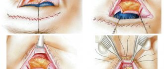
In nasal tip plastic surgery, there are simpler methods, but less effective and less predictable, which simply involve removing cartilage. They do not give the same results as more technically complex methods. But with the help of the latter it is possible to achieve a greater narrowing of the tip, moreover, they are more predictable and “high quality”. In our practice, we always use more complex and reliable types of correction. In general, tip rhinoplasty is easier to tolerate than full rhinoplasty. It doesn't cause bruising, and if it does, it's minimal.
Hospital
In the postoperative period, the patient remains in the hospital under medical supervision for an average of 2-3 days. At his service:
- 1-2 bed rooms, with shower, functional beds, to which oxygen is supplied.
- Each patient has an individual vital signs monitor.
- Delicious meals are provided, taking into account all the recommendations of the attending physician.
- Each patient has an individual TV with headphones.
After discharge, the patient should visit the doctor as often as recommended by him.
Plastic results
With the help of correction, the tip of the nose can be made narrower, more defined, and also raised. Moreover, you can raise the tip of the nose provided that the bridge of the nose is low. Since simply narrowing in such a situation will not give anything. Quite often, patients with a high back come to us and want to lift the drooping tip of the nose, but in this case, plastic surgery of only the tip of the nose will only visually increase the “sail”. Therefore, the approach to aesthetic correction should be comprehensive.
Rehabilitation period
After the operation, the patient is given a plaster cast for 5-7 days. After its removal, swelling will persist for some time (up to 1 month), but it will be insignificant. For 1.5 months, restrictions on physical activity (sports, fitness), warming procedures (baths, saunas, solarium) are prescribed. For the first month, sleep only on your back.
Indications and contraindications
Rhinoplasty of the nose can be done if there are visual defects, which include:
- The presence of a hump;
- Saddle shape;
- Hooked tip;
- Excessive length;
- Asymmetrical wings;
- Other aesthetic defects that cause psychological discomfort to the client.
Rhinoplasty of the nose is performed only when the patient reaches the age of 18: until this time, the cartilage continues to grow and form, i.e. plastic surgery on the nose will not give a stable result. Contraindications to the procedure are standard:
- Metabolic disorders (diabetes mellitus);
- Blood diseases;
- Acute infections and exacerbations of chronic diseases;
- Pregnancy and breastfeeding.
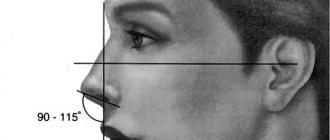
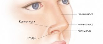
Correcting the shape of the nose is possible only after consultation with a surgeon, who measures the main anatomical indicators and compares them with the average statistical data.
Rhinoplasty of the tip of the nose prices:
The price of nasal tip plastic surgery is slightly lower than the cost of full rhinoplasty due to the more delicate work. The difference is about 20-30 thousand rubles and can vary from 120,000 to 180,000 rubles for surgical correction.
Nose surgery photos and reviews:
You can view photos before and after nose tip correction Read the reviews in the “About Us – Reviews” section.
FAQ:
Is it possible to change only the tip of the nose (raise it, or make it narrower) separately from the other “parts” of the nose?
Yes, it is indeed possible to change the tip of the nose separately from other “parts” of the nose. But this is only feasible if the back of the bow is “good” - not high, in other words, if you look in profile, the back does not have a large “sail”.
Are patients who come for rhinoplasty of the tip of the nose most often those who have never had rhinoplasty, or have the majority already had this operation and their tip has drooped?
In our practice, there are both cases. Some have never had rhinoplasty, and we see that the problem can be solved not with classical surgery, but with plastic surgery of the tip of the nose only. But there are also patients who have already had rhinoplasty previously, and they only want to raise the tip, which has sank over time.
Is it true that after rhinoplasty the tip of the nose always droops? Or is this a myth?
No, this is not a myth. The tip of the nose can indeed droop over time, and we have such patients. And in most cases, surgery on the tip of the nose gives an excellent aesthetic result; this small correction completely changes the patient’s appearance.
If a patient comes in with a problem of a drooping tip, is it possible to lift it once and for all?
In any case, the tip of the nose drops to a certain extent, so when we perform the operation, we lift it a little more than necessary. This is called overcorrection. Therefore, at first the tip of the nose may be raised a little, taking into account the fact that when it goes down, it will be at the level it needs to be.
We care about your health
Epithelial tumors
Papillomas. There are papillomas of the vestibule and the nasal cavity. The first ones are gray in color, dense, villous surface, small in size, not malignant. It is believed that papillomas are caused by viruses from the family of papillomoviruses types 6, 11 and 16. Papillomas of the nasal cavity primarily affect the mucous membrane. They often recur after surgical removal. With anterior rhinoscopy or endoscopy, papillomas are gray-white in color, soft in consistency, single or multiple, bleed when touched, and resemble cauliflower. Treatment of nasal papillomas is predominantly surgical or cryodestruction. Surgically removed material should be sent for histological examination due to the possibility of malignant degeneration (transitional cell carcinoma). Another problem in the treatment of these patients is the frequent recurrence of papillomas. To prevent it, it is recommended to prescribe interferon drugs - domestic Laferon or foreign intron A. The method of treatment with Laferon consists of inhalation administration at the beginning of therapy (1 million Laferon is diluted in 20 ml of 0.9% NaCl solution). Inhalations continue for the first 10 days, then Laferon is prescribed intramuscularly once a day, at a dose of 1 million units, for 20 days. Some clinics determine the titers of antibodies to it to monitor the presence of the virus in the blood serum. When the levels increase above 1:200 in the blood of patients, antiviral drugs (Zoverax or others) are prescribed. Papilloma viruses also infect the cervix, which leads, in less pathogenic types, to the occurrence of condylomas.
Inverted papilloma. Called due to the ability of squamous epithelium to invaginate in the form of a wide ribbon into the connective tissue, these tumors have destructive growth, recur and become malignant. They are also known as columnar cell papillomas.
Adenoma is a benign tumor of the glandular epithelium of the nasal mucosa. It mainly affects the lower parts of the nasal cavity on a broad base with a smooth surface. Characterized by slow growth. The tumor is erysipelas-gray in color, no more than 2 cm in size, and is accompanied by rapid growth, germination into adjacent tissues, and changes in the histological structure. Its treatment is surgical. The removed material should be examined histologically.
Nonepithelial tumors
Hemangioma is a tumor developing from blood vessels, similar in nature to developmental defects. What it has in common with other tumors is rapid growth, during which hemangioma destroys surrounding tissue and causes a cosmetic and sometimes functional defect. In 1863, Vikhrov published a macro-microscopic classification of hemangiomas, dividing them into capillary, cavernous and racemose (branched). Subsequently, this principle was included in many classifications.
Today, this classification is used, which includes additional data on tumor growth, its spread, blood supply characteristics and relationship with large blood vessels.
True hemangiomas: 1) capillary (simple): exophytic (venous and arterial type); 2) cavernous: mucous-submucosal; diffuse (spreading into depth); 3) branched (angiodysplasia); 4) mixed (angiofibromas, hemlymphangiomas, branched with cavernous, branched with Barre-Mason glomusangiomas, etc.).
Diagnosis of tumors in the nose
- Routine external examination of the patient. Palpation of the tumor.
- Anterior and posterior rhinoscopy.
- Examination of the nasal cavity and paranasal sinuses using an endoscope. Diaphanoscopy.
- Comprehensive X-ray examination.
- CT and MRI.
- Cytological examination of discharge obtained during puncture, sinus lavage, as well as nasal discharge.
- Histological examination of pieces of the neoplasm.
Treatment of nasal tumors
- Surgical removal of the tumor (if it is of significant size with preliminary embolization of the vessels feeding the tumor).
- Cryodestruction of the tumor.
- Sclerosis therapy.
- Radiation therapy in combination with surgical removal (for tumor malignancy).
Bleeding polyps of the cartilaginous part of the nasal septum are red, round in shape with a smooth surface. They are characterized by frequent bleeding. Treatment is surgical.
Fibromas, myomas, lipomas are rarely found in the cavity and paranasal sinuses. Angiofibroma, fibromyoma, osteofibroma, adenofibroma, neurofibroma, histiocytoma may occur. The treatment of these tumors is surgical.
Osteomas. They are most common among benign tumors of the paranasal sinuses, in people 20-40 years old. According to some authors, osteomas are localized in the area of bone sutures in 50% of patients. Histologically, three forms of osteomas are distinguished: compact (ivory), spongy and compact-spongy. Osteomas are extremely slow growing and predominantly asymptomatic. They are often discovered incidentally during X-ray or CT scanning. With large tumors or localization in the main sinus, a headache may be observed; with osteomas of the frontal sinus, exophthalmos, diplopia, and visual impairment may be observed.
Treatment of osteomas is exclusively surgical. The sooner it is carried out, the better success will be achieved. Mandatory histological examination of the removed tumor. Sometimes, with compact osteomas of the main sinus, when there is a risk of serious complications (damage to the cavernous sinus, internal carotid artery), surgical intervention should be carried out jointly with neurosurgeons.
Chondromas. They develop from the remains of the primordial cartilaginous skeleton and can be attributed to developmental anomalies. Tumors are characterized by slow expansive growth with penetration into the orbit or cranial cavity. They often recur and, in addition, can become malignant. Osteochondromas can occur in the nasal cavity. Neurogenic tumors
Paragangliomas (glomus tumors, chemodectomas) grow from small cell groups of the nasal mucosa. Rarely found in the nasal cavity. The tumor has infiltrative growth, often recurs, and malignantly degenerates. In such cases, it often grows into the skull and brain.
Tumor-like lesions
Tumor-like diseases are described in the relevant sections of the reference book, but fibrous dysplasia deserves special attention.
Fibrous dysplasia. Refers to tumor-like lesions of the nose and paranasal sinuses and is a defect in the formation of osteogenic mesenchyme. Characterized by the replacement of bone with fibrous tissue. Some authors consider the disease to be a developmental anomaly of unknown origin. Diagnosis of the disease is radiological but mainly histological confirmation is the main motive for choosing treatment. This lesion is also known as fibrous osteodystrophy, osteitis fibrosa. The bones of the upper jaw, main and frontal bones are predominantly degenerated. Treatment is surgical. The affected bone areas are removed. Relapses are possible. Sometimes such patients are treated in dental hospitals. V.
Malignant tumors of the nose and paranasal sinuses
The incidence of malignant tumors is steadily increasing by 2.6-3.0% per year. Malignant tumors of the nose and paranasal sinuses account for about 0.5% of cancer incidence statistics. In 2002, per 100 thousand population of Ukraine, the incidence of malignant tumors of this localization was 0.59, 287 patients were diagnosed for the first time. For comparison, in Belarus in the same year it was 0.7, 70 patients were identified. In Russia, 832 patients were detected for the first time, the incidence was 0.6. In the United States, 2,000 patients with cancer of the nasal cavity and paranasal sinuses are diagnosed annually.
Localization of tumors in the nose and paranasal sinuses, which are a system of narrow thin-walled cavities rich in nerves, blood and lymphatic vessels, promotes growth and rapid spread to adjacent formations, which significantly complicates treatment and leads in many cases to death.
Among the factors that cause malignant growth in this area are wood dust, petroleum products, professions associated with exposure to potent compounds - benzenes, acids, alkalis, nickel ores, varnishes and smoking. Among the factors that contribute to the growth of these tumors, chronic diseases are also noted - rhinitis, polypous sinusitis, ethmoiditis, frontal sinusitis, viral agents. The Epstein-Bar virus is considered the etiological factor of malignant lymphomas, papillomovirus types 11, 16, leads to the occurrence of transitional cell carcinoma and papillomas in the area of the nasal epithelium.
In most cases, the disease affects people of working age. Early clinical manifestations in people with tumors of this localization are insignificant and therefore the disease is more often detected in stages III – IV, when the walls of the cavities are already destroyed and penetration into adjacent anatomical areas begins.
According to most authors, malignant tumors are most often localized in the maxillary sinus (75-80%), in second place are the cells of the ethmoidal labyrinth and the nasal cavity (10-15%), less often the sphenoid and frontal sinuses are affected (1-2%).
When treating people with malignant tumors of the nose and paranasal sinuses, surgical, radiation and chemotherapy methods are used, but the results remain unsatisfactory, while the number of patients with this pathology does not decrease.
It should be noted that the palette of morphological forms of malignant tumors of the nose and paranasal sinuses is diverse.
Basal cell carcinoma occurs more often in various areas of the skin of the nose. Trichoepithelioma also belongs to basal cell malignant neoplasms.
Melanoma is observed in 2-3% of malignant tumors of this location. Most often it is located in the nasal cavity. The tumor is characterized by polymorphism. However, the sarcomatous form predominates. Early symptoms include bleeding and difficulty breathing. Melanoma metastasizes in 60% of cases to the cervical lymph nodes.
Clinical picture of tumors in the nose
Clinical manifestations of malignant tumors depend on the stage of the disease, localization, growth form, as well as the morphological structure of the tumor.
Symptoms:
- Pain. They appear sharply when localized in the upper jaw along the n. trigeminus. When the tumor is localized or grows into the area of the pterygopalatine fossa, shooting pain appears in the area of the eyeball or temple, which indicates irritation of the n.auriculotemporalis. Pain as the first sign of the disease was observed in 17% of patients.
- Impaired nasal breathing. When the tumor was localized in the nose, similar complaints were noted in 62.8% of patients. Patients whose tumor did not spread beyond the paranasal sinuses, or those whose tumor grew towards the cheek, temporal bone, alveolar process, or orbit did not complain of nasal breathing disorder.
- Nasal discharge. More often they are unilateral mucopurulent with an admixture of blood.
- Headache of various types, often with various paresthesias in the facial area on the side of the tumor. Often in such cases neuralgia is diagnosed. If they are not treatable, you should always remember about the possible development of a malignant disease.
- Pain in the teeth, deformation of the face, nose, lacrimation, exophthalmos, deformation of the hard and soft palate - these symptoms can occur to varying degrees with malignant tumors of the nose and paranasal sinuses.
Malignant tumors of the mucous membrane of the maxillary sinus occur without symptoms for a long time. Clinical manifestations depend on the starting point and size of the tumor. Tumors arise on all walls of the sinus, from the superomedial part of the sinus they quickly cause bleeding, swelling of the eyelids, lacrimation, exophthalmos; when they grow forward, they grow into the anterior wall and then in the cheek area they can be identified by palpation. As the tumor grows downward, it grows into the hard palate and changes its configuration. The growth of a tumor from the medial wall into the nasal cavity makes breathing difficult and leads to the formation of polyps. If the tumor grows predominantly in the direction of the posterior wall with germination into the fossa, the nasal part of the pharynx, then it causes pain.
When a tumor destroys the orbital wall, the process spreads to the orbital tissue, which is manifested by exophthalmos, decreased visual acuity and diplopia. Malignant tumors of the ethmoid labyrinth. Isolated lesions of the labyrinth cells are observed in 10% of cases.
When the tumor spreads towards the orbit, the thin wall of the latter is destroyed and tumor growth into the orbit is observed.
From the cells of the labyrinth, neoplasms can also spread to the maxillary, sphenoid sinuses, anterior and middle cranial fossa, and nasal pharynx.
Tumors of the sphenoid (main) sinus. Primary cancer of this location is extremely rare. Typically, these are tumors that extend from the maxillary sinus or ethmoid. The earliest symptoms are ocular, often damage to the efferent nerve, less often - decreased vision. Patients seek help from an ophthalmologist or neurologist. Characteristic orbital-superior syndrome (damage to the vessels and nerves that pass through the optic foramen and the superior orbital fissure), visual impairment, up to blindness, ptosis, diplopia, pain in the occipitotemporal and occipital regions.
Tumors of the frontal sinus are rare and have symptoms when affected, which do not extend beyond the sinus, similar to the symptoms of chronic frontal sinusitis. In recent years, CT and MRI have made it possible to differentiate this disease from mucocele, osteomyelitis and sinusitis. Symptoms depend on the direction of tumor growth. Germination downwards and medially leads to penetration into the orbit, ethmoidal labyrinth, exophthalmos appears, germination posteriorly - the anterior cranial fossa is affected, then a sharp headache, convulsions, and mental disorders are possible. Penetration of the tumor through the anterior wall leads to swelling and discoloration of the skin in the frontal area of the head.
Overview video of ENT surgery in Moscow, Kurkino and Khimki:


