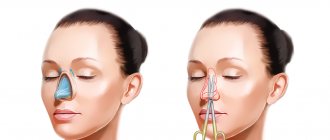
Many of us, trying to get a beautiful and pleasant appearance, resort to various cosmetic surgeries. Rhinoplasty is considered the most common surgical procedure to correct the shape of the nose.
However, the human body tends to provide lost bone strength after surgery, so neoplasms often arise at this site. This causes the development of what is known as a “callus.” Such a neoplasm is quite rare, however, as a result of such a procedure, almost every patient undergoes it. Bone callus has nothing in common with ordinary callus; it does not represent hardened epithelium.
The growth is formed in the event of improper fusion of bones after surgery or nasal fractures, and therefore represents a normal regeneration process.
Why is “bone callus” dangerous?
Such an education does not mean anything serious for life, however, diagnosis and prevention must be carried out in a timely manner. In its absence, serious consequences may occur, including constant severe pain in the patient.
According to statistics, complications occur in 12 percent of victims. In 30 percent of cases, it is necessary to perform a second operation to prevent the proliferation of bone tissue.
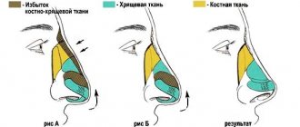
When performing rhinoplasty, part of the bone and skin is removed. Operations of this type are always associated with changes in the structure of the nose. During interventions, the human body gives a protective reaction.
Unlike other body systems, bone tissue has a special healing process, which includes 3 successive stages:
- The first is accompanied by the appearance of neoplasms around the site of connective tissue damage;
- At the second stage, the development of bone tissue fibers is noted;
- Then the bone tissue thickens due to fiber calcification. Over time, it completely replaces the soft one.
When passing the third stage, a small growth forms right at the site of fusion. If grinding is done during rhinoplasty surgery, then rehabilitation almost always takes place with the appearance of such a callus. The size of the growth always depends on the individual characteristics of the human body, the depth of damage to the bone and surrounding tissues.
Among the main reasons for the development of such a callus are the following factors:
- The presence of individual characteristics of the body, which have a fairly high degree of tissue restoration;
- An equally important factor to ensure a successful operation is the experience of the surgeon. Professional specialists with extensive experience know several secrets that help to carry out the operation in such a way as to prevent bone tissue from growing in the future.
Causes
The peculiarities of the occurrence of callus lie in the properties of bone tissue, which differs from other organs and systems in the special course of regeneration and restoration processes.
The healing process goes like this:
- Connective tissue forms around the damaged area.
- It forms soft bone tissue from thin fibers.
- These fibers ossify and become stronger. A growth appears at the site of tissue fusion. Its size depends on the severity of the injury received as a result of the operation and the regenerative properties of the patient’s body, which are individual for everyone.
The reasons for the appearance and growth of callus after plastic surgery include the following::
- The individual ability of the body to restore bone tissue.
- Experience of a plastic surgeon - after the work of a real professional, hypergrowth of bone fibers does not occur.
More often, the growth of callus after rhinoplasty is observed after removal of the hump and with complete rhinoplasty, when a global correction of the shape of the nose is performed.
What does callus consist of?
Callus has a special structure, which appears as a result of the restoration of bone tissue when it is damaged, and is a natural process. We can say that this neoplasm consists of connective tissue growing at the site of bone fusion.
Bone tissue also grows in three stages:
- The appearance of a provisional callus;
- Formation of osteoid tissue;
- Replacement of connective fibers with bone ones.
Almost always, cartilage tissue is formed first, which eventually turns into bone. This process takes about one year.
Pathogenesis
of connective tissue occurs . After about a week, osteoid tissue begins to form, undergoing transformation either into bone itself, or initially into cartilage, and later into bone. This is usually caused by the mobility of bone fragments, which leads to trauma and disruption of the microcirculation of the resulting regenerate. The formation of initially cartilage tissue is due to the fact that this process requires less oxygenation and fewer biologically active compounds.
The formation of periosteal callus occurs as a result of processes in the bone-forming cells of the periosteum (periosteum) and endosteum. These mechanisms play a critical role in the process of healing of injured bone. The periosteum plays a fundamental role in the fusion process, from which the periosteal callus is formed.
Stages of callus formation
The formation of callus during a fracture is a complex regeneration process that occurs in several stages, although there may be variations in different locations; let’s look at the example of ribs:
- The formation of a primary connective tissue callus takes approximately 10-14 days - first, blood accumulates at the fracture site and a hematoma , hyperemia and serous impregnation are formed, initiating the influx of leukocytes that cause swelling, as well as fibroblasts - cells capable of producing connective tissue structures, against the background of which the the process of alteration - osteoclastosis, designed with the help of osteoclasts to destroy necrotic and damaged cells of the bone and adjacent soft tissues, while the process of formation of vessels (granulating tissue) occurs.
- The formation of young mesenchial tissue plays a critical role in filling bone defects and hematomas; hyaline or fibrocartilage grows between and around fragments.
- The formation of osteoid callus - the process of transformation of connective tissue into osteoid within 3-4 months is caused by decalcification - processes of deposition of inorganic compounds present in normal bone, sometimes it is mistakenly mistaken for a “cartilaginous callus” and it resembles a soft (primary) callus, forming approximately after 5 weeks, during which time reverse processes occur at the fracture site - reduction of blood vessels, elimination of edema and normalization of blood flow against the background of disappearance of signs of inflammation.
- The bone callus itself is a neoplasm enriched with apatites (hydroxyapatites) in the form of a conglomeration of amorphous osteoid tissue turning into bone, which is first distinguished by a loose structure, and then, decreasing in size, acquires the features of normal architectonics in the phase of reverse development of bone tissue structures (formation of trabeculae, restoration of the bone marrow canal), the process can last for years.
Callus does not form when bone fragments are tightly juxtaposed, when the size of the gap is fixed to 500 microns, there is good blood supply, there is no periosteum, as well as in fractures of the flat bones of the skull, sternum, pelvis and scapula, since fusion occurs due to connective tissue due to the peculiarities bone embryogenesis.
In the layers of skeletogenic cells of the periosteum and endosteum, bone beams can immediately, initially form, bypassing the fibrocartilaginous stage of bone formation, so the callus can be small in size or not expressed at all.
Layers of callus
Callus can have a different layered structure depending on the tissue source of its formation and usually differs in:
- periosteal layer;
- endosteal layer;
- intermediary layer or otherwise called intermediate, which can develop from elements of the Haversian canals and occupies the space between the endo and periosteal layers;
- the final - paraosseous layer covers from the outside all of the above layers, the source of its formation is the surrounding soft tissue;
- At the base of the entire layered structure is osteoid tissue.
What is the best way to remove a callus after rhinoplasty?
Every person, in case of formation of a callus on the nose, tries to choose the safest methods for its removal.
With hypergrowth of such a formation, the following consequences are possible:
- Ugly hump on the nose;
- Swelling and edema;
- Deformation of the normal shape of the nose.
All of these phenomena not only noticeably worsen the appearance of the face, but also require repeated surgical interventions. Therefore, if the first suspicious symptoms appear, you should visit a plastic surgeon.
Diagnosis of the disease
The development of callus can be diagnosed using radiography. In the picture it will be visible as a small shell at the site of tissue damage. Rehabilitation is a fairly long and complex process. Its main goal is to stop the growth of bone tissue.
Callus cannot be called a disease, because there is no treatment for it as such. However, there are many different techniques that can improve the patient’s nutrition, reduce the inflammatory process at the site of injury and prevent further growth of bone tissue.
In order to improve the healing of bone tissue, specialists prescribe the following therapy:
- Physiotherapy;
- Surgery to remove bone tissue;
- Treatment with medications.
Removal of callus is considered a radical method and is carried out only in cases where other therapy has not been effective.
Surgical treatment of patients with pseudarthrosis
Surgical treatment of patients with pseudarthrosis has been used for a long time, and its methods are being improved as science develops. For pseudarthrosis that formed after a closed fracture, at one time the method of choice was metal osteosynthesis with bone grafting.
After exposing the area, the pseudarthrosis is freed from scars and the bone fragments are refreshed, which, after reposition, are firmly fixed with a metal rod killed by the intramedullary. Then the area of pseudarthrosis is covered with a bone autograft, which is taken from the proximal metaepiphysis of the tibia or iliac wing; allografts (preserved cadaveric bones) or xenografts (bovine bone) are used. The graft is closely fitted with a spongy surface to the exposed layer of the pseudarthrosis area and firmly fixed with wire or bolts. The operation is completed by applying a plaster cast, which immobilizes the limb until the bone heals.
With tight pseudarthrosis without displacement of fragments, good results are achieved using a less traumatic operation - bone grafting with Khakhutov. After exposing the area of pseudarthrosis from the side of the subperiosteal wound, grafts of the same width are cut out from both fragments. Their length in one of the fragments should be 2/s, and in the second - 1/s of the total length of the graft. The grafts are moved so that the longer part covers the pseudarthrosis gap, and the shorter part fills the resulting defect after movement. After surgery, the limb is fixed with a plaster cast until the bone heals.
decortication surgery , has proven its worth . After opening all the soft tissues in the area of pseudarthrosis, thin plates of the cortex are knocked down with a subperiosteal chisel so that they are contained on the periosteum with the adjacent soft tissues. After performing circular decortication, the wound is sutured and a plaster cast is applied.
To stimulate reparative osteogenesis and improve blood supply to the pseudarthrosis area, some surgeons use a chisel to make incisions into the callus and bone to a depth of 2-3 mm in the form of a fir cone. The treatment of patients with infected pseudarthrosis complicated by osteomyelitis and after open fractures was very problematic. Treatment was delayed for many months and even years, since open surgical treatment can be carried out no earlier than 6 months after the healing of the festering wound or closure of the fistula.
To accelerate the healing of infected pseudarthrosis, the Steward-Bogdanov operation, or extrafocal bypass polysynostosis, was used, and for defects of the tibia, the Hahn operation was used - moving the fibula under the tibia.
Ilizarov apparatus into traumatological practice opened a new era, which radically changed the treatment tactics for pseudarthrosis, including those complicated by osteomyelitis and bone defects.
The use of hardware osteosynthesis eliminates deformation, creates stable fixation of the damaged segment, provides movement in adjacent joints, and allows loading the limb. However, with hypovascular pseudarthrosis, the process of bone fusion even in the apparatus remains slow, and therefore it is necessary to additionally apply bone grafting.
Patients with suppurative processes in the area of pseudarthrosis are treated according to the general rules of purulent surgery under conditions of hardware osteosynthesis.
For pseudarthrosis complicated by osteomyelitis , even when there is a fistula, the use of the device and the creation of stable fixation leads to increased regeneration, attenuation of the inflammatory process, closure of the fistula and bone fusion. If there is a formed sequestration, sequestrectomy is performed in the device or before its application. With the help of hardware osteosynthesis, it is possible to shorten the treatment period for patients and achieve bone fusion.
For bone defects, a 4-ring (or more) compression-distraction device is applied, a unipolar, and for large defects, a bipolar osteotomy (compactotomy) is performed in the metaphyseal (cancellous) area of the bone. After the formation of the primary cellular regenerate (7-10 days), the middle bone fragment begins to be lowered towards the defect. Lowering is carried out very slowly, 1 mm per day (in one or two steps of 0.5 mm), bringing the middle rings of the device closer together. As the space in the osteotomy area expands, it is filled with new regenerate and gradually grows.
When the ends of bone fragments approach each other at the site of the former defect, some compression is created to cause necrobiosis and stimulate the local reparative process and fusion of the fragments. For complete bone fusion, the device should be kept in a neutral position for 2.5-4 months. This treatment method allows you to eliminate bone defects over a significant area (15 cm or more).
Features of treatment
The goal of treatment is to eliminate the possible complication. For this, various techniques and methods are used. It is important to use them immediately after surgery. This approach makes it possible to get the desired result faster.
But if there are individual contraindications, effective procedures may be limited. At the first stage, the most important thing is to reduce inflammation at the site of injury using medications.
The tumor can also be removed through surgery. Surgical intervention is a last resort; it is used when other methods have not brought the desired result.
Often this defect after taking medications can manifest itself as the following unpleasant sensations:
- Increase in temperature;
- The appearance of severe pain and redness of the bridge of the nose;
- Difficulty breathing.
Recommendations from specialists are always aimed at effective and safe restoration of damaged tissue. Failure to comply with these rules can lead to serious complications for the patient. Regular visits to the surgeon will help you avoid such complications and undergo rehabilitation faster.
It is important to remember that the body's natural defense system can cause some problems as a result of rhinoplasty. But timely use of physical therapy and medications prevents the need for repeat surgery. As a result of a set of procedures and taking certain medications, the callus quickly resolves.
Taking good care of your body always allows you to maintain a beautiful appearance of your face and the functionality of your nose.
Taking medications
Drug therapy is considered a fairly common method of treating callus. It is most often used by surgeons as a treatment, as well as for prevention purposes.
In order to prevent the occurrence of hypertrophied callus, it is recommended to use drugs that contain glucocorticoid hormones that can eliminate swelling and ensure rapid bone recovery.
- The most common drug is Diprospan, which is administered by injection. It can increase scarring, relieve swelling and reduce inflammation;
- Another, no less well-known drug is “Kenalog”, the drug is administered intramuscularly, providing an excellent anti-inflammatory effect;
- Also highly popular is a homeopathic drug that has a complex effect on the skin - Traumeel S. The products are used externally or internally; the manufacturer makes them in the form of drops, ointments and tablets.
Physiotherapeutic procedures
Many people prefer to use various procedures that can help restore the nose to its previous shape. Even experienced specialists recognize them as highly effective. With physiotherapy, it is possible to ensure gradual resorption of the callus and also activate its regeneration.
The most common methods of such treatment are the following:
- Using electrophoresis simultaneously with lidase and hydrocortisone preparations. This method can bring a good effect not only at the early stage of callus development, but also with significant compaction;
- Thermotherapy or thermotherapy is also considered effective;
- A good result can be obtained with the help of ultrasound, which is carried out using steroid ointment or phonophoresis;
- It is also possible to use UHF or magnetic therapy.
Everyone understands that it is best to prevent the development of a callus on the nose. It is much easier to carry out thorough prevention with all precautions; in this case, you can avoid such unpleasant consequences, which will then be much more difficult to get rid of.
As preventive measures, experts advise following several rules:
- Contact a surgeon as soon as possible if initial signs or some symptoms of growth development appear;
- Strictly follow all doctor’s recommendations during treatment;
- Choose the right clinic and specialist who will perform rhinoplasty. Such surgical intervention is quite common nowadays, it has a very optimal cost, so you should carefully navigate among the numerous offers in order to choose the most optimal option for yourself.
Recommendations from experienced specialists
To prevent the occurrence of callus, experienced professionals give several tips; by adhering to these rules, you can maintain your appearance as before.
- In the first few days after the procedure, it is recommended to remain in bed; try to comply with this requirement, because this will allow you to get a good result not only regarding your appearance, but also your well-being;
- In the first 2-3 weeks, it is best to stay at home, and you should also try to avoid strenuous physical activity;
- It is not recommended to blow your nose at first; to clean it, it would be much better to use regular cotton swabs;
- Complex strength exercises are not performed; heavy lifting can be carried out no earlier than after 2 months;
- If you wear glasses, you should discard them during rehabilitation. If necessary, they can be worn for a short time;
- Try not to eat foods that are too cold; it is much better to warm everything up;
- For a month you should forget about beaches, saunas, steam baths, and solariums. This will help avoid severe overheating in the sun and under strong steam.
By following all of the above recommendations, a patient who has undergone rhinoplasty will be able to recover quickly and also prevent the occurrence of “callus.”
Diet for callus
Diet for fractures
- Efficacy: therapeutic effect after a month
- Timeframe: 2 months
- Cost of food: 1600-1800 rubles per week
The diet after a fracture and during the formation of callus should be complete and balanced. It is extremely important that every day your body receives:
- a sufficient amount of proteins, the source of which should be veal, poultry, rabbit, legumes, cottage cheese and cheeses, as well as bone decoctions;
- mineral compounds essential for osteogenesis are found in fish, seafood, eggs, dairy products and yoghurts such as “Rastishki”;
- antioxidants and vitamins - can be taken in the form of additional complexes, but it is better to enrich the diet with fruits, berries, vegetables and herbs, drink green tea with ginger, smoothies and fruit drinks;
- healthy fats - we must not forget about their benefits, so unrefined oil, nuts, seeds and seeds should be added to vegetables and other dishes;
- carbohydrates - porridge and whole grain baked goods - are essential components of a complete diet.
