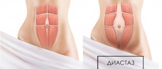
Separation of the rectus abdominis muscles, or diastasis, is a common problem affecting mostly women. According to statistics, 70-100% of women develop this pathology in the third trimester of pregnancy. Normally, after childbirth, the structures should return to their original position, but in approximately 30% of cases this does not happen. A gap remains between the muscle tissues, which causes a bulging, flabby abdomen and provokes more serious consequences. That is why it is important to know how to determine diastasis of the abdominal muscles at home, which doctor can help, and what treatment methods modern medicine offers.
Diastasis: definition of the concept
The abdomen has powerful muscles and the following main groups can be distinguished: • external - oblique and rectus muscles, the latter forms the much-desired “cubes” on the abdomen; • internal - transverse and oblique muscles, their main task is to maintain the position of the organs. The rectus muscles are separated by a thin tendon membrane - also known as the linea alba. A lot depends on her postpartum condition. Normally, the distance between the muscles is no more than 20 mm. But under the influence of certain reasons, the tendon membrane is weakened, overstretched, and neighboring muscles diverge. This condition is called diastasis recti. Several types of diastasis have been identified. The classification is based on the area of divergence: above or below the navel, or a mixed version. Symptoms and prognosis largely depend on this.
Suturing of diastasis without incisions using the SCOLA method
The SCOLA (subcutaneous onlay laparoscopic approach) technique is suitable for patients who have diastasis, local fat deposits, but no large excess skin.
During the operation, a special device is used - a laparoscope, which is inserted into the abdominal cavity through punctures of 5 mm in size in the bikini line area. The device is equipped with a miniature video camera, the image from which is transmitted to the screen in an enlarged size, which allows the doctor to perform complex manipulations with the highest precision. The surgeon sutures the rectus muscles using special threads, and a soft mesh implant made of inert material is used. Made in 3D weaving, the mesh implant grows with connective tissue, and its external coating prevents the adhesion of organs. Thus, the sutured tissues remain in the desired position, and the development of relapse is excluded.
As a result of such an intervention, the patient receives a stomach without traces of the incision, without wrinkling of the skin, with a corrected shape of the navel.
Causes
The diagnosis of “diastasis” is made to 2-3 out of 10 women after the birth of a baby.
Risk groups have also been identified: the likelihood of rectus muscle divergence is higher in women with a fragile physique who do not play sports. Mothers of large families with children with a small age difference are also at risk. Statistically, diastasis is recorded less often in active women, with developed and trained rectus abdominis muscles and normal weight.
There is a direct connection between improperly functioning muscles of the thoraco-abdominal diaphragm, pelvic floor and abdomen, and disruption of their coordinated work, which leads to their dysfunction. This is one of the reasons for the increase in intra-abdominal pressure and, as a result, leads to diastasis. Pregnancy and childbirth are other obvious causes of increased intra-abdominal pressure. There are several explanations for this: the growth of the fetus and uterus. A role is played by the production of the hormone relaxin, which helps to “soften” the ligamentous apparatus of the pelvic bones for the possibility of natural childbirth. But relaxin also acts on the linea alba, which can provoke its weakness and, as a consequence, the formation of diastasis.
Predisposing factors in the development of diastasis with its possible complications can be considered pregnancy with twins or triplets, high fetal weight at the time of birth, difficult and prolonged delivery.
Diastasis is quite possible in men and even children. Therefore, pregnancy and childbirth are not the only reasons, there are others: • Congenital features The white line (aponeurosis) is formed by connective tissue. With its congenital weakness, the likelihood that the muscles will begin to diverge significantly increases. To prevent complications, it is important to identify a predisposition to discrepancy even before pregnancy, taking preventive measures: strengthening them using therapeutic exercises. • Diseases and dysfunctions of the intestines Chronic constipation is one of the causes of increased intra-abdominal pressure. Its prolonged existence in combination with other factors increases the risk of divergence. In general, rapid weight gain, obesity, a sedentary lifestyle and a number of chronic diseases are dangerous, one of the symptoms of which is increased intra-abdominal pressure. • Strength training People who practice heavy strength training are at risk of encountering diastasis. Coaching mistakes and excessive load are not only a risk factor for diastasis, but also its complications, for example, umbilical hernia.
It is extremely important to identify muscle discrepancies in a timely manner, otherwise an incorrectly selected set of exercises that train the abdomen and its muscles, especially during the postpartum recovery period, will lead to serious complications - increased discrepancies between the muscles.
Self-diagnosis of diastasis
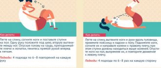
If you suspect you have diastasis, we suggest you perform a self-diagnosis using video and record the results. To record, you can use the table in the infographic below or any medium convenient for you.
Diastasis is measured using three parameters:
- Divergence length . Measure how many centimeters the muscles extend up and down from the navel.
- Width of divergence . Measure how many centimeters there are between the muscles above and below the navel.
- Finger insertion depth . Evaluate how much your fingers sink when performing the test. During the recovery process, you will learn to engage the transverse abdominis muscle and the depth of immersion of your fingers during the test will become less.
Record your self-test results in a table to track changes.
During each test, observe the following aspects:
- Assess whether you experience pain in the pubic symphysis (the joint of the pubic bones).
- Try to feel the boundaries of the muscles along the middle of your abdomen and evaluate whether it is a soft tear or a clear separation of the muscles.
- See if you see a bulge or ridge.
- Listen to the sensations - do you experience discomfort in the pelvic floor (involuntary release of urine, release of gas, air from the vagina).
If something alarms you while performing these points, you should consult with your doctor to fully assess the condition of the abdominal cavity and/or pelvic organs before starting a cycle of exercises to correct diastasis.
Self-diagnosis of diastasis
How to independently determine diastasis?
When preparing for pregnancy, it is recommended to visit a number of doctors and identify diastasis and predisposition to it - one of the points of mandatory examination. At the Quality of Life clinic, specialists will not only be able to identify, but also eliminate the problem using non-surgical methods, helping to recover faster after childbirth, avoiding complications. The discrepancy of the rectus muscles is detected independently, and complex examination methods are not needed - if you tense the abdomen, then a longitudinal ridge will be noticeable in its center, just in the area of the white line. There are other tests available at home: • take a lying position with your knees slightly bent; • the right or left hand should be located in the back of the head; • with the fingers of the other hand, focusing on the navel, find the midline; • lifting up and simultaneously twisting the torso, you need to look for the “failure” with your fingers. If there is a dip in the stomach of no more than two centimeters, approximately 1-1.5 fingers, there is no reason to worry. But as part of pregnancy planning, a doctor’s consultation is not included. Depending on the size of the discrepancy, the following degrees of diastasis are distinguished: • I – does not exceed 22-50 mm; • II - about 51-70 mm; • III – more than 71 mm. In this case, the protrusion is constant, does not depend on straining, and its configuration changes.
Immediately after the birth of the baby, the muscles may diverge by up to 3 cm - this is normal. To shorten the recovery period without causing complications, medical care is needed in the early postpartum period.
Diastasis of the rectus abdominis muscles - symptoms and treatment
Diastasis of the rectus abdominis muscles does not always require surgical correction. For women who are less than 12 months postpartum and overweight patients who have not previously tried to correct diastasis with exercises, it is recommended:
- control weight: body mass index (BMI) should not exceed 26 with an average build;
- do exercises to strengthen the anterior abdominal wall [1][8].
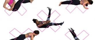
Exercises for diastasis
There is no single set of exercises; different trainers use different programs. It should be noted that all exercises are aimed at strengthening the muscular frame of the abdominal wall, but they do not always help reduce the width of the white line of the abdomen [8][10]. For diastasis, it is recommended to perform the following exercises:
- Supination (outward rotation) of the knee in a position on all fours . Starting position - emphasis on your knees and straightened arms. From this position, bend your left leg and bring your knee towards your sternum, then do the same with your right leg. You can perform 10 repetitions on each leg, 5-6 approaches per day.
- Exercise " plank " and its variations. The emphasis can be placed on your knees or toes, on your elbows or straightened arms. This is a static exercise; it does not lead to excessive muscle stretching. The execution time gradually increases from 20 seconds to 1 minute. Perform 3–5 approaches per day.
- Exercise "to the eye " . In a position on all fours, as you exhale, arch your back upward, rounding it like a cat, while straining and drawing in your stomach as much as possible. Keeping your muscles tense, exhale and slightly bend your back in the opposite direction. Do 10–15 repetitions, 3–4 sets per day.
- A set of Kegel exercises. They are aimed at strengthening the abdominal and pelvic floor muscles. Technically, these exercises are not difficult, but they must be performed carefully. If the technique is incorrect, the effect can be the opposite. It is recommended to do approaches 3-4 times a day for 10-15 minutes. As a rule, the effect appears after 4–6 weeks.
Classic exercises for strengthening the abdominal muscles with lifting and twisting the torso, including wall bars, can only increase diastasis and should be excluded. Young mothers can start exercising only 4–6 months after giving birth. During this time, connective tissue structures restore their strength.
According to some authors, up to 87% of patients remain dissatisfied with the results of training programs and require surgical treatment [11]. This is probably due to irreversible damage to the integrity of the tendon bundles of the white line of the abdomen.
Surgery
The goal of all diastasis surgeries is to eliminate the enlarged gap between the rectus abdominis muscles and strengthen the newly formed, thinner linea alba. Open classical or laparoscopic techniques, as well as interventions from mini-approaches, can be used. The results for all techniques are similar, so the choice of operation, as a rule, depends on the individual experience of the surgeon. All techniques can be used for the combination of diastasis recti and umbilical hernia [1][6][8][9].
Robotic surgery is more expensive and does not offer advantages over laparoscopic techniques, so it is not used as widely.
Open methods. Access to the extended linea alba is performed through an incision on the anterior abdominal wall: longitudinal above the midline or transverse below the navel if excess fatty tissue needs to be removed. The “fat apron” is removed to prevent suppuration of exfoliated fiber and improve appearance. The surgeon frees the edges of the internal oblique abdominal muscles from fatty tissue, brings them together using numerous individual or several continuous sutures, and then installs a mesh implant over the formed suture.
The operation is traumatic, after which the patient remains in the clinic for 5–7 days. Some authors report that there are no relapses when using open techniques, others report 4% or more relapses [9]. This operation is performed if it is necessary to remove the “fat apron”, and also if the surgeon does not have the skills to perform laparoscopy. In addition, this technique may be preferable when there is a combination of diastasis recti and an incisional midline hernia.
Laparoscopic techniques. The operation is performed through separate punctures on the abdominal wall. The surgeon and assistant, using special manipulators and a video camera, separate the linea alba from the preperitoneal fatty tissue from the abdominal cavity, and then strengthen it with one continuous or several separate sutures. When the diastasis is eliminated, the posterior surface of the abdominal wall can be further strengthened with a mesh implant. This technique can be used for a combination of diastasis and hernia of the white line of the abdomen. The recurrence rate of diastasis after laparoscopic plastic surgery of the linea alba does not exceed 2% [9]. However, using this technique it is impossible to eliminate the “fat apron” if it exists. This operation is carried out if the surgeon has the necessary skills and the technical capabilities of the medical institution.
Techniques using mini-accesses. This operation combines the positive qualities of open and laparoscopic methods for eliminating diastasis. A 4–5 cm long transverse incision is usually made below the navel at the level of the bikini line or directly above the navel (depending on the extent of the diastasis). Then, a channel is formed in the subcutaneous fatty tissue above the linea alba for the surgeon to work with. Viewing in the canal is usually carried out using a special video camera, as in laparoscopy. The surgeon also approximates the inner edges of the rectus abdominis muscles and then strengthens the anterior abdominal wall with a mesh implant [12]. The recurrence rate using the mini-approach is approximately the same as with open surgery. The technique is used if there are contraindications for laparoscopy (severe adhesive disease in the abdomen after surgery, severe chronic heart failure).
If the clinic has the necessary equipment and a qualified plastic surgeon, each technique can be supplemented with liposuction and liposculpture, in which excess fat is removed from certain areas of the abdomen and thus the relief of subcutaneous fat is formed.
Possible complications after surgery:
- Bleeding with the formation of a hematoma of the surgical wound or in the abdominal cavity. If the bleeding stops on its own, the hematoma does not increase, no blood comes from the wound and the patient feels normal, the hematoma is removed. If the bleeding continues, it must be stopped: remove several stitches and stitch or cauterize the bleeding vessel.
- Seroma of a postoperative wound. This is an accumulation of serous fluid in the thickness of the abdominal wall, namely in the area where the surgeon freed the linea alba from fatty tissue. It manifests itself as swelling and thickening of the subcutaneous fatty tissue. The seroma is removed by a surgeon in the dressing room. If the seroma suppurates, urgent hospitalization is required in the purulent surgery department. This complication is less common during laparoscopy [9].
- Suppuration of the postoperative wound and mesh implant. The operation site turns red and hurts, body temperature rises, chills and sweating appear, especially in the evening. With this complication, it is necessary to remove the pus from the wound and prescribe antibacterial therapy. It is often necessary to remove the mesh implant. It is not known exactly how often surgical wounds fester; according to some data, the complication rate can reach 2% or more [9]. Suppuration occurs due to the addition of infection, especially if there are foci of infection in the body: carious teeth, chronic tonsillitis, chronic inflammatory diseases of the urinary tract (cystitis, pyelonephritis) and female genital organs (salpingitis, endometritis).
- Severe pain in the wound area in the first month after surgery or longer than three months. The patient is concerned if nerve fibers are damaged or compressed during surgery. With such a complication, a second operation may be required: release of the nerve from compressive tissue, removal of the mesh implant, excision of the postoperative scar, removal of the sutures fixing the mesh implant [1][9]. Some authors report that chronic pain syndrome after open surgery can be detected in 17% of patients; laparoscopy can reduce this figure to zero [9].
It is not known exactly how often complications develop after surgery, since in most cases treatment is carried out in private clinics and complications are not included in registries. On average, the incidence of complications is up to 10–15% [9].
Why is divergence dangerous?
The most striking consequences of diastasis are a bulging belly and a change in its configuration and overall figure. Symptoms become most pronounced 2-3 months after delivery. In an attempt to regain their previous shape, women take up active physical exercise. But the problem remains and even becomes more serious and pronounced. Diastasis is far from just an aesthetic problem. When the integrity of the abdominal wall is violated, caused by the divergence of the rectus muscles, typical complaints arise: pain in the lumbar area, disorder of venous blood flow in the pelvis, congestion in the veins of the legs and thighs, which contributes to the development of varicose veins and the threat of thrombosis. This is also one of the reasons for the prolapse of internal organs, which is a predisposing factor and the cause of urinary incontinence and frequent cystitis (inflammation of the bladder).
Problems with the digestive tract are also common - chronic constipation, dyspeptic disorders: pain or discomfort, a feeling of rapid satiety, stomach turmoil, nausea, even vomiting, belching and heartburn.
The best treatment for diastasis and its complications is prevention, because it is possible to reduce its likelihood. The main thing is to strengthen the muscles even at the stage of pregnancy planning. Then even the formed problem will go away on its own and painlessly.
Symptoms
The clinical picture depends on the degree of development of the disease. So in the initial stages, when the stretching of the aponeurosis is insignificant, there may be no symptoms. The first thing that patients usually pay attention to is a cosmetic defect in the form of a protrusion located along the anterior abdominal wall.
As the disease progresses, complaints such as:
- Back pain, especially in the lumbar spine.
- Discomfort or pain in the area of the stomach projection.
At later stages, there is a disturbance in the functioning of the gastrointestinal tract. The patient will draw the doctor's attention to constipation, flatulence, and nausea. Possible urinary incontinence when coughing or sneezing.
If these symptoms occur, it is recommended to consult a specialist. You can always undergo a consultation in our clinic, where experienced doctors will determine the presence of the disease and carry out all the necessary diagnostic examination and treatment.
Treatment
If you notice diastasis recti that occurs several weeks after giving birth, it is important not to worry. The main thing is to identify the problem in a timely manner and begin treatment, under the supervision of specialists. Typically, recovery lasts 10 weeks, during which time the muscles begin to shift and changes are not enough; restoration of the tissue structure is also necessary, which takes much longer - 8-12 months. At the Quality of Life clinic, specialists are ready to help significantly shorten the recovery period and avoid surgical treatment (results within 7-10 weeks), even if the discrepancy is 6-8 cm. There are successful cases of recovery. And practice shows that more often surgical intervention, even with stage II diastasis, is a last resort. It is impossible to get rid of the discrepancy on your own, and an incorrectly selected set of measures only aggravates the condition. Supervision by a specialist and individual selection of exercises that take into account the clinical picture are necessary. Treatment of diastasis can be carried out in 2 ways, and the choice is determined by its degree and the patient’s well-being: • Conservative treatment A complex that includes regular therapeutic and breathing exercises, a course of massage, wearing a bandage and tapes and other recommendations regarding lifestyle and nutrition. • Surgical treatment This is necessarily followed by a period of rehabilitation, including exercise therapy methods, kinesiotaping, etc.
If the cause of muscle separation is illness of the digestive tract, lungs, then they must be treated as the root cause. Otherwise, it is more difficult to get rid of the problem, and the likelihood of relapse is high. Achieving a flat stomach is also impossible.
Prevention of the problem
There are a number of recommendations and several exercises that will reduce the risk of muscle divergence.
First of all, the osteopath notes, it is necessary to carefully prepare for pregnancy and childbirth. Further, you should not ignore breathing exercises, especially those aimed at retracting the abdomen.
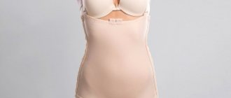
To correct the situation, starting from 22-24 weeks, you should wear a bandage. It should also be used for another two months after childbirth. Afterwards, it should be replaced with shapewear for up to six months.
It is also necessary to pay attention to the diet. “It should include animal proteins and fats, as they improve the quality of connective tissue. It is important to take vitamin D3 on a regular basis, especially for pregnant women and those planning to conceive and give birth to a child. Vitamin D helps strengthen connective tissue. This is a very important and necessary preventive measure,” says the osteopath.
Is belly fat dangerous for women? More details
Physical therapy methods
The main task of physical therapy (physical therapy) is to activate the transverse muscles of the abdomen, as well as the pelvic floor and diaphragm, this helps to normalize intra-abdominal pressure, thereby eliminating the main provoking factor. In many cases, exercise therapy is preceded by osteopathic treatment. It creates a foundation, a frame for further work, “softens” the body, facilitating further work with the help of therapeutic exercises. Physical therapy is certainly important and useful, but with diastasis, not all exercises are allowed. Some of them may be dangerous and cause harm, that is, aggravate the situation.
Exercise therapy and a set of gymnastic activities must be pre-approved by a doctor and carried out under his supervision; only a doctor can give the correct and safe load to the muscles.
There are exercises that are strictly prohibited in case of discrepancy: • exercises with a fitball while lying on your back; • some yoga asanas that involve working the abdominal muscles; • abdominal exercises, torso twists; • exercises with lifting weights are strictly prohibited, and in everyday life you should not lift a load weighing more than 4-5 kg. Therapeutic gymnastics is carried out according to an algorithm and has several goals: • the first stage of treatment is preparatory, its main task is to prepare the muscles for further work with them, which can last up to 2 months; • strengthening and training the transverse muscle ensures the maintenance of internal organs in the correct position, which can be considered as the prevention of numerous diseases and conditions. The recommended set of exercises should be performed regularly and on an ongoing basis. A diagnostic test is required once a week to evaluate the results of treatment.
Conservative treatment is a long and complex work, so you should not expect instant results. But the main advantage is the ability to avoid surgical intervention and minimize the consequences.
What will help correct diastasis
Previously, it was believed that to correct diastasis, it was enough to pump up the oblique muscles: they would become stronger and “tighten” the discrepancy along the white line. In fact, everything is much more complicated.
The abdominal muscles are a complex multi-layered system, and not just abs on the stomach. The deepest layer is the transverse abdominis muscle. Above it are the rectus muscle, internal and external oblique muscles.
Reducing diastasis is the coordinated work of all these muscles and the ability to safely include them in work.
Before including abdominal exercises in the training process, you need to work on your posture and learn to engage the deep muscles.
Correct posture
One of the most important exercises is developing the skill of correct posture. When the body is aligned, the chest is raised, the shoulders are separated and lowered, the abdominal muscles are given the correct tone. They work to maintain this position, which makes the stomach visually flatter and helps balance intra-abdominal pressure.
For some time, you will need to constantly monitor your body position: shoulders are straightened, located exactly above the pelvis, the stomach is moderately tucked, the hips are level, the feet are shoulder-width apart, the toes are spread for stability.
At first it will be unusual, but over time you will form the habit of standing and moving without breaking your upright position.
At first, monitoring your body position will not be easy, but gradually it will become a habit. Photo: www.freepik.com
Try not to tense your facial muscles, so as not to cause tension in your body. When moving your ribs and shoulders, you should not feel any discomfort in your stomach or hold your breath.
Make sure that the tailbone does not move forward, and that the weight is distributed evenly and does not shift to one hip. Skewed hips indicate that the transverse abdominis muscle is not working. To engage the transverse muscle, pull your belly toward your lower back, concentrating on the area below your belly button.
Exercise to form the correct body positionApproach the wall, turn your back to it and place your feet approximately 20-25 centimeters from the wall, buttocks, shoulder blades, shoulders and the back of your head touching the wall. Having adjusted the position, perform 15–20 breathing cycles:
|
These breathing exercises work to correct diastasis in a comprehensive manner:
- Breathing engages the deep core muscles and teaches them to work in harmony. This helps balance intra-abdominal pressure.
- Alignment against the wall gives the body the correct position and the muscles the correct tone.
Perform this complex daily for 2-3 weeks and observe the results.
Breath
Breathing exercises help the diaphragm return to proper function after childbirth. During pregnancy, the diaphragm is pushed upward by the growing uterus and loses the ability to fully descend during breathing (inhalation phase).
The work of the thoracic diaphragm is closely related to the work of the pelvic diaphragm - they, like a vertical piston, always work together. Their well-functioning work has a beneficial effect on the functioning of all internal organs and the condition of the muscles of the anterior abdominal wall. Therefore, it is extremely important to set up this mechanism in a timely and correct manner.
Below you will find breathing techniques that will help your diaphragm return to proper function.
Belly breathing. Lie on the floor on your back, bend your knees, bring your knees and feet together, place one hand on your stomach, the other on your chest.
As you inhale, gently inflate your stomach so that your hand on your stomach moves upward. As you exhale, gently lower your stomach down, pulling your pelvic floor muscles inward.
Try to avoid upward movements of the chest, controlling this with the other hand.
Lateral (costal) breathing. Take a comfortable sitting position with a straight back, place all 5 fingers of your palm on the lower ribs under the chest, point your elbows to the sides.
Try “breathing into your hands” by pushing with the ribs of your palms, and feel the lateral expansion of your chest. The shoulders should remain motionless.
Alternate upper and lower breathing, sitting or lying down. Lie on your back, bend your knees, place your feet hip-width apart, parallel to each other.
Slowly begin to inhale, filling the stomach first and then the chest. When exhaling, push the air out first from the abdomen, then from the chest.
As you exhale, additionally pull your stomach towards your lower back, direct the pubic bone towards the ribs (you can slightly twist the pelvis towards yourself and pull the pelvic muscles inward).
Pelvic floor muscles
Strengthening the pelvic floor muscles prevents the prolapse of the internal organs during the recovery of the abdominal muscles. Perform them daily, devoting 5-10 minutes a day to your health. You can see a list of exercises in the article “Strengthening the pelvic floor muscles.”
It is important to train your pelvic floor muscles not only with Kegel exercises. The pelvic diaphragm is involved in the breathing process and must be able to relax and contract when we breathe and move.
Incorporate your breathing during exercises for your abs, back, and buttocks as you perform them. This will help keep your pelvic floor toned.
Kinesio taping
Tapes applied to the abdomen according to a special pattern are an effective method of treatment, which, in addition to other methods, allows you to get results faster. Kinesio taping of the abdomen allows you to achieve the following effects: • stabilize muscles; • relieve unpleasant and even painful sensations; • eliminate hyperextension and prevent its progression; • provide the muscles with the necessary tone; • stimulate metabolic processes.
To achieve maximum effect, tapes must be installed by a specialist according to the scheme. But the therapeutic effect is achieved only with a combination of taping and therapeutic exercises lasting several months.
Prohibited exercises when diagnosing diastasis after childbirth
Before you start working on eliminating diastasis, you should pay attention to exercises that are strictly prohibited. These include the following types of loads:
- Exercises that require you to lie on your back on a fitness ball.
- Yoga, namely poses that stretch the abdominal muscles.
- Using heavy objects or doing barbell exercises.
- A load that involves being on all fours.
Correction of nutrition and lifestyle
Nutrition should always be varied and meet the needs for nutrients, vitamins and minerals, regardless of health status. But with diastasis, it is necessary to avoid foods that contribute to gas formation and flatulence (legumes, cabbage, large amounts of sweets, etc.).
The basis of the diet should be products that stimulate normal intestinal function: dairy and fermented milk containing fiber, fresh fruits and vegetables. If you follow the recommendations, the risk of diastasis is 40% lower.
Sleeping position also matters. For diastasis, sleeping on your side or back is considered the most optimal. The habit of sleeping on your stomach must be abandoned. The fact is that it increases the load on the linea alba, causing the muscles to be subject to additional stretching. It is recommended to get out of bed in the morning from a position on your side, and the main muscle load should be distributed specifically to the leg muscles.
Changes in posture that occur during pregnancy aggravate the discrepancy of the abdominal muscles, so the effect must be complex. It is important to act quickly so that a vicious circle does not form: diastasis worsens posture, and posture worsens diastasis.
Breathing exercises for diastasis
Breathing exercises help retrain the diaphragm, or rather, return it to its normal position and functioning. During pregnancy, the growing uterus seems to push the diaphragm up and it loses the ability to return to its normal position. Since the diaphragm forms the upper part of the core muscles, it is important to retrain it and return it to its original position. Here are some good exercises:
- Lying on your back, with a pillow under your buttocks, raised slightly above chest level, you should take short, shallow breaths into your stomach. This exercise should be done for a minute.
- While sitting, you need to put your hands on your chest and take short and shallow breaths for a minute.
The distance between the two abdominal muscles is not the most important aspect. It is much more important that all the abdominal muscles can work together again, providing quality support to the spine.
Treatment after caesarean section
The scar that forms after a cesarean section interferes with the full functioning of the thoraco-abdominal diaphragm and pelvic floor muscles. Taken together, it is these factors that predispose to increased intra-abdominal pressure, which contributes to the divergence of the abdominal muscles. During rehabilitation and treatment, it is important to work with the scar, to make it softer and more pliable. Soft osteopathic techniques make it more pliable, which will make it possible to work with the muscles of the abs and pelvic floor, the diaphragm, but also to prevent adhesions and increase the likelihood of natural childbirth in the future.
Working with the scar after a cesarean section will create the right prerequisites for physical therapy. This allows you to remove the “stiffness” of the body and the main areas of the body necessary for the treatment of diastasis.
Diastasis removal price
The price of an operation to remove diastasis is formed from the cost of the intervention, which is considered quite complex, payment for anesthesia and hospital, medical personnel, etc. Compression garments are also necessary after surgery.
For an experienced surgeon who works in a private plastic surgery clinic, the price will not be low, and this is understandable: the doctor has been working with a variety of modern techniques for many years, gets excellent results, and is in demand among patients.
Also, usually the clinic provides full management of the patient during the rehabilitation period: the clinic assumes responsibility for monitoring the healing of the suture and the patient’s condition, dressings and antiseptic treatment, and responsibility for health.
| Name | Price |
| Endoscopic abdominoplasty | 120,000 rub. |
| Abdominoplasty with one-stage waist modeling, category 1 | 220,000 rub. |
| Excision of the abdominal fold due to deformation of its lower part (miniabdominoplasty) category 1 | 120,000 rub. |
| Primary appointment (examination, consultation) with a plastic surgeon | FOR FREE |
Rehabilitation after surgery
If the muscles diverge by more than 10 cm, surgical treatment may be required, which has strict indications. But even after such treatment there is a period of rehabilitation. Already 10-14 weeks after surgical treatment, patients are advised to return to their normal lives, and exercise is allowed. But therapeutic exercises, that is, a set of exercises aimed at speedy recovery, are allowed much earlier. The recovery scenario and set of exercises are selected individually, and this depends on a number of factors: • surgical treatment method: endoscopic, classical surgical; • degree of diastasis and discrepancy in centimeters; • patient's condition; • etc. But even after surgical treatment, the probability of relapse is high, so there is no way to do without therapeutic exercises. Only a correctly selected set of exercises, supervision by a specialist and their regular implementation are the key to complete recovery and normalization of the condition.
It is important that a set of preventive exercises be compiled by a specialist. Some of them require some training. It is important to perform them correctly, otherwise there will be more harm than good.
Rehabilitation
Rehabilitation is an important stage in the completion of the patient’s treatment and his full recovery. It depends on many factors: age, the presence of chronic diseases, the development of complications of the underlying pathology and many other reasons.
Our main goal is the health of our clients. Therefore, in the early postoperative period, the patient is under round-the-clock supervision of doctors and nursing staff who provide all the necessary assistance.
We strive to ensure that our patients begin mobilization as early as possible in order to prevent thromboembolic complications, such as stroke, myocardial infarction, and pulmonary embolism. It must be remembered that heavy physical activity is prohibited for up to 6 weeks after surgery.
It is important to follow a diet for 2-4 weeks, which will contribute to the normal functioning of the gastrointestinal tract. Exclude foods rich in fiber from your diet, as they promote active peristalsis and gas formation.
After discharge from the clinic, you must remember and follow all the recommendations of your doctor. Your doctor may recommend that you take pain medications and also stop some medications that you take regularly during the recovery period.
Prevention measures and benefits of the “Quality of Life” clinic
“It is much easier to prevent any disease or condition than to cure it” is an unwritten rule of modern medicine. It is quite possible to avoid diastasis in the postpartum period, the main thing is an integrated approach and timely initiation of action. Ideally, you should visit a doctor at the stage of preparation for pregnancy. Preventative measures will help to avoid a number of problems during the period of carrying a long-awaited baby and after childbirth. Recommended measures to prevent diastasis include: • you should not lift heavy objects, you need to enlist the support of loved ones; • when standing up, you need to lean on your hands, and try to shift your body weight to your feet; • exclude exercises that train the rectus abdominis muscles; • permitted exercises are aimed exclusively at strengthening the deep-seated muscles of the pelvis and abdomen.
Having a child is one of the main life missions of a woman. But everything has its price. There is no need to panic if problems are discovered immediately after the birth of the baby. The specialists of the Quality of Life clinic have extensive experience in the treatment of diastasis and will develop an effective set of exercises that will help quickly, painlessly and safely solve the problem and get rid of possible consequences. The main thing is to seek help in a timely manner and begin recovery as early as possible.
Similar articles:
- Directions
- Specialists
- For visitors
- Articles and videos
ONLY UNTIL January 31st!
20% DISCOUNT ON CLASSES ON THE REDCORD SUSPENSION SYSTEM
Classes are conducted by instructor-methodologist Osipova Maria.
REDCORD allows you to eliminate muscle imbalance by relaxing some muscles and stimulating others. This allows you to resume the motor pattern and return the patient to normal functioning!
Exercise "Vacuum"
Vacuum can be combined with posture and breathing exercises. Carry out sequential mastery of the exercise in the starting positions lying on your back, then on all fours, and then standing.
We talk more about vacuum and the technique of performing it in the article “Will vacuum exercise help make your stomach flat?”
Often after childbirth, women are faced with the fact that diastasis does not converge on its own. Our body has a resource for recovery, but sometimes it needs help. The postpartum period is one such case. #sekta has a course for mothers to recover after childbirth, which we recommend to anyone who has complaints about diastasis.
In the course, complexes for correcting diastasis are part of the training load. They are performed three times a week or more often and combine work with breathing, back muscles and the inclusion of the abdominal muscles: rectus and oblique. The result of this work is a reduction in diastasis and a decrease in the depth of immersion of the fingers along the entire length of the white line of the abdomen.
Regularity is important in the process of correcting diastasis. Recovery will be slower if you train occasionally or do training constantly, but do not monitor your movements in everyday life.
The course complexes for mothers are suitable for the correction of first and second degree diastasis. For third-degree diastasis, consultation with a surgeon is necessary to choose a correction method.
In this article we shared only a small part of our knowledge and gave direction for work. This is not an instruction for action, but a basis for developing a plan to combat diastasis.
Authors: obstetrician-gynecologist Valeria Pushkina, Department of Women's Diseases and Reproductive Health at the National Medical Center named after. N.I. Pirogova, Daria Knyazeva, group program instructor, postpartum recovery specialist, trainer-methodologist at the Ideal Body School #Sekta
Literature:
1. https://dianelee.ca/article-diastasis-rectus-abdominis.php. 2. Lee DG 2004 The Pelvic Girdle 3rd edn. Elsevier SyntaxError. 3. Lee DG 2007 Clinical Reasoning and Pelvic Girdle Pain: Show me the Patient! In: Proceedings of the 6th World 4. Congress on Low Back and Pelvic Girdle Pain, Barcelona, Spain, p 27. 4. https://dianelee.ca/article-diastasis-rectus-abdominis.php#sthash.ACQZUzfw.dpuf . 5. https://www.befitmom.com/diastasis_recti.php. 6. https://www.physiotherapy-treatment.com/diastasis-recti.html. 7. Fit Healthy Moms. 3 Ab Exercises to Heal Diastasis Recti. Available from: https://www.youtube.com/watch?v=Q6SfiH2-TEQ. 8. MomsIntoFitness. Diastasis Recti Exercises 5 min Core Workout. 9. Lee D, Lee LJ, McLaughlin L 2008 Stability, continuity and breathing: the role of fascia following pregnancy and delivery. JBMT 12(4):333–348. 10. Lee D 2011 Chapter 6 Pregnancy and its potential complications. 11. Lee D 2011 The Pelvic Girdle - An integration of clinical expertise and research, Elsevier.