White spots on legs and arms: 5 reasons
Often women and others notice the appearance of small white spots on their legs and arms (hypopigmentation). Accordingly, this begs the question: What could it be and what actions should be taken?
This phenomenon is provoked by a number of factors, but it is worth identifying the most common:
- Vitiligo is a skin disease whose etiology is still unknown, and therefore cannot be cured. The only thing that can be done is to improve the condition of the skin as much as possible.
- Pityriasis versicolor is a fungal infection that is present on the dermis of any person and is activated in a favorable environment: prolonged exposure to ultraviolet radiation, increased sweating, and a humid climate. The fungus is treated with special drugs (Clotrimazole, Terbinafine and others).
- Guttate hypomelanosis is another disease, but it is genetic and usually inherited. The exact reasons for its origin have also not been identified, but since it occurs more often after 40 years, it is also associated with aging. Small spots sometimes itch and peel off.
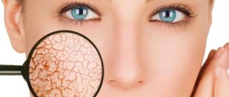
- Lichen alba is a dermatological pathology characterized by changes in skin color. It most often occurs in preschool and teenage children living in regions with a warm and humid climate (you can clearly see this manifestation in the photo below). The skin becomes dry and very flaky. To be sure that it is lichen and to find out how to treat it, you should consult a doctor.
- An adverse reaction to certain pharmaceutical drugs - if they are taken for a long time, skin cells begin to produce pigment (melanin) poorly, resulting in an uneven tan. One such drug is birth control pills.
Discoloration of certain areas of the skin often occurs due to a lack of melanin in the body. Then white spots on the legs and other parts of the body appear after tanning naturally or in a solarium. They do not cause much discomfort.
Cosmetologist Rita Rakus
Representatives of the fair sex with extremely sensitive and pale skin and children under 13 years of age who have not yet fully developed their immune system are more susceptible to this disease.
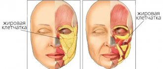
Women also face a similar problem during pregnancy, after 40 years and during menopause. But this does not mean that others are immune from the appearance of such spots on the body.
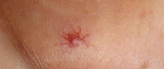
Skin vasculitis
Skin vasculitis is a group of diseases of a multifactorial nature, in which the leading symptom is inflammation of the blood vessels of the dermis and subcutaneous tissue.
The difficulty in covering this topic is that until now there is no generally accepted classification or even agreed upon terminology of vasculitis. Currently, about 50 different nosological forms have been described, and understanding this diversity is not easy. The diversity of clinical manifestations and insufficiently studied pathogenetic mechanisms have led to the fact that under different names only a variant of the main type of skin lesion may be hidden. Also, in addition to primary vasculitis, which is based on inflammatory damage to the blood vessels of the skin, there are also secondary vasculitis (specific and nonspecific), developing against the background of a certain infectious (syphilis, tuberculosis, etc.), toxic, paraneoplastic or autoimmune (systemic lupus erythematosus, dermatomyositis etc.) process. It is possible to transform skin vasculitis into a systemic process with damage to internal organs and the development of severe, sometimes life-threatening complications.
Skin vasculitis is a polyetiological disease. The most common connection is with a focal infection (streptococci, staphylococci, mycobacterium tuberculosis, yeast, viruses, etc.). Hypersensitivity to a number of drugs, in particular to antibiotics and sulfonamide drugs, is of particular importance. Often, despite a carefully collected anamnesis and examination, the etiological factor remains unclear. Among the risk factors for vasculitis, one should take into account: age (children and the elderly are most vulnerable), hypothermia, excessive insolation, severe physical and mental stress, trauma, surgery, liver disease, diabetes, hypertension. The pathogenetic mechanism for the development of skin vasculitis is currently considered to be the formation of circulating immune complexes with their subsequent fixation in the endothelium, although this has not been definitively proven for all diseases of this group.
Skin vasculitis is a heterogeneous group of diseases, and their clinical manifestations are extremely diverse. However, there are a number of common features that unite these dermatoses:
1) inflammatory nature of skin changes; 2) symmetry of rashes; 3) tendency to edema, hemorrhage and necrosis; 4) primary localization on the lower extremities; 5) evolutionary polymorphism; 6) connection with previous infectious diseases, medication, hypothermia, allergic or autoimmune diseases, and impaired venous outflow; 7) acute or worsening course.
Skin lesions with vasculitis are diverse. These may be spots, purpura, nodules, nodes, necrosis, crusts, erosions, ulcers, etc., but the main clinical differential sign is palpable purpura (hemorrhagic rash that rises above the surface of the skin and is felt on palpation).
There is no generally accepted classification of vasculitis. Vasculitis is systematized according to different principles: etiology and pathogenesis, histological picture, severity of the process, features of clinical manifestations. Most clinicians use predominantly morphological classifications of cutaneous vasculitis, which are usually based on clinical changes in the skin, as well as the depth of location (and, accordingly, the caliber) of the affected vessels. There are superficial (damage to the vessels of the dermis) and deep (damage to the vessels at the border of the skin and subcutaneous tissue) vasculitis. Superficial ones include: hemorrhagic vasculitis (Henoch-Schönlein disease), allergic arteriolitis (polymorphic dermal angiitis), leukoclastic hemorrhagic Miescher-Storck microbiota, as well as chronic capillaritis (hemosiderosis): Majocchi's annular telangiectatic purpura and Schamberg's disease. To the deep: cutaneous form of periarteritis nodosa, acute and chronic erythema nodosum.
Hemorrhagic vasculitis is a systemic disease that affects small vessels of the dermis and manifests itself as palpable purpura, arthralgias, gastrointestinal (GIT) lesions and glomerulonephritis. It occurs at any age, but boys aged 4 to 8 years are at greatest risk. Develops after an infectious disease, after 10–20 days. The acute onset of the disease, with fever and symptoms of intoxication, is most often observed in childhood. The following forms of hemorrhagic vasculitis are distinguished: cutaneous, cutaneous-articular, cutaneous-renal, abdominal-cutaneous and mixed. The current can be lightning fast, sharp and protracted. The duration of the disease varies - from several weeks to several years.
The process begins symmetrically on the lower limbs and buttocks. The rashes are papular-hemorrhagic in nature, often with urticarial elements, and do not disappear with pressure. Their color changes depending on the time of appearance. The rashes occur in waves (once every 6–8 days); the first waves of the rash are the most violent. Articular syndrome appears either simultaneously with skin lesions or after a few hours. Large joints (knees and ankles) are most often affected.
One of the variants of the disease is the so-called necrotic purpura, observed during the rapid course of the process, in which necrotic skin lesions, ulcerations, and hemorrhagic crusts appear.
The greatest difficulties are caused by the diagnosis of the abdominal form of hemorrhagic vasculitis, since skin rashes do not always precede gastrointestinal phenomena (vomiting, cramping pain in the abdomen, tension and pain on palpation, blood in the stool).
The renal form is manifested by impaired renal activity of varying degrees of severity, from short-term unstable hematuria and albuminuria to a pronounced picture of acute glomerulonephritis. This is a late symptom and never occurs before the skin is affected.
The fulminant form of hemorrhagic vasculitis is characterized by an extremely severe course, high fever, widespread rashes on the skin and mucous membranes, viscerapathies, and can result in the death of the patient.
Diagnosis of the disease is based on typical clinical manifestations; in atypical cases, a biopsy is performed. In the abdominal form, surgical supervision is necessary. Observation by a nephrologist is recommended for three months after resolution of purpura.
The term “allergic arteriolitis” Ruiter (1948) proposed to name several related forms of vasculitis, differing in clinical manifestations, but having a number of common etiological, pathogenetic and morphological features.
The pathogenetic factors of the disease are considered to be colds and focal infections. The rashes are usually located symmetrically and are polymorphic in nature (spots, papules, vesicles, pustules, necrosis, ulcerations, telangiectasia, blisters). Depending on the predominant elements, three forms of the disease are distinguished: hemorrhagic type, polymorphic-nodular (corresponds to three-symptomatic Gougerot-Duperre disease) and nodular-necrotic dermatitis (corresponds to Werther-Dümling nodular-necrotic dermatitis). When the rash regresses, cicatricial atrophies and scars may remain. The disease is prone to relapse. Often before the rash, patients complain of malaise, fatigue, headache, and at the height of the disease - pain in the joints (which sometimes swell) and in the abdomen. Diagnosis of all types of the disease is difficult due to the lack of typical, characteristic symptoms. Histological examination reveals fibrinoid lesions of small-caliber vessels with the formation of infiltrative accumulations of neutrophils, eosinophils, lymphocytes, plasma cells and histiocytes.
The clinical course of hemorrhagic leukoclastic microbid Miescher-Storck A sign that makes it possible to distinguish this disease as an independent one is the presence of a phenomenon - leukoclasia (disintegration of the nuclei of granular leukocytes, leading to the formation of nuclear dust) during histological examination. Thus, hemorrhagic leukoclastic microbide can be interpreted as a dermatosis caused by chronic focal infection (intradermal tests with streptococcal antigen are positive), occurring with severe leukoclasia.
Chronic capillaritis (hemosiderosis), in contrast to acute purpura, is characterized by a benign course and is exclusively a skin disease.
Schamberg's disease is a lymphocytic capillaritis characterized by the presence of petechiae and brown purple spots, occurring most often on the lower extremities. Patients are concerned solely as a cosmetic defect.
Majocchi purpura is characterized by the appearance on the lower extremities of pink and liquid-red spots (without previous hyperemia, infiltration), slowly growing to form ring-shaped figures. In the central part of the spot, slight atrophy and achromia develop, and vellus hair falls out. There are no subjective sensations.
Periarteritis nodosa is characterized by necrotizing inflammation of small and medium-sized arteries of the muscular type, followed by the formation of vascular aneurysms and damage to organs and systems. Most common in middle-aged men. Of the etiological factors, the most important are drug intolerance (antibiotics, sulfonamides), vaccination and persistence of HbsAg in the blood serum. The disease begins acutely or gradually with general symptoms - increased body temperature, rapidly increasing weight loss, pain in the joints, muscles, abdomen, skin rashes, signs of damage to the gastrointestinal tract, heart, peripheral nervous system. Over time, polyvisceral symptoms develop. Particularly characteristic of periarteritis nodosa is kidney damage with the development of hypertension, which sometimes becomes malignant with the occurrence of renal failure. There are classic and cutaneous forms of the disease. Skin rashes are represented by nodules - single or in groups, dense, mobile, painful. The formation of nodes along the arteries is typical, sometimes they form strands. Localization on the extensor surfaces of the legs and forearms, on the hands, face (eyebrows, forehead, corners of the jaw) and neck. They are often not visible to the eye and can only be determined by palpation. Necrosis may develop in the center with the formation of long-term non-healing ulcers. Periodically, the ulcers may bleed for several hours (a symptom of a “bleeding subcutaneous node”).
Sometimes the only manifestation of the disease may be reticular or branched livedo (persistent violet-red spots), localized on the distal parts of the extremities, mainly on the extensor surfaces or lower back. It is typical to detect nodules along the course of livedo.
Diagnosis of the disease is based on a combination of damage to a number of organs and systems with signs of significant inflammation, fever, changes primarily in the kidneys, heart, and the presence of polyneuritis. There are no laboratory parameters specific for this disease. Dynamic clinical observation of the patient is crucial for diagnosis.
Acute erythema nodosum is a panniculitis that is characterized by the presence of painful pink nodules on the extensor surface of the lower extremities. Accompanied by fever, malaise, diarrhea, headache, conjunctivitis and cough. Among adults, erythema nodosum is 5–6 times more common in women, with a peak age of 20–30 years. The disease is based on hypersensitivity to various antigens (bacteria, viruses, fungi, neoplasms and connective tissue diseases). Half of the cases are idiopathic. Diagnosis is based on history and physical examination. A complete blood count, chest X-ray (detects bilateral adenopathy in the hilar region), a throat swab, or a rapid streptococcal test should be performed.
Chronic erythema nodosum is a group of different types of dermohypodermatitis nodosum. Women aged 30–40 years are most often affected. Nodes of various sizes appear on the legs with reddened skin over them, without a tendency to necrosis and ulceration. Inflammatory phenomena in the area of the rash and subjective sensations (arthralgia, myalgia) are mild. Clinical variants of chronic erythema nodosum have their own characteristics, for example, the tendency of the nodes to migrate (Beferstedt's erythema migrans) or the asymmetry of the process (Vilanova-Pinol hypodermitis).
Tactics for managing a patient with skin vasculitis
- Classify the disease (characteristic clinical picture, anamnesis, histological examination).
- Search for an etiological factor, but in 30% of cases it cannot be established (search for foci of chronic infection, microbiological, immunological, allergological and other studies).
- Assessment of the general condition and determination of the degree of disease activity: general blood and urine analysis, biochemical blood test, coagulogram, immunogram. Degree of vasculitis activity: I. Rashes are not abundant, body temperature is not higher than 37.5, general symptoms are insignificant, ESR is not higher than 25, C-reactive protein is not more than ++, complement is more than 30 units. II. The rash is abundant (extends beyond the lower leg), body temperature is above 37.5, general symptoms are headache, weakness, symptoms of intoxication, arthralgia; ESR is higher than 25, C-reactive protein is more than ++, complement is less than 30 units, proteinuria.
- Assessment of signs of systemicity (research according to indications).
- Determination of the type and regimen of treatment depending on the degree of activity: Art. I. — treatment on an outpatient basis is possible; II Art. - in the hospital. In all cases of exacerbations of skin vasculitis, bed rest is necessary, since such patients usually have pronounced orthostasis, which should be observed until the transition to the regressive stage. A diet excluding irritating foods (alcoholic drinks, spicy, smoked, salty and fried foods, canned food, chocolate, strong tea and coffee, citrus fruits) is recommended.
- Etiological treatment. If it is possible to eliminate the causative agent (drug, chemicals, infection), then resolution of the skin lesions follows quickly and no other treatment is required. But we must remember that when sanitizing foci of infection, an increase in the vascular process may be observed.
- Pathogenetic treatment.
- Preventive measures: medical examination, prevention of provoking factors (infections, hypothermia, insolation, stress, etc.), rational use of medicines, employment, physical therapy, sanatorium treatment.
Treatment of hemorrhagic vasculitis
- Glucocorticosteroids (prednisolone up to 1.5 mg/kg) alleviate the manifestation of skin-articular syndrome, but do not shorten the disease or prevent kidney damage. Prescribed in severe cases and under the cover of heparin, because they increase blood clotting.
- Nonsteroidal anti-inflammatory drugs (NSAIDs) in usual therapeutic dosages. The choice of a specific drug is not of fundamental importance (indomethacin, diclofenac, acetylsalicylic acid).
- Anticoagulants and antiplatelet agents. Heparin for a common process: 300–400 units/kg/day. The duration of the course should be at least 3–5 weeks. Under coagulogram control.
- Therapeutic plasmapheresis, when the manifestations of the disease are not eliminated by the listed means.
- Nicotinic acid in tolerable doses intravenously.
- You should not use: antihistamines (possibly only at the very beginning of the disease), calcium supplements, all vitamins.
Treatment of skin vasculitis
1) NSAIDs (naproxen, diclofenac, Reopirin, indomethacin, etc.); 2) salicylates; 3) Ca preparations; 4) vitamins P, C, antioxidant complex; 5) vasodilators (xanthinol nicotinate, pentoxifylline); 6) 2% solution of potassium iodide, 1 tbsp. l. 3 times a day (erythema nodosum); 7) anticoagulants and antiplatelet agents; detoxification methods IV drip; 9) glucocorticosteroids (GCS) 30–35 mg/day for 8–10 days; 10) cytostatics; 11) ultra-high frequency therapy, diathermy, inductothermy, ultrasound with hydrocortisone, ultraviolet irradiation.
External treatment. For erosive and ulcerative rashes
1) 1–2% solutions of aniline dyes; 2) epithelializing ointments (solcoseryl); 3) ointments containing glucocorticoids, etc.; 4) lotions or ointments with proteolytic enzymes (Chymopsin, Iruksol); 5) Dimexide applications;
For knots - dry heat.
Treatment should not end with the disappearance of clinical manifestations of the disease. It continues until laboratory parameters are completely normalized, and in the next six months to a year, patients are given maintenance treatment
Literature
- Adaskevich V.P., Kozin V.M. Skin and venereal diseases. M.: Med. lit., 2006, p. 237–245.
- Kulaga V.V., Romanenko I.M., Afonin S.L. Allergic diseases of the blood vessels of the skin. Lugansk: “Etalon-2”, 2006. 168 p.
- Berenbein B. A., Studnitsin A. A. et al. Differential diagnosis of skin diseases. M. Medicine, 1989. 672 p.
I. B. Mertsalova, Candidate of Medical Sciences RMAPO, Moscow
Contact information about the author for correspondence
Other reasons
If we exclude common causes, the appearance of white spots on the skin may be associated with the following situations:
- use of low-quality cosmetics (creams, tonics, lotions);
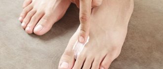
- after shaving the skin;
- leading a sedentary lifestyle (for example, when you stand on your feet or sit for a long time), which leads to poor circulation in the lower part of the body;
- wearing tight clothing, which causes excessive sweating;
- unbalanced diet, lacking vitamins and microelements.
Any of these factors can lead to a decrease in melanin synthesis, which inevitably causes hypopigmentation.
Do not immediately panic when you discover white spots on your legs and arms. The first thing you need to do is visit a dermatologist to rule out any serious diseases after the examination.
Only after finding out the cause can you begin treatment, which will be prescribed by a specialist.
Separately, it is worth highlighting information about the appearance of white spots on the legs and other parts of the body in children. In most cases, this is caused by the following diseases:
- Hypomelanosis . It makes itself felt in the first years of life after infectious diseases.
- Tumorous sclerosis . It is a signal of more serious problems in the body (epilepsy, mental retardation, damage to some internal organs).
Is it possible to cure white pigmentation on the skin and how?
If you notice the appearance of white spots on your legs after sunbathing, then you cannot diagnose yourself and start getting rid of them in all possible ways.
What it really is and how to treat it should be clarified exclusively by a dermatologist after an examination. Subsequent therapy will depend on the diagnosis.
Drug intervention
When a certain skin disease is diagnosed, the doctor usually prescribes the following measures:
- using ointments containing melatonin and taking it in tablet form;
- ultraviolet irradiation;
- laser stain leveling;
- hormone therapy;
- restoring the functioning of the endocrine system by taking appropriate medications.
If hypomelanosis is detected in children (very often seen in photos on the Internet), it often causes the appearance of white spots on the skin, then doctors usually prescribe drugs that inhibit the development of this disease. Epidermis peeling sessions also help.
Cosmetologist Christina Brecht
If the skin is damaged by a fungus, you can treat it yourself using antifungal ointments and sprays. They are sold at the pharmacy. Clotrimazole and Lamikon are considered the most effective.
Cosmetology procedures (3 options)
White spotting on the legs and arms can be dealt with by resorting to certain cosmetic procedures. But this is provided that this phenomenon is not caused by any skin diseases.
After diagnosis, a professional cosmetologist can offer the following methods to solve the problem:
- Superficial peeling involves the use of fruit acids, due to which dead cells are carefully removed from the epidermis. This method is effective for relatively fresh stains.
- Deep peeling – a stronger acid is used for this procedure, so it should not be done by people with particularly sensitive skin. After it, minor damage is possible, which will heal for about a week.
- Ozone therapy is used when more thorough cleansing of the skin from pollution and pathogenic microorganisms is required.
It is better not to carry out the listed procedures in the summer, since after them sun exposure to the dermis is undesirable.
Folk remedies
It is not possible to completely cure white spots on the legs and arms using folk remedies, but it is possible to make them less noticeable. Here are some productive recipes collected from reviews and forums:
- Squeeze the juice from one lemon and combine it with kombucha tincture (1:2). Soak a cotton swab in the resulting solution and treat white spots on the skin. Do this every 3-4 hours until the pigmentation becomes less noticeable.
- Another effective method: separate the white from a chicken egg, beat it with a mixer and gradually add lemon juice (2 tablespoons) to it. Then this mixture is heated until it thickens, remembering to stir constantly. After this, turn it off and add lavender oil. After cooling, the product is applied to problem areas. Wash off when it hardens.
- Mix natural honey, rice flour, sandalwood powder and turmeric (all components are taken in equal proportions). The result should be a composition of homogeneous consistency, which is distributed evenly over the stains.
- Grind sesame seeds in a coffee grinder and take 1 tsp of them orally. twice a day. After 2 weeks, the pigmentation will not be so pronounced.
If white spots appear after sunbathing, cosmetologists recommend wiping the skin with fresh cabbage leaves or ginger.
Proper care
Facial care, where there is also a high risk of white spots after tanning, involves using masks and creams with a whitening effect.
A few very effective recipes:
- Combine freshly squeezed lemon juice and hydrogen peroxide, taken in equal parts. Then a piece of gauze or cotton cloth is soaked in the solution and applied to the face. After a quarter of an hour, it is removed and washed with lukewarm water. This compress is done 2 times a week.
- Twist oatmeal flakes in a coffee grinder or blender and mix with ground coffee (take 1 tbsp of each ingredient). Add 10 ml of lemon or cranberry juice. This mixture is applied to problem areas and washed off with water after 10 minutes. After this, lubricate the skin with nourishing cream.
You can get rid of white pigment spots using pharmaceutical products. For example, the drug Achromin has a healing effect and quickly returns the skin to a healthy appearance. They treat stains every time they go outside and before going to bed.
Skin diseases in diabetes mellitus
Severe metabolic disorders underlying the pathogenesis of diabetes mellitus (DM) lead to changes in almost all organs and tissues of the body, including the skin. The etiology of skin lesions in diabetes is certainly associated with impaired carbohydrate metabolism and the accumulation of corresponding products of impaired metabolism, which leads to structural changes in the dermis, epidermis, follicles and sweat glands. In combination with diabetic polyneuropathy, micro- and macroangiopathies, impaired local and general immunity, this leads to the appearance of various types of rashes, age spots, ulcerations, as well as purulent-septic complications.
The skin of patients with diabetes undergoes peculiar general changes. In severe cases of the disease, it becomes rough to the touch, its turgor decreases, and significant peeling develops, especially on the scalp. Hair loses its shine. Calluses and cracks appear on the soles and palms. A pronounced yellowish coloration of the skin often develops. Nails become deformed and thicken due to subungual hyperkeratosis. Diffuse hair loss may be a symptom of poorly controlled diabetes.
Often, dermatological manifestations can act as “signal signs” of diabetes: itching of the skin, dry mucous membranes and skin, recurrent skin infections (candidiasis, pyoderma).
Currently, more than 30 types of dermatoses have been described, which either precede diabetes or develop against the background of a manifest disease. Conventionally, they can be divided into 3 groups:
- Primary - caused by diabetic angiopathy and metabolic disorders (diabetic dermatopathies, necrobiosis lipoidica, diabetic xanthomatosis, diabetic blisters, etc.).
- Secondary - fungal and bacterial infections.
- Dermatoses caused by drugs used in the treatment of diabetes (eczematous reactions, urticaria, toxicoderma, post-injection lipodystrophy).
As a rule, diabetic skin lesions have a long and persistent course with frequent exacerbations and are difficult to treat.
Diabetic dermatopathy. The most common lesion in diabetes is the appearance on the anterior surface of the legs of symmetrical reddish-brown papules with a diameter of 5–12 mm, which then turn into pigmented atrophic spots (more often detected in men with a long duration of diabetes). There are no subjective symptoms, the course is long, and they can disappear on their own within 1–2 years. Pathogenesis is associated with diabetic microangiopathy. There is no specific treatment for dermatopathy.
Diabetic bladder. Refers to rare skin lesions in diabetes. Blisters appear suddenly, without redness, on the fingers, toes, and feet. Dimensions vary from a few millimeters to several centimeters. The vesicular fluid is clear, sometimes hemorrhagic and always sterile. In most cases, blisters heal without scarring after 2–4 weeks of symptomatic treatment.
Rubeose. In childhood and adolescence, in patients with insulin-dependent diabetes, hyperemia in the form of a slight blush is observed on the skin of the forehead, cheeks (less often, chin), which is sometimes combined with thinning of the eyebrows.
Diabetic erythema. It occurs as ephemeral erythematous spots, which are observed mainly in men over 40 years of age who have had diabetes for a short time. These spots are characterized by large sizes, sharp boundaries, rounded outlines and a rich pink-red color. They are localized mainly on open skin - the face, neck, dorsum of the hand. Subjective sensations are either absent, or patients complain of a slight tingling sensation. The spots have a very short lifespan (2–3 days) and disappear spontaneously.
Acanthosis nigricans. It is characterized by villous hyperpigmented growths, mainly in the folds of the neck and armpit. Patients complain of “dirty skin” that cannot be washed. There are sometimes also small papules on the most prominent points of the finger joints. The pathogenesis is based on the liver's production of insulin-like growth factors, which interact with epidermal receptors and cause thickening of the epidermis and hyperkeratosis.
Diabetic xanthoma. It develops against the background of hyperlipidemia, with the main role played by an increase in triglycerides in the blood. Yellowish plaques are localized mainly on the flexor surfaces of the limbs, on the chest, face, neck and consist of an accumulation of triglycerides and histiocytes.
Necrobiosis lipoidica. A relatively rare chronic dermatosis characterized by focal disorganization and lipid degeneration of collagen.
Insulin-dependent diabetes is the most common cause of necrobiosis lipoidica and occurs in 1–4% of such patients. Skin manifestations may be the first - and for a long time the only - manifestation of diabetes. It is believed that in 18–20% of patients, necrobiosis lipoidica can occur 1–10 years before the development of typical symptoms of diabetes, in 25–32% of patients it develops simultaneously with this disease, but in the majority (55–60%) diabetes precedes skin lesions. There is no direct relationship between the severity of clinical manifestations of necrobiosis lipoidica and the severity of diabetes.
The disease can occur at any age, but more often it affects people between 15 and 40 years of age (mainly women). It occurs against the background of insulin-dependent diabetes and is characterized by large single lesions on the skin of the legs. The disease usually begins with the appearance of small bluish-pink spots or smooth flat nodules of rounded or irregular shape, prone to peripheral growth, followed by the formation of clearly demarcated, elongated oval or polycyclic indurative-atrophic plaques. Their central part (yellowish-brown) is slightly sunken, and the marginal part (bluish-red) is slightly raised. The plaques have a smooth surface, sometimes flaking along the periphery. Gradually, the central part of the plaques atrophies, telangiectasias, mild hyperpigmentation, and sometimes ulcerations appear on it. As a rule, there are no subjective sensations. Pain occurs with ulceration.
The appearance of the lesions is so characteristic that usually no additional research is required. In atypical forms, a differential diagnosis is made with granuloma annulare, sarcoidosis, and xanthomatosis.
There is currently no effective treatment. Drugs that normalize lipid metabolism are used (Lipostabil, Clofibrate, Benzaflavin); improving microcirculation (Curantil, Trental, Teonicol). Drugs such as Aevit, Dipromonium, Nicotinamide, Angiotrophin are indicated. Intralesional administration of corticosteroids, insulin, and Heparin is effective. Externally: applications of 25–30% Dimexide solution, application of Troxevasin, Heparin ointments, application of occlusive dressings with fluoride-containing corticosteroid ointments. Physiotherapy: phonophoresis of hydrocortisone, electrophoresis of Aevit, Trental. Laser therapy: for ulceration, surgical intervention is sometimes used (removal of lesions followed by skin grafting).
Itchy dermatoses (skin itch, neurodermatitis). They are often the first signs of diabetes. There is no direct relationship between the severity of diabetes and the intensity of itching. On the contrary: it has been noted that the most severe and persistent itching is observed in latent and mild forms of diabetes. In most patients, skin itching precedes the development of not only skin lesions in diabetes, but also the diagnosis itself (from 2 months to 7 years). Less commonly, itching develops against the background of established and treated diabetes.
The predominant localization is the folds of the abdomen, inguinal, intergluteal, and ulnar folds. The lesions are often unilateral.
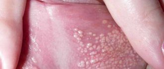
Fungal skin lesions. The most common candidiasis is usually caused by Candida albicans. It is more common in old age and in obese patients with predominant localization of lesions in the genital area and large folds of skin, interdigital folds, mucous membranes (vulvovaginitis, balanopastitis, angular cheilitis). Candidomycosis may play the role of a “signal symptom” of diabetes.
Candidiasis of any localization begins with severe and persistent itching, which is later accompanied by objective signs of the disease. First, a whitish strip of macerated stratum corneum appears deep in the fold, and surface cracks and erosions form. The surface of the erosions is moist, shiny, bluish-red in color, bordered by a white rim. Around the main focus, “dropouts” appear, represented by small superficial vesicles and pustules. Opening up, these elements turn into erosions, also prone to growth and fusion. The diagnosis is confirmed by microscopic or cultural examination.
For local treatment, time-tested, simple and affordable means are used - alcohol or aqueous (the latter is better for large folds) solutions of aniline dyes: methylene blue (2-3%), brilliant green (1%), as well as Castellani liquid, ointments and pastes , containing 10% boric acid. Almost any local antimycotic can be used in the form of 1–2% creams, ointments, and solutions. External agents are used until the skin lesions are completely resolved, and then for another week. Systemic antimycotics include fluconazole, itraconazole or ketoconazole. Fluconazole is prescribed at a dose of 150 mg/day once; in case of torpid course, 150 mg/day once a week for 2–3 weeks. Itraconazole is prescribed at 100 mg/day for 2 weeks or 400 mg/day for 7 days. Ketoconazole is prescribed 200 mg/day for 1–2 weeks. The advisability of prescribing systemic antimycotics is determined by the effectiveness, previous therapy, the motivation of the patient who wants to get rid of the manifestations of the disease as soon as possible, as well as the availability of drugs.
Infectious diseases. Bacterial skin lesions occur in patients with diabetes much more often than in the general population and are difficult to treat. Diabetic foot ulcers are the most serious complication and can lead to amputation and even death.
Pyoderma, boils, carbuncles, cellulitis, erysipelas, paronychia and panaritium are most often caused by staphylococcal and streptococcal flora. The addition of infectious and inflammatory skin diseases, as a rule, leads to severe and prolonged decompensation of diabetes and increases the body's need for insulin. The diagnosis must be confirmed by obtaining a culture to determine sensitivity to antibiotics. The patient is prescribed oral dicloxacillin or erythromycin (if allergic to penicillin). Taking dicloxacillin is the main method of treating outpatients, since 97% of microorganisms are sensitive to it. Non-festering lesions can also be treated by applying heat locally. When fluctuating, the boil must be opened and drained. Large abscesses sometimes require incision and drainage.
In conclusion, it should be noted that skin lesions in diabetes are common conditions today, quite often encountered in clinical practice. Their treatment has certain difficulties and should begin with effective control of blood sugar levels and development of an adequate regimen for taking antidiabetic drugs. Without correction of carbohydrate metabolism in this group of patients, all therapeutic measures are ineffective.
Literature
- S. G. Lykova, O. B. Nemchaninova. Skin lesions in diabetes mellitus (pathogenesis, pathomorphology, clinical picture, therapy). Novosibirsk: Novosibirsk Medical Institute. 1997. 44 p.
- A. S. Mashkilleyson, Yu. N. Perlamutrov. Skin changes in diabetes mellitus // Bulletin of Dermatology and Venereology. 1989. No. 5. pp. 29–31.
- A. Yu. Sergeev, Yu. V. Sergeev. Fungal infections. Guide for doctors. M., 2003.
- I. I. Dedov, V. V. Fadeev. Introduction to Diabetology: A Guide for Physicians. M., 1998. 404 p.
- M. I. Martynova, E. E. Petryaykina, V. F. Pilyutik. Features of skin disorders in insulin-dependent diabetes mellitus. "Attending doctor".
I. B. Mertsalova, Candidate of Medical Sciences RMAPO, Moscow
Tanning rules (recommendations)
What to do if, after a thorough medical examination, no dermatological problems are identified, but the tan still does not lie evenly.
It is recommended to change your approach to skin care and tanning sessions. Here are some useful tips:
- Avoid low-quality and cheap cosmetics. It is better to buy one cream or lotion, but such that it fully provides the dermis with all the necessary components. It is especially important that the composition contains vitamins (A, E) and minerals, as well as natural oils.
- Drink plenty of water. Dehydration negatively affects the skin, increasing its sensitivity to UV radiation. It is advisable to exclude pickles and smoked foods from your daily diet, which retain water in the body.
- Clean your skin regularly. Dead skin cells, clogged pores, sebum and other impurities cause uneven tanning. Therefore, take wet wipes to the beach and exfoliate more often.
- Follow basic hygiene rules. In the solarium, make sure that the cabin is pre-treated with disinfectants. During the session, use personal household items: mat, footprints, glasses. This way you can protect yourself from contracting any infection.
- Use special SPF creams with a protection index of more than 30. It is recommended to renew the cream layer several times a day and each time after leaving the water. Use such creams in urban areas with the onset of summer.
- Stick to a proper diet. If the skin is healthy, then the tan will lie evenly on it and the risk of pigmentation is minimal.
Do not sunbathe for more than 2 hours, especially during the sun's peak activity (from 11 a.m. to 4 p.m.). If the spots do not disappear, but, on the contrary, their number and size increase, then completely avoid sunbathing and visiting the solarium.
Dermatological surgeon Karin Litani