REMOVAL OF NEW PLANTS
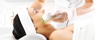
LASER REMOVAL OF NEW TUMORS
Duration of the procedure:
5-30 min.
Number of procedures:
1
Price:
from 100 rub.
Recovery period: 7-14 days
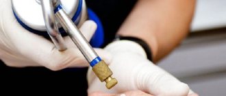
Equipment: CO2 laser.
Laser removal of tumors using a CO2 laser painlessly relieves the patient of benign formations on the skin. Most importantly, after healing there are no scars or scars left on the skin.
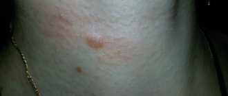
Types of skin neoplasms
Laser removal of skin tumors is effective for the following pathologies: atheroma, basal cell carcinoma, keratoma, botryomycomoma, cutaneous horn, warts, mole, fibroma, papilloma, condyloma, xanthelasma, molluscum contagiosum, nevus, etc.
Laser removal of tumors - Before / After Photos
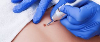
How is the procedure performed?
Laser removal of tumors on the face or body takes no more than 5-10 minutes. During the procedure, nearby tissues are not affected. Local anesthesia is used for patient comfort.
Before removing the tumor, the area is first treated with a disinfectant. After that, the pulse power and exposure time of the laser device are selected (depending on skin type). Then the evaporation process begins.
After removing the tumors, an antiseptic and a patch are applied to the treated area. Next, the doctor gives recommendations on caring for the wound.
Dermatoscopy is also performed before the procedure. If necessary, the tumor is sent for histological examination.
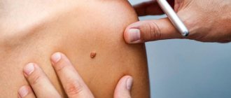
Ways to remove moles
Nevi are removed exclusively surgically. Removing moles using traditional methods is not only ineffective, but also extremely dangerous - thereby you can provoke tumor degeneration.
The main methods of removing moles (similar techniques are used to remove warts):
- Surgical removal.
- Cryodestruction.
- Electrocoagulation.
- Laser removal of moles.
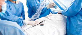
Let's figure out which method is better? The oldest method is surgical removal of moles and warts. The nevus is excised with a scalpel within the healthy skin, after which sutures are applied to the wound (optional, depending on the size of the formation), and the removed tissue is sent for histological examination.
Cryodestruction and electrocoagulation are similar techniques. In both cases, the nevus or wart is completely destroyed, only with cryodestruction, liquid nitrogen (cold) is used to remove the formation, and with electrocoagulation, electric current is used. The disadvantage of the methods is that the destroyed tissue cannot be sent for histological examination, which needs to be carried out.
Laser mole removal is the most modern technology. Instead of a scalpel, a laser beam is used to remove the formation. The nevus is not destroyed, which allows for histological diagnosis. The technique also has other advantages, which will be discussed in the next section.
Post-procedure care
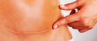
After removal of the tumor, a small wound remains, which heals within 1.5-3 weeks (depending on the size of the removed tumor).
During healing, a crust forms on the wound, then the crust falls off and a pink spot remains - new skin, which, depending on the area of the body, lasts from 3 weeks to six months. If the wound was small (up to 1 cm), then the place where the tumor was removed will be impossible to distinguish from areas of skin that are nearby. But if the wound was large, then a light spot may remain on the skin in the future, this is explained by the fact that there was not enough healing tissue and scar tissue formed.
After the laser procedure, it is not recommended for two weeks:
- prevent skin overheating or hypothermia;
- overexert yourself physically;
- go to the pool, solarium, sauna;
- contact with household chemicals;
- rub the wound with a towel;
- peel off the crust after excision;
- treat with alcohol.
The doctor will give recommendations for care and prescribe an antiseptic to treat the area where the tumor was removed.
What moles need to be removed?
Let's start with the fact that from a histological point of view, a mole, or nevus, is a neoplasm on the skin with a high content of melanin (pigment).
Moles are formed as a result of changes in hormonal balance, in particular, an increase in the secretion of melanotropic hormone and some other pituitary hormones. Although a mole should be considered a benign tumor, it does not pose any particular health risk. On the human body you can count dozens and even hundreds of small moles, and many nevi of medium and large size. The probability of malignancy (malignant degeneration) of a small mole is close to zero, but in the case of medium or large nevi, the risk increases, so caution should be exercised.
When is mole removal necessary? Everything is quite simple here. There is a system for assessing the degree of risk of malignant degeneration, which in the English-language literature is called ABCDE, and in the Russian-language literature - AKORD. The criteria for this evaluation system are as follows:
- Asymmetry . Harmless moles are symmetrical. (Perhaps they are not absolutely symmetrical, but they can be called as such.) Severe asymmetry of the nevus is a risk factor and an indication for mole removal with a laser or other method.
- Edge . A safe neoplasm has smooth edges. If the edge of the nevus becomes jagged, you need to consult a dermatologist or dermato-oncologist.
- Coloring . Benign nevi are evenly colored. Dangerous moles have inclusions, areas of darker or lighter color. Uneven coloring is another alarming symptom.
- Size . If the diameter of the mole does not exceed 6 cm, the risk of degeneration is minimal. Large nevi (7 cm or more) degenerate more often, and therefore it is better to think about removing the mole before this happens.
- Dynamics. A very simple criterion: if the appearance of the nevus begins to change, it is better to remove it. Removal of moles is indicated if a new growth on the skin begins to enlarge or bleed, or its density, texture, surface, border or color have changed.
Laser removal of moles is also carried out at the request of the patient, that is, for aesthetic reasons. In such a situation, it is enough to make sure that the neoplasm is benign. (Malignant tumors are also removed, but according to different protocols.)
Benefits of the procedure
Laser removal of skin tumors is considered the safest and most effective method. The positive qualities of the technique include:
- safety;
- efficiency;
- can be done at any time of the year (however, if you have returned from a hot vacation, cosmetologists recommend resting the skin for several days before the removal procedure);
- no bleeding;
- absence of pain (since it is done with anesthesia);
- sterility;
- exclusion of relapses;
- does not leave scars or scars;
- short rehabilitation period.
Relapse after removal of skin basalioma WILL BE CURED without surgery!
Afanasyev Maxim Stanislavovich.
Doctor of Medical Sciences, Professor of Sechenov University, oncologist, surgeon, oncogynecologist, gynecologist-immunologist, expert in the treatment of recurrent basal cell carcinoma.
"I am 51 years old. 4 years ago, a basal cell carcinoma was removed with a laser on my face. A year ago she appeared again, in the same place. I really don’t want to cut it out again, the scar is very noticeable, on the cheek near the nose. The size is about 1 cm. I work every day, constantly communicate with people, I need to remove it, preferably in the least traumatic way.”
Patients with repeated basal cell carcinomas after their removal are increasingly turning to me. Indeed, this is a huge problem that is becoming more acute year after year.
In addition to the fact that the incidence of basal cell carcinoma is increasing, most often this tumor occurs on the face, neck or head, where repeated use of any radical treatment method is associated with the loss of an organ or part of it. For this reason, treating relapse with traditional methods is often simply impossible.
The eyelash growth area is one of the most difficult. Recurrence of basal cell carcinoma in this area will raise very serious questions for the patient and his surgeon. Source: www.tabanmd.com
In my practice, I use photodynamic therapy to treat relapses after radiation or relapses after surgery.
It specifically eliminates altered cancer cells and does not affect healthy tissue. Even against the background of a significant tissue deficit in the relapse area, this allows for almost non-invasive and maximally aesthetic interventions.
In this article I will analyze the causes of relapses. I will give statistics on the effectiveness of different treatment methods and tell you how to save the situation if basal cell carcinoma develops again.
Basalioma of the chin after excision and healing. Source: metro.co.uk
Risks of recurrence of basal cell carcinoma - what is the likelihood of encountering it again after removal?
Cutaneous basal cell carcinoma, or cutaneous basal cell carcinoma, or BCCC, is a skin cancer. It extremely rarely metastasizes, but has a high tendency to relapse.
According to various sources, the incidence of relapses within 3-12 months after treatment is 21%. And over a period of 10 years, the tumor recurs in 40-46% of cases.
Unfortunately, when deciding to remove basal cell carcinoma (and doing the right thing), few doctors and patients plan intervention tactics taking into account possible relapse.
When agreeing to one or another method of treating basal cell carcinoma, you need to take into account one fact that is well known to doctors: any treatment is a provoking factor. The therapeutic effect on the tumor itself stimulates its activity and growth if the intervention is not radical enough.
This approach leads to the fact that treatment of recurrent basal cell carcinoma in the future either turns out to be impossible, or is associated with crippling and disfiguring loss of part of the nose, cheek, eye, ear... And such cases of long-term ineffective treatment of basal cell carcinoma are not uncommon. See for yourself.
The photo was taken immediately after removal of the basal cell carcinoma and after healing. Source: todddunn.files.wordpress.com
Therefore, when deciding on the choice of treatment method for basal cell carcinoma, it makes sense to consider several factors:
- The method guarantees a high degree of radicality of treatment - it allows you to completely remove all tumor cells, including the “root” of basal cell carcinoma.
- Preserves the integrity of the organ - eye, nose, ear, lips, etc.
- Provides minimal cosmetic defect (for example, all surgical methods require excision of an additional 2-5 mm of tissue from the tumor border).
- There are no long-term complications from using the method itself (it is believed that several years after radiation therapy, irradiated cells sometimes degenerate into cancer cells).
These are the properties that the photodynamic therapy (PDT) method has. It allows you to specifically eliminate all tumor cells without affecting healthy tissue, and in the vast majority of cases, obtain a relapse-free result in one procedure.
When treating a relapse, PDT does not require the removal of healthy tissue and preserves the integrity of the organ.
Risk factors for relapse
The high frequency of recurrence is associated with the characteristics of the tumor - basal cell carcinoma does not have a capsule, its real boundaries exceed the visible ones, and it is very difficult to determine the depth of invasion.
Here is a far from complete list of factors for relapse of basal cell carcinoma:
- Tumor shape. The nodular-ulcerative form of BCC recurs more often.
- The depth of tumor “growth” into the tissue. The surgeon sees only the affected area on the skin, but does not understand how deep the tumor goes. Tumors with deep invasion recur more often. Tumors with intradermal spread have the best prognosis.
- Tumor size. Relapses more often develop after treatment of tumors larger than 2 cm, so you should not “grow” moles on your face. In this case, basalioma cells can be detected at a distance of up to several centimeters from the lesion on healthy skin.
- Age and health conditions. Concomitant diseases can reduce immunity, which aggravates the course of the malignant process.
- “Inconvenient” location of basal cell carcinoma in anatomical folds and in close proximity to vital organs. These are the nose, nasolabial folds and ears. The tumor recurs slightly less often in the area around the eyes and in the eyelash growth area. Complete radical removal in these areas is simply impossible due to the risk of organ dysfunction or facial disfigurement.
- Different areas of the face have significantly different thicknesses of the skin and subcutaneous fat, which also affects the nature of the tumor and has a great influence on the choice of treatment method.
- Inadequate choice of treatment method or repeated treatment with different methods. This is the most important risk factor.
Many different methods are used to treat basal cell carcinomas. Unfortunately, none of them guarantee a 100% cure. Below I present a forecast for the development of relapse within 5 years after treatment, drawn from various sources:
- Close-focus X-ray irradiation (close-focus X-ray therapy). Recurrence of basal cell carcinoma after irradiation occurs in 46% of cases.
- PDT produces relapse in 6% of cases.
- Surgical excision. Relapse after removal of basal cell carcinoma develops in 30% of cases.
- Cryotherapy. The inability to control the depth of impact causes the largest number of relapses – 35%.
- Laser coagulation – 31%.
- Micrographic surgery according to Mohs. This is a relatively new method, which is still poorly represented in Russia, so it is still difficult to show the frequency of relapses.
- After previous combination treatment, repeated relapses occur in 47% of cases.
According to research results, vaporization, which is popular today (a method of treating basal cell carcinoma with a laser), turns out to be one of the least effective methods. According to this indicator, it is approaching cryodestruction.
The most effective method is still surgical excision of basal cell carcinoma. But, as we see, the effectiveness of the procedure is accompanied by a noticeable loss of healthy tissue.
What are the dangers of recurrent basal cell carcinoma on the face and head?
Recurrence of basalioma on the nose, as well as recurrence of basalioma on the lip, is fraught with the need for mutilating surgery. Since any surgical excision is carried out with the obligatory capture of 6 mm of healthy tissue, repeated removal of basal cell carcinoma on the nose usually ends with the removal of part of the nose and requires complex plastic surgery of the nose with tissues from other parts of the body.
Scope of surgery for nasal basal cell carcinoma. Source: www.ohniskin.com
The location of basal cell carcinoma in the corners of the eye is generally a contraindication to surgery, since in most cases it ends in the loss of the organ.
Source: www.tabanmd.com
Surgical excision of basal cell carcinoma on the forehead and scalp is complicated by the lack of subcutaneous fat in these areas. Excision of basal cell carcinoma involving healthy tissue leads to the impossibility of suturing the wound and requires the application of a skin flap from the patient's thigh.
Close focus X-ray therapy (FRT) cannot be performed on the ear. This leads to perichondritis - painful suppuration and swelling of the ear. The same procedure cannot be performed on the upper and lower eyelids, or exclusively using a special ocular prosthesis. Many patients report decreased vision after BPRT.
The bottom line, as we see, is that repeated treatment is often simply impossible.
Signs of relapse
80% of relapses occur between 3 months and 1 year after treatment. The remaining 20% - within 5-10 years. Therefore, recovery can be judged no earlier than 5 years after the procedure.
A relapse appears in the same place where the primary basal cell carcinoma was removed. Thus, recurrence of basal cell carcinoma after excision occurs directly in the scar.
What symptoms suggest a relapse? Repeated basal cell carcinoma looks like a non-healing wound or a new formation similar to the previously removed one.
If the formation appears on a new area of skin, it is a new basal cell carcinoma, which is not considered a relapse.
Is relapse treatable?
In the vast majority of cases, basal cell carcinoma is completely curable. The PDT procedure allows me to provide aesthetic and disease-free treatment of basal cell carcinoma in one procedure.
Treatment of recurrent basal cell carcinoma using PDT is performed with tissue preservation and has almost no risk of relapse.
I treat recurrent basal cell carcinoma using PDT. Based on my experience, I can say that basal cell carcinoma can be treated, and is treated extremely successfully.
I strongly recommend that all my patients always choose PDT as a priority treatment method. And that's why:
- This is a gentle, non-surgical and effective organ-preserving method of treating relapses.
- Selectively destroys the tumor while preserving healthy tissue.
- Does not require hospitalization.
- Well tolerated even by older people.
- There are practically no contraindications for people with concomitant diseases, for whom traditional treatment is associated with the development of complications.
- PDT is a non-toxic method: the photosensitizer does not cause significant changes in blood parameters.
- Has no serious complications.
- Can be used multiple times.
- Perhaps the only method of treating patients with a frequently recurrent form of the disease.
- One of the few methods for treating hard-to-reach neoplasms and tumors located in close proximity to vital organs.
- Allowed if there are contraindications to other methods.
- The author's development makes it possible to treat even complex localization of basal cell carcinoma in the eyes without the risk of weakened vision.
- Low risk of relapse.
- In the vast majority of cases, one PDT session is sufficient to eliminate the tumor.
Of course, it is better to use photodynamic therapy as the first method of treating basal cell carcinoma, but even for the recurrent form, my author’s protocols show very good results. Even if the blood flow in the scar is impaired, photodynamic reactions have a reserve in terms of the depth of impact: tumors with a scar of up to several millimeters will still fall under photochemical reactions indirectly through ischemic necrosis around the scar.
To get advice on whether photodynamic therapy is indicated for you, and to calculate the price of the procedure, send your medical history and tests by e-mail or call by phone in Moscow.
The reception is conducted by Afanasyev Maxim Stanislavovich - oncologist, immunologist, oncogynecologist, doctor of medical sciences, professor and member of the academic council of the First Moscow State Medical University named after. THEM. Sechenov Ministry of Health of the Russian Federation, expert in the treatment of basal cell carcinoma.
Reception is carried out in two clinics in Moscow, as well as in St. Petersburg, Makhachkala, Kursk, Stavropol, Barnaul, Samara, Naberezhnye Chelny, Salavat, Chelyabinsk, Surgut and other regions of Russia. You can check the date, location of the appointment in your city and sign up for a consultation with the administrator by calling 8-800-555-77-26.
After treatment, I maintain feedback with all patients and resolve any issues that arise. Hepatitis and positive HIV status are not contraindications for treatment with PDT.
References:
1. Chuprov I.N. Features of recurrence of basal cell skin cancer // Materials of the All-Russian conference with international participation: 100th anniversary of the Russian Society of Pathologists. St. Petersburg, 2009. pp. 345-347.
2. Panova I.E. Recurrence of basal cell cancer of the skin of the eyelids // Breast Cancer. Appendix Clinical ophthalmology. 2006. T. 7, No. 1. P.11-14.
3. Savelyeva A.E. Risk factors for the development of relapses of basal cell skin cancer: abstract. dis. ...cand. honey. Sci. M., 2004. 24 p.
4. Kurdina M.I., Tymchishina M.V. Detection of malignant skin tumors during clinical examination // Abstracts of the 1st Congress of Oncologists. St. Petersburg; M., 1996. Part II. P. 402.
5. Panova I.E., Usova R.A., Semenova L.E. and others. Treatment tactics for various clinical forms of basal cell carcinoma of periocular localization // Standardization in Oncology. Chelyabinsk, 2002. pp. 44–46.
6. Panova I.E. Recurrence of basal cell cancer of the skin of the eyelids // Breast Cancer. Appendix Clinical ophthalmology. 2006. T. 7, No. 1. P.11–14.
7. Chuprov I.N. Features of recurrence of basal cell skin cancer // Materials of the All-Russian conference with international participation: 100th anniversary of the Russian Society of Pathologists. St. Petersburg, 2009, pp. 345–347.
8. Matveeva O.V. Results of photodynamic therapy of basal cell skin cancer with local use of the photosensitizer Radachlorin // Radiation and risk. 2016. T. 25, No. 2. P.79-90.
Contraindications
Laser therapy is not allowed if:
- malignant neoplasms;
- allergies to ultraviolet radiation;
- high skin sensitivity;
- weakened immune system;
- skin inflammation;
- elevated sugar levels;
- epilepsy;
- elevated body temperature;
- also during pregnancy and lactation.
The procedure is non-traumatic, with minimal side effects.
Medical ]SkinLazerMed[/anchor] employs doctors with extensive experience in removing any tumors. On the day of your visit before the procedure, you will receive a free consultation with a specialist! You can make an appointment by calling: 317-78-21. You can also leave a request on our website skinlazermed.ru and our administrators will contact you! Take care of your health with SkinLazerMed!
Reviews
Alina K.
I had a birthmark right under my eyebrow. I thought it was a monstrosity and dreamed of getting rid of it. True, for a long time I didn’t have the courage to go to the doctor. First I saw a dermatologist. He examined me for oncology and reassured me that nothing bad would happen. I made an appointment for surgery. They gave me several injections around the spot and it was removed in 10 minutes. The doctor gave me a bottle of medicine and told me to wipe the sore every day. Morning and evening I wet the bandage and treated the skin. The brown crust fell off after 6 days. Everything was clean under the eyebrow, no marks remained. It should have been done a long time ago.
Irina S.
Our entire family has growths all over our bodies. I have experience with cutting using different methods, but laser turned out to be the most successful. I've already removed about twelve of them. The last time there were two - hanging on the shoulder and on the stomach near the zipper of the jeans. These must be cut off to avoid scratching. They can turn into cancer. True, you need a lot of money if you need to bring together several. Histology is normal. I spent about half an hour in the office while they injected me and then cauterized me. The one on the stomach left a hole. The doctor said that the roots were deep and they were completely cut out. This means it won't grow again. At home I wiped it with peroxide and did bandages every day. I covered my shoulder from the sun for another 2 months. Everything healed completely within a month. There are no scars left. Cool procedure, I recommend it to everyone.