To remove papillomas in modern medicine, several methods of physical influence are successfully used, including traditional surgery, radio wave method, electrocoagulation, etc. Regardless of the chosen method, proper care after removal of a papilloma, especially if the appendage was large, will help avoid complications and the occurrence of cosmetic defects ( scars) and relapse.
Medical clients can receive multi-level professional assistance - from diagnosis to removal of pathological tumors and consultations on rehabilitation. Our specialists are ready to receive you in Moscow, on Kashirskaya metro station, and in Ramenskoye, on st. Chugunova, 21-A.
Possibility of unforeseen consequences
Laser correction of moles is one of the safest ways to get rid of nevus, avoiding complications and unforeseen consequences. A beam of directed light radiation literally evaporates the pigmented cells of moles, which is visually manifested by the complete removal of the epidermal formation in just a few minutes. Minimization of negative consequences occurs due to the following objective factors.
- The laser beam acts in a strictly targeted and selective manner, without injuring healthy skin structures, which allows the technique to be used even to remove the smallest nevi in the eyelid area and mucous membranes;
- There is virtually no risk of infection, since the laser exhibits a powerful antibacterial and antimicrobial effect, protecting the wound surface from infection;
- Laser destruction is not accompanied by bleeding or hemorrhage, since during the manipulation process all blood capillaries supplying the body of the mole undergo thermal coagulation;
- The laser activates regenerative processes in the skin, which significantly accelerates wound healing;
- The technique is low-traumatic, which avoids the formation of connective tissue and all the negative consequences associated with this factor.
Despite the safety of the method, it should be borne in mind that before the procedure it is necessary to carefully study the list of contraindications, since the consequences and complications of laser correction most often result from neglect of this list, which includes the following conditions:
- Pregnancy;
- Acute infectious process;
- Oncology of any localization;
- Inflammation in the intended area of laser exposure;
- Period of menstruation in women;
- Immunodeficiency;
- Exacerbation of chronic pathologies;
- Hyperthermia.
Removal of papillomas using an electrocoagulator
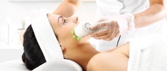
The effect of smooth, clean skin without papillomas can be achieved in one session using electrocoagulation. This is a procedure for removing superficial formations on the skin using cauterization with electric current. The treatment is carried out using an electrocoagulator - a medical device that generates current.
The procedure is available at the Clinic of Dr. Samoilovskaya near the metro station “Chkalovskaya” (or “Petrogradskaya”) on Petrozavodskaya street, 13 in St. Petersburg.
Features of the procedure
Electrocoagulation is a modern promising technique that allows you to eliminate tumors from the skin. The procedure is carried out in specialized medical institutions. During the session, papillomas are quickly removed due to the effect of direct or alternating current on the affected area. Setting up the device and choosing a nozzle depend on the degree of localization and number of tumors.
During the treatment process, intense heating of tissue occurs in a certain area. Local exposure to high temperature leads to coagulation of proteins - they coagulate, the liquid evaporates, and the tissues that form the papilloma burn. Drying the wound under the influence of current prevents infection and eliminates bleeding. In most cases, one procedure is sufficient to eliminate tumors, even if there are several papillomas.
The duration of the procedure depends on the number of formations. Timely removal of papillomas helps prevent their degeneration into malignant ones and improves the aesthetics of appearance.
For your information! In addition to aesthetic medicine, electrocoagulation is used in gynecology, proctology, ENT practice, surgery and other areas.
Indications and contraindications
Papillomas are formations on the surface of the skin that cause physical and psychological discomfort. The cause of their appearance is the human papillomavirus. Elimination of formations using electrocoagulation is carried out by a doctor after examining the patient. Indications for the procedure are the presence of papillomas on open and closed areas of the body, including the face, décolleté and neck.
But electrocoagulation is not possible in all cases. The following conditions are contraindications:
- chronic diseases in the acute stage;
- herpes rash;
- bleeding disorders;
- photodermatosis;
- presence of a pacemaker;
- serious cardiovascular disorders;
- oncological diseases;
- unhealed skin lesions in the treatment area;
- infectious diseases (including respiratory);
- mental disorders;
- pregnancy;
- serious endocrine diseases.
For your information! Removal of papillomas can be performed not only in adults, but also in children.
Current issues
Most patients, after undergoing a laser mole removal procedure, observe some characteristic reactions of the body during the first two weeks, some of which are normal responses that are compensatory in nature. Let's consider several pressing issues regarding the consequences of removing nevi on the face and body.
Can cancer occur after mole removal? Skin cancer, or melanoma, usually develops as a consequence of the malignant transformation of a melanoma-prone mole. Moreover, if during the laser correction process the specialist incorrectly determined the required depth of the beam and did not completely remove the mole, the risk of developing melanoma increases.
After removal, a tubercle appeared. Despite the low-invasiveness of the technique, in the process of removing the nevus, healthy surrounding tissue remains, which will have to undergo the healing process of the wound surface that has formed in place of the mole. In this case, this zone can form a small tubercle, which will be covered with a dense crust in a few days. This is considered a normal reaction to manipulation.
Inflammation after removal of a birthmark. The inflammatory process after laser correction usually lasts for the first days, after which it goes away. If inflammatory signs persist for a long time, you must immediately contact the specialist who performed the procedure.
Can a scar or scar form after removal of a nevus? The formation of scars can only be observed in the case of forced premature removal of the protective crust from the wound surface.
The scar itches and hurts after mole removal. Itching, burning, peeling and redness around a mole after removal are characteristic symptoms of a burn, which can be caused by unprofessional work of a doctor. This unpleasant complication develops if the specialist incorrectly determined the depth of exposure and subjected healthy skin cells to laser burning.
We care about your health
If you want to remove a mole quickly and without pain, contact the Medkvadrat clinic. The procedures are performed by qualified surgeons and dermatologists. In their work, specialists use effective methods: laser and radio wave surgery, electrocoagulation. Modern equipment allows treatment of adults and small children.
Moles are benign skin growths that occur in people of any age with different skin types. They form on all parts of the body: on the face, neck, arms and legs. They can be convex and flat, vascular and pigmented.
The most common types of moles are:
- Pigmented nevi are clusters of melanocyte cells that form the pigment “melanin”. They have the form of a spot, nodule or plaque. Their size and number largely depend on heredity.
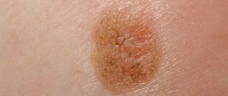
- Papillomas are thread-like or cone-shaped warts on a thin stalk. Their appearance is associated with carriage of the HPV virus, which is most often transmitted by contact. The virus lies dormant in the body and manifests itself when immunity decreases.
- Warts – usually look like flat, “flattened” flesh-colored plaques. May be single or multiple. The cause of warts is the HPV virus.
- A keratoma is a flat, brown plaque with a rough surface that can crumble and crack.
- Atheroma or wen is a flesh-colored formation that protrudes above the surface of the skin in the form of a hemisphere. Formed when the excretory duct of the sebaceous gland is blocked.
- Vascular moles (hemangiomas) are red spots or nodules of varying intensity. The cause of its appearance is vascular pathology.
If the formations on the skin are single, small in size and do not bother you, there is no need to touch them. However, there are cases when surgical treatment cannot be avoided.
When is mole removal performed: papillomas, nevi, warts?
It is necessary to get rid of formations if:
- The bulging formation is bothering you. You constantly injure him with accessories, clothes or a razor. In this case, a mini-operation is required, since against the background of damage, the nevus cells degenerate and become malignant.
- A mole spoils the appearance. Modern hardware surgery allows you to completely get rid of growths of any location: face, neck, eyelid skin. You will not feel pain, and not even a pigment spot will remain at the site of formation.
What methods are used to remove moles and papillomas?
The most popular methods:
- cryodestruction (cauterization with liquid nitrogen);
- mechanical removal with a scalpel or tweezers;
- electrocoagulation (exposure to electric current);
- laser treatment;
- radio wave surgery (radio knife).
The most progressive and gentle methods are laser and radioknife treatment. The procedures are well tolerated, so in 1 session you can get rid of several formations at once.
At the Medkvadrat clinic, doctors use both methods. Each of them has its own characteristics and advantages.
Laser mole removal
The doctor acts on the formation with a powerful laser pulse for several seconds. The thermal energy that is generated in the skin leads to the gradual destruction of pigment or vascular formation. The doctor acts on the mole locally: damage to neighboring areas of the skin does not occur.
Advantages:
- Safety. The laser “seals” small vessels, so no blood is released during the procedure and there is no risk of infection.
- Minimal pain. The area of influence of the laser beam is so small, and the effect occurs so quickly that the person does not experience significant pain.
- Good cosmetic effect. Laser treatment is performed on the face, neck and other open areas of the body. Since no open wound is created, the skin heals very quickly and without scarring.
Radio wave surgery.
The radio wave method involves local exposure of living tissue to high frequency electric current (100-300 Hz). Under the influence of radio waves, cells literally evaporate from the surface of the skin.
The device that uses the radio wave treatment method is called “Surgitron”.
During the procedure, the doctor cuts off the mole with a non-contact scalpel. As with laser exposure, the mini-operation is bloodless, since the radioknife “solders” the walls of the capillaries.
Advantages:
- Relative painlessness.
- Minimum exposure time.
- Fast healing.
- There is no risk of complications (if performed correctly).
- It is possible to conduct a histological examination to confirm the benignity of the formation.
Removal of moles and papillomas in “Medkvadrat”: in questions and answers.
Which doctors perform this procedure?
Only qualified specialists, surgeons, dermatologists and cosmetologists treat benign skin tumors in our center.
At the clinic on Kashirskoye Highway, appointments are provided by:
Sheptiy O.V. – surgeon, dermatologist, candidate of medical sciences. Specializes in the treatment of adults and children with vascular pathology: port-wine stains, hemangiomas. Work experience – 28 years.
Epikhin N.V. – dermatologist, laser surgeon. Specializes in laser treatment of vascular disorders: telangiectasia, hemangiomas, port-wine stains. Medical experience – 18 years.
Tamazova L. A. – dermatovenerologist, cosmetologist, candidate of medical sciences. Performs tumor removal using laser, surgitron, and liquid nitrogen. Medical experience – 15 years.
Melnikova Yu. G. – dermatovenerologist, cosmetologist, candidate of medical sciences. Diagnosis of skin tumors using a dermatoscope. Removal of moles (nevi, papillomas, warts, keratomas) using cryodestruction methods, electrocoagulation, and radioknife.
At medical centers in Kurkino, receptions are provided by:
Russkov S. Yu. – dermatologist, doctor of the highest category. Diagnosis and laser surgical treatment of warts, papillomas, keratomas, basal cell carcinomas, kandilomas, keratomas, hemangiomas, nevi. Medical experience – 29 years.
Markova E. A. – dermatovenerologist, cosmetologist, candidate of medical sciences. Removes warts, papillomas and other benign skin formations. Total experience – 17 years.
Can the patient choose his own treatment method?
The method of exposure - laser, radio knife, mechanical removal - is determined by the doctor. The method of treatment depends on the type of formation. Thus, laser treatment is used for flat pigmented moles and vascular spots. Convex nevi and flesh-colored papillomas can be easily removed using the radio wave method.
If a surgeon or cosmetologist has doubts about the benign quality of the formation, before the intervention, the specialist refers the patient to a consultation with an oncologist or performs a dermatoscopy.
What device is used in the clinic for laser treatment?
Doctors at our clinic work with a modern VbeamPlatinum laser device. It is intended for the correction of vascular changes and pigmented formations of the skin, warts, post-acne spots, scars and scars.
VbeamPlatinum differs from similar devices in the increased length of the laser pulse. For this reason, treatment is faster, and the doctor has the opportunity to influence formations located deep in the skin.
The device is equipped with a built-in dynamic cooling system, so you feel virtually no pain during mole removal .
Does the center perform histological examination?
Yes, histological examination is carried out. The biomaterial is sent to the laboratory immediately after treatment. There is no need to return for the results: when the analysis is ready, the conclusion will be sent to you by email.
Does the doctor provide pain relief?
Laser and radio wave treatments are usually well tolerated without pain relief. If the patient has a low sensitivity threshold, the doctor uses an anesthetic: gives an injection, applies a cream or spray.
Our specialists perform removal of large formations and formations of a large area under general anesthesia. The clinic has comfortable rooms where the patient can recover after the procedure.
Young children undergo mild sedation (short anesthesia).
What benign skin lesions are removed at the center?
Our doctors treat any benign skin tumors: warts, nevi, keratomas, atheromas, hemangiomas, papillomas, etc.
Make an appointment with a specialist right now! Contact phone number.
Care after laser mole removal
If the laser ablation procedure was performed by a qualified specialist, the risk of side effects is minimal, but the patient will also have to follow certain post-procedure rules, listed below.
- It is necessary to provide rest to the wound surface, without injuring it by friction of clothing or shoes, and also by taking care of the protective crust;
- Contact of the wound with water should be avoided for two weeks after manipulation;
- It is recommended to protect the skin surface from direct sunlight, and also use creams with a high UV protection factor. To the question of when you can sunbathe after removing a mole, the answer will be at least three weeks;
- You cannot use ointments, creams and gels for healing on your own without a doctor’s prescription;
- The procedure area should be protected from any mechanical damage, carefully protecting the integrity of the protective scab.
Laser removal is one of the simplest and least traumatic methods of mole destruction. Compliance with simple rules of rehabilitation will allow you to avoid scars, cicatrices and other unpleasant consequences and obtain an ideal physiological and aesthetic result.
Is a scar a reason to go to the doctor?
Reasons for visiting a doctor:
- aesthetic dissatisfaction;
- itching and pain in the scar area;
- the formation of deformities and contractures, which can lead to disability.
If scar tissue begins to develop pathologically and causes a keloid scar, it is better to consult a doctor.
There are factors that increase the risk of developing a scar:
- genetic predisposition;
- race (black patients are more likely to develop keloids);
- localization of injury (when removing tumors in the chest, neck and shoulders, the likelihood of keloid scars is higher);
- age (the formation of keloid scars is typical for young people). [3]
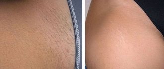
Before and after. How we remove scars
Characteristic
Spider veins are dilations of the capillaries of blood vessels. On the face they appear in the area of the nose and cheeks. Spider veins are also called telangiectasia.
According to studies, more than 90% of the world's population are “owners” of telangiectasia, which, in turn, combines all types of vascular dilatation on the human body.
The name vascular network appeared due to the fact that dilated capillaries become quite noticeable on the body and form the appearance of a mesh. It is worth noting that such expansion of capillaries does not form without reason. This disease mainly affects women in labor and young mothers; telangiectasia can also be a consequence of a disease such as venous insufficiency.
In the course of the studies, it was revealed that the appearance of the vascular network is not affected by any individual factors or diseases. It has become known that those who are susceptible to chronic varicose veins may not have vascular networks on their body. Thus, we can conclude that the vascular network is not the cause or consequence of any individual diseases, it is a separate problem of the body in each specific case and is often caused by hormonal imbalance.
What research is done before removing skin tumors?
The main optical device used by a dermatologist to examine moles and other skin growths before their removal is a dermatoscope. A dermatoscope consists of a magnifying lens and a light source to improve the visibility of subsurface structures of the skin.
The examination procedure using a dermatoscope is called dermatoscopy. This diagnostic method allows an experienced specialist to visually assess and diagnose the nature of a skin tumor under tens of times magnification.
Removal of nevi in Israel
Traditionally, in Israel the safest and most effective method of mole removal is used - surgically, using a scalpel. The operation is most often performed under local anesthesia and does not require hospitalization of the patient. The cosmetic skin defect that remains after surgery is practically no different from similar marks after cryodestruction or laser removal of moles. The main advantage of this approach is that as a result of the operation, the doctor receives a tissue sample of the mole, which is then subject to histological testing. When using other methods, the substrate of the mole is “burned out”, and it is not possible to establish its origin, as well as prevent the development of possible consequences.
In Israeli clinics, they approach the removal of nevi very responsibly. An indispensable condition for the operation is subsequent histological examination of the material. Thus, even with the slightest pathological changes in the nevus, timely measures can be taken and the development of possible complications can be prevented.
In Israel there are quite a few specialized departments for the treatment of melanoma. In this regard, the prevention of this disease is given special importance. Careful monitoring of moles, diagnosis of suspicious nevi and timely removal of pathological pigment spots is the priority of Israeli specialists. Modern international protocols are used for treatment.
Mole removal in Israel is carried out in most cases on an outpatient basis. For nevi of atypical localization, hospitalization may sometimes be necessary. For example, removal of an eye nevus is classified as a complex case, and performing this operation requires a surgeon with extensive experience and high qualifications.
The advantage of treating nevi in Israel, in addition to the use of modern surgical techniques, is the highly accurate pathohistological diagnosis of the material. The study of a removed nevus is carried out using many parameters, due to which the likelihood of a diagnostic error is practically reduced to zero.
In case of diagnostically difficult and doubtful cases, in order to avoid errors, the pathohistological sample is additionally sent for examination to another pathologist.
Advantages of electrocoagulation:
- The manipulation is carried out locally, in the area of formation without traumatizing the surrounding healthy skin.
- Speed of manipulation.
- The possibility of conducting a histological examination of the formation to exclude skin cancer.
- Safety and minimal risk of complications (simultaneous cauterization (soldering) of blood vessels, exclusion of bleeding and infection).
- Relative painlessness, because Painkillers are used during manipulation.
- Rapid regeneration of the skin (7 – 14 days).
- A wide range of uses of this method (for removing skin formations: warts, condylomas, papillomas, seborrheic keratomas, nevi, soft dermatofibromas).
- One of the inexpensive methods for removing skin lesions.
Preparation for the procedure
It doesn’t matter for what reason it is necessary to remove moles with an electric knife, you must undergo an examination before the procedure.
Mandatory indications for excision are:
- location of the nevus in a place of constant friction and pressure;
- increase in size;
- change in shape, clarity of edges;
- bleeding, inflammation, pain.
It is impossible to determine by eye whether a benign growth is present or whether cancer cells are present. It is necessary to conduct a histological examination. The oncologist gives the go-ahead for removal and saves the excised tissue for further medical procedures.
Do not go to beauty salons for the procedure! The cosmetologist does not have the necessary education and after unprofessional intervention melanoma may develop!
The preparation process takes a few minutes:
- local anesthesia is applied using ointment or gel. It will protect the area exposed to current from overheating and prevent burns to healthy tissue;
- the patient is placed on the couch in a comfortable position, allowing access to the nevus.
What if a scar appears?
Once scar tissue has formed, unfortunately, it will not be possible to completely get rid of it. All medical actions in this case will be aimed at minimizing the manifestations of the scar. The following methods are used to treat formed keloids:
- GCS injections are a popular and effective method for treating keloid and hypertrophic scars. A GCS solution is injected into the scar tissue 1–2 times a month until the expected results appear. Unfortunately, with excessive use of the drug, skin atrophy may develop at the injection site, telangiectasia or an atrophic scar may appear. [4]
- Surgical excision of the scar - you should not resort to this method as the only remedy, since it always ends in a relapse that exceeds the existing scar in area. This method can be resorted to after previous long-term therapy carried out in various ways. [4]
- Cryotherapy is the treatment of scars with liquid nitrogen. There are various methods of its use: spray, contact method and intralesional cryoprobe. The latter method is more effective. In addition, the combination of cryotherapy with GCS injections gives the best result compared to others. [4]
- Laser treatment – various lasers are used. Nd:YAG laser with a wavelength of 1,064 nm is used to target the vessels of scar tissue. In addition, CO2 and Er:YAG lasers are actively used in fractional mode. But it is not recommended to use ablative lasers (CO2 and Er:YAG) as the only treatment method, since in this case there is a high probability of relapse. [5]
A dye laser (PDL) with a wavelength of 585 nm is also considered effective. [6]
There is evidence of the effective use of CO2 laser in fractional mode during surgery to improve the cosmetic outcome of healing. [7]
- Ointment with 5-fluorouracil for the treatment of scars, but it is not sold in Russia. [4]
- Botulinum toxin type A is used to weaken the tension of the wound edges by reducing muscle activity. Injections are given immediately after surgery. [8]
- Imiquimod cream is recommended for use after surgical excision of a scar to minimize the likelihood of recurrence. [9]
- Other methods - bleomycin, verapamil, TGF-β, interferon, tacrolimus, sirolimus, tamoxifen, epidermal growth factor, retinoic acid, tamoxifen and other drugs are also considered effective for the treatment of scars. [10]
How to remove a mole with an electric knife
Electrocoagulation is a surgical process that has its own peculiarities. The set of actions and sequence depends on the type of growth and size.
Procedure in each individual case:
- The doctor examines the birthmark, determines the diameter and depth of location in the skin. This allows you to select the size of the nozzle for the main device.
- The area around and the mole are treated with an antiseptic solution.
- Anesthesia is performed. Large formations are chipped, and small ones are lubricated with a local anesthetic cream or ointment.
- Bringing an electric knife to the mole, the specialist applies an electric current and burns out the growth.
- If there are large wounds, stitches are applied.
- Small traces are disinfected with a solution of potassium permanganate.
Electric knives differ in the type of electrode. It is selected according to the type of neoplasm - papilloma, nevus, keratoma, wart. To remove growths on the cervix in women, a type of method called electrical excision or excision for short is used. The electrode affects a depth of 3-4 mm. The procedure is performed by a gynecologist surgeon.
What is tumor removal using electrocoagulation?
The Surgitron device was invented and put into mass production in 1973 in the USA, and approved for use in the Russian Federation in 1995. The essence of its action is the emission of high-frequency (3.8-4.0 MHz) radio waves with a certain energy reserve by surgical electrodes with a diameter of only 50-150 microns. Tissue cells, upon contact with radio waves, absorb energy, resulting in the evaporation of intracellular fluid and achieving precision (high-precision) tissue dissection.